Embed Size (px)
Citation preview

MMO12-1
Phenotype and Genotype of Cryptococcus neoformans in Khon Kaen Province, Thailand
ฟีโนไทป์และจีโนไทป์ของเช้ือ Cryptococcus neoformans ในจังหวดัขอนแก่น ประเทศไทย
Jeerapar Noiseeluang (จีรภา นอ้ยสีเหลือง)* Sukanya Srigulbutr (สุกญัญา ศรีกลุบุตร)** Phuangphaka Sadee (พวงผกา สาดี)** Dr.Kunyaluk Chaikumpar (ดร.กญัญลกัษณ์ ชยัค าภา)*** Dr.Arunee Sangka (ดร.อรุณนี สงักา)**** Dr.Maitree Pakarasang (ดร.ไมตรี ปะการะสงัข)์****
Dr.Chularu Prariyachatigul (ดร.จุฬารัตน์ ปริยชาติกลุ)*****
ABSTRACT Cryptococcosis is an opportunistic disease in immunocompromised patients by Cryptococcus neoformans.
The differentiation and classification of pathogenic Cryptococcus species provide useful data for molecular epidemiological studies. The objective is to study the molecular epidemiology of URA5 gene in C. neoformans infections. Seventy isolate of C. neoformans from clinical samples were typing by PCR-RFLP technique. Sixty-nine isolates were identified as molecular type VNI (98.57%) and one isolate were VGII (1.43%). It was susceptible 100% with 3 antifungals as follow: fluconazole, voriconazole and posaconazole. The susceptibility of itraconazole, 5-flucytosine, amphotericin B were 97.22% (MIC90= 0.25 µg/ml), 94.44% (MIC90= 8 µg/ml) and 80.56% (MIC90= 1 µg/ml) respectively. C. neoformans (VNI) is the most wide spread species throughout the world, as the same situation in Khon Kaen, Thailand.
บทคดัย่อ โรคคริปโตคอคโคซีส เป็นโรคท่ีเกิดจากการติดเช้ือราฉวยโอกาสในผูป่้วยท่ีมีภูมิคุม้กันบกพร่องจากเช้ือ
ราคริปโตคอคคสันีโอฟอร์แมนส์ท่ีซบัซอ้น ความแตกต่างและการจ าแนกสายพนัธ์ุจะให้ขอ้มูลท่ีเป็นประโยชน์ส าหรับการศึกษาระบาดวิทยาระดบัโมเลกุล การศึกษาน้ีมีวตัถุประสงค์เพ่ือศึกษาระบาดวิทยาระดบัโมเลกุลและลกัษณะท่ีแสดงออกต่อความไวของยาชนิดต่างๆ ของเช้ือราคริปโตคอคคสันีโอฟอร์แมนส์ การคดักรองรูปแบบทางพนัธุกรรมจ านวน 70 สายพนัธ์ุท่ีแยกไดจ้ากผูป่้วยดว้ยวิธี URA5-RFLP พบวา่เช้ือมีรูปแบบทางพนัธุกรรมเป็น VNI ร้อยละ 98.57 (69/70) และเป็น VGII ร้อยละ 1.43 (1/70) การศึกษาความไวของยา 3 ชนิด คือ fluconazole voriconazole และ posaconazole ใหผ้ลความไวต่อยาร้อยละ 100 ในขณะท่ียา itraconazole 5-flucytosine และ amphotericin B ใหผ้ลความไวร้อยละ 97.22 ร้อยละ 94.44 และร้อยละ 80.56 ตามล าดบั ค่า MIC90 ของยาดงักล่าวน้ีคือ 0.25 8 และ 1 ไมโครกรัมต่อมิลลิลิตรตามล าดบั ในการศึกษาน้ีไดข้อ้สรุปเช้ือราวา่คริปโตคอคคสันีโอฟอร์แมนส์มีรูปแบบพนัธุกรรมชนิด VNI เป็นสายพนัธ์ุท่ีแพร่หลายมากท่ีสุดทัว่โลก รวมทั้งในประเทศไทยดว้ยซ่ึงสอดคลอ้งกบัการศึกษาส่วนใหญ่
Keywords: Cryptococcus neoformans, Molecular Epidemiology, PCR-RFLP ค ำส ำคญั: เช้ือราคริปโตคอคคสันีโอฟอร์แมนส์ ระบาดวทิยาระดบัโมเลกลุ พีซีอาร์ อาเอฟแอลพี * Student, Master of Science Program in Medical Sciences, Faculty of Associated Medical Sciences, Khon Kaen University ** Supportive Staff, Division of Clinical Laboratory, Srinagarind Hospital, Faculty of Medicine, Khon Kaen University, *** Assistant Professor, Department of Microbiology, Faculty of Medicine, Khon Kaen University **** Assistant Professor, Department of Clinical Microbiology, Faculty of Associated Medical Sciences, Khon Kaen University ***** Associate Professor, Department of Clinical Microbiology, Faculty of Associated Medical Sciences, Khon Kaen University
- 690 -
MMO12-2
Introduction Cryptococcus neoformans and C. gattii species complex cause cryptococcosis in humans and animals worldwide. Moreover, C. neoformans is an opportunistic pathogen that mostly infected in HIV infections or cancer, primarily affecting the central nervous system. The difference of varieties have been based on molecular typing by the polymerase chain reaction (PCR) and follow with fingerprinting, restriction fragment length polymorphism (RFLP) (Boekhout et al., 2001; Currie et al., 1994; Latouche et al., 2003; Meyer et al., 2003), amplified fragment length polymorphism (AFLP) and multi locus sequence typing (MLST) (Bovers et al., 2008; Meyer et al., 2009). To identification of concordant genotype in C. neoformans and C. gattii species complex, these technique could distinguish for each clearly species. Previous studies showed the outbreak strain in the worldwide as molecular type VNI, which are also the major case of infection with HIV-positive patients (Dromer et al., 1996). In Thailand, C. gattii, serotype B and C were reported the main cause of disease before the AIDs era (Sukroongreung et al., 1996). In 2013, Kaocharoen and coworker that were reported molecular type VNI more than 90% from clinical and environmental isolates in Thailand. In addition, they had reported molecular type VNII (2.45% in clinical isolates) and VNIV (0.2% from environmental isolates) and VGI (0.2% clinical isolate), VGII (2.4% clinical isolate) (Kaocharoen et al., 2013). Moreover, two species of C. neoformans and C. gattii has been numerous differences geographical distribution, clinical presentation and molecular characters. Treatment of the fungal pathogen is difficult, due to the many similarities between the cellular machinery and humans. In the current study amphotericin B is
the main treatment of choice and often complement with 5-flucytosine. Azole, such as fluconazole and itraconazole are frequency used in the management of cryptococcosis. In addition, In Africa had reported of resistance to fluconazole that primarily used therapy in AID patients (Manosuthi et al., 2006). Nowadays, the focus of monitoring molecular epidemiology, drug resistant against C. neoformans species complex and in vitro each other drug, which are important to provide treatment. Objective of the study To study the epidemiology of Cryptococcus neoformans infection in patients in Khon Kaen, Thailand by phenotypic (antifungal susceptibilities) and genotypic (PCR-RFLP). Materials and methods
Patients and ethics statement The cryptococcosis patients were recruited
from Khon Kaen Hospital and Srinagarind Hospital, Khon Kaen, Thailand. The study was reviewed and approved by the Office of The Khon Kaen University Ethics Committee in Human Research (HE561236). Written informed consent was provided by patients’ legal guardians. The epidemiological profiles of the patients (age, sex, immunological status, ward, biological sample and residence) were obtained by analysis of the examination requisitions.
Sample size The sample sizes were determined from
Yamane (Yamane, 1967). This formula assumes a degree of variability of 0.05 and a confidence level of 95%. Population size is the number of C. neoformans infected patients from May 2012 to May 2013, in which 84 cases were reported from Khon Kaen
- 691 -
MMO12-3
Hospital and Srinagarind Hospital. Therefore the sample size would consist of at least 70 isolates as substituted by the formula.
The formula from Yamane is: 𝑛 =
𝑁
1 + 𝑁(𝑒2)=
84
1 + 84(0.052)= 69.42
Where: n = sample size
N = population size e = the level of precision This formula assumes a degree of
variability (i.e. proportion) of 0.05 and a confidence level of 95%.
Isolation of C. neoformans from clinical specimens
C. neoformans isolates were isolated from clinical specimens of patients in Khon Kaen Hospital and Srinagarind Hospital during October 2013 to December 2014 which was identified by medical technologists. Forty-one isolates from Khon Kaen Hospital and 29 isolates from Srinagarind Hospital. The isolates from both hospitals were reconfirmed by conventional methods. The 48 h single colony of 35 °C from Sabouraud dextrose agar (SDA, HiMedia, India) were picked and stained with Nigrosin (Sigma, Singapore) and examined for capsules. The isolates were also confirmed by using urease activity on Christensen (BD, USA) urea agar medium.
References strains The reference strains of eight molecular
types of the C. neoformans and C. gattiis species complex were provide by Medical Doctor Popchai Ngamskulrungroj, Department of Microbiology, Faculty of Medicine Siriraj Hospital, Mahidol University, Thailand. The original reference strains were kindly permitted from the research of Dr. Wieland Meyer (Meyer et al., 2003). A set of
Laboratory standard C. neoformans reference strains representing each molecular type were WM 148 (serotype A, VNI), WM 626 (serotype A, VNII), WM 628 (serotype AD, VNIII), WM 629 (serotype D, VNIV), WM 179 (serotype B, VGI), WM 178 (serotype B, VGII), WM 175 (serotype B, VGIII) , WM 779 (serotype C, VGIV).
DNA extraction and purification Genomic DNA was extracted by glass bead-
phenol:chloroform method (Yamada et al., 2002) slightly modified with CTAB (Appold, 2008; Ros-Chumillas et al., 2007).
URA5 gene RFLP analysis All 70 isolates of C. neoformans were test
for URA5 gene RFLP analysis. The URA5 gene was amplified with the two flanking primers: URA5 (5'ATGTCCTCCCAAGCCCTCGACTCCG3') and SJO1 (5'TTAAGACCTCTGAACACCGTACTC3'). PCR of the URA5 gene was performed in a final volume of 50 µl containing 50 ng DNA. 1x PCR buffer (500mM KCl, 100mM Tris-HCl (pH 9.1 at 20 °C) and 0.1% TritonTM X-100) were used. The buffer was optimized with 0.2mM of each dNTP (Vivantis, Molecular laboratory grade, Malaysia), 0.2mM each of dATP, dCTP, dGTP and dTTP (Vivantis, Malaysia), 0.3mM MgCl2 and 1.5 U taq DNA polymerase (Vivantis, Malaysia). PCR of the URA5 gene was performed for 35 cycles in a thermal cycler at initial denaturation at 94 °C, 2 min, denaturation at 94 °C, 45 s, annealing at 61 °C, 1 min and extension at 72 °C, 2 min and followed by a final extension cycle at 72 °C, 10 min. Amplification products were visualized by 1.5% agarose gel electrophoresis in 1x TBE buffer at 75 V for 1 h and strained with 0.3 µg/ml ethidium bromide. Then, each of the amplified products 15 µl were mixed with 3 ml buffer and were
- 692 -
MMO12-4
digested with Sau96I (10 U/µl) and HhaI (20 U/µl) for overnight at 37 °C and separated by 3% agarose gel and run electrophoresis at 100 V for 3 h by using a 50 bp DNA ladder. The molecular types were determined with comparison with the profile from the standard strains of the eight principle genotype (VNI-VNIV and VGI-VGIV) of C. neoformans and C. gattii species complex (Meyer et al., 2003). Antifungal susceptibility testing
The In vitro susceptibility profiles of 36 isolates of C. neoformans against antifungal agent were determined by the reference methods of broth microdilution in accordance with document M27-A3 of the CLSI (Park et al., 2008; Pfaller et al., 2007; Pfaller et al., 2014). Stock inoculum suspensions of yeast were obtained from 24 h cultures on SDA at 35 °C. Picked several well-isolate colonies of >1 mm diameter from pure culture of yeast isolate and added in sterile saline to turbidity equal to 0.5 McFarland standard (Oxford) and were adjusted with spectrophotometer in saline suspension to match transmittance at 530 nm. Then, suspensions were transferred 20 µl of the inoculum into 11 ml of YeastOne® inoculum broth (1.5-8x10³ CFU/ml). The antifungal drugs were used such as The Sensititre® YeastOne™ Test Panel (Trek Diagnostic Ltd., West Sussex, England). The wells were reconstituted by the addition of 100 µl of inoculum suspension. A check of colony count should be done by removing 10 µl from the positive control well and plating onto SDA and cover all wells with the adhesive seal. After incubation at 35 °C for 72 h, MICs were determined
by observe the lowest antifungal concentration preventing to develop of red colour (first blue well) and quality control using Candida spp.
Results All of 70 clinical isolates were positive for C. neoformans by Nigrosin staining, microscopic characteristic and urease production. The cryptococcal cells were generally appearing as round shape cells, surrounded by a large polysaccharide capsule. After testing by the urease enzyme of C. neoformans isolates showed that all C. neoformans create enzyme urease and hydrolyses urea to ammonia, which leads to higher pH and color change indicator (Phenol red), from yellow to pink. Among the 70 C. neoformans clinical isolates from both Hospitals, 41 (58.57%) clinical isolated from Khon Kaen hospital were derived into male 27 (38.57%) and female 14 (20%) and 29 (41.43%) clinical isolated from Srinagarind hospital divided into male 18 (25.71%) and 11 (15.71%) from female as shown in table1. To examine the molecular typing by using PCR-RFLP, all the isolates were amplified with primer pair URA5-SJ01. A fragment of 750 bp was showed in figure 1. The specific 750 bp was digested with the restriction enzyme Sau96I and Hhal in double digestive condition. From 70 C. neoformans clinical isolates, 69 (98.57%) isolates were presented molecular type VNI and 1 isolate was presented molecular type VGII (1.43%) show in figure 2.
- 693 -

MMO12-5
Table 1 Epidemiological characteristic and molecular group of patients with C. neoformans
Molecular type
Sex HIV
status Hospital
Age (range)
Biology sample
Total (n)
Cerebrospinal fluids
(CSF) Blood Pus
Bone marrow
Tracheal suction
VNI Male positive Khon Kaen 0-15 16-30 31-45 46-60 >60
- 6 6 - -
- 1 3 - -
- - - - -
- 1 - - -
- - - - -
- 8 9 - -
Srinagarind 0-15 16-30 31-45 46-60 >60
- 1 - 1 -
- 1 3 - -
- - - - -
- - - - -
- - - - -
- 2 3 1 -
Male Negative Khon Kaen 0-15 16-30 31-45 46-60 >60
- - 3 1 -
1 - 3 1 -
- - - - -
- - - - -
- - - - -
1 - 6 2 -
Srinagarind 0-15 16-30 31-45 46-60 >60
- 1 1 3 -
- 2 3 2 -
- - - - -
- - - - -
- - - - -
- 3 4 5 -
Female Positive Khon Kaen 0-15 16-30 31-45 46-60 >60
- 2 1 1 1
- 1 - - -
- - - - -
- - - - -
- - - - -
- 3 1 1 1
Srinagarind 0-15 16-30 31-45 46-60 >60
- - - - -
- - - - -
- - - - -
- - - - -
- - - - -
- - - - -
Negative Khon Kaen 0-15 16-30 31-45 46-60 >60
- 1 1 - -
- 1 1 1 3
- - - - -
- - - - -
- - - - -
- 2 2 1 3
Srinagarind 0-15 16-30 31-45 46-60 >60
- 1 1 1 -
- - 1 3 1
- - 1 - 1
- - - - -
- - - - 1
- 1 3 4 3
VGII Male Negative Khon Kaen 31-45 1 - - - - 1 Total (%) 34 (48.57) 32(45.71) 2(2.86) 1(1.43) 1(1.43) 70(100)
- 694 -
MMO12-6
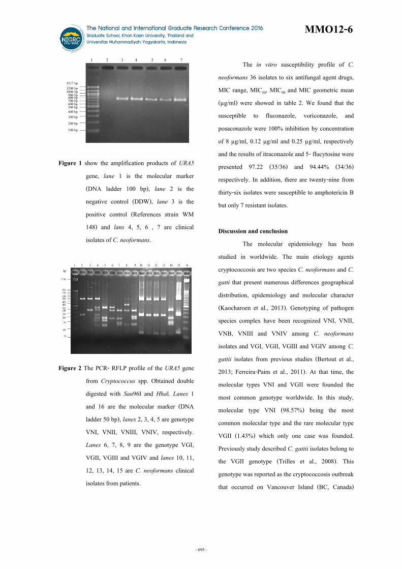
Figure 1 show the amplification products of URA5 gene, lane 1 is the molecular marker (DNA ladder 100 bp), lane 2 is the negative control (DDW), lane 3 is the positive control (References strain WM 148) and lans 4, 5, 6 , 7 are clinical isolates of C. neoformans.
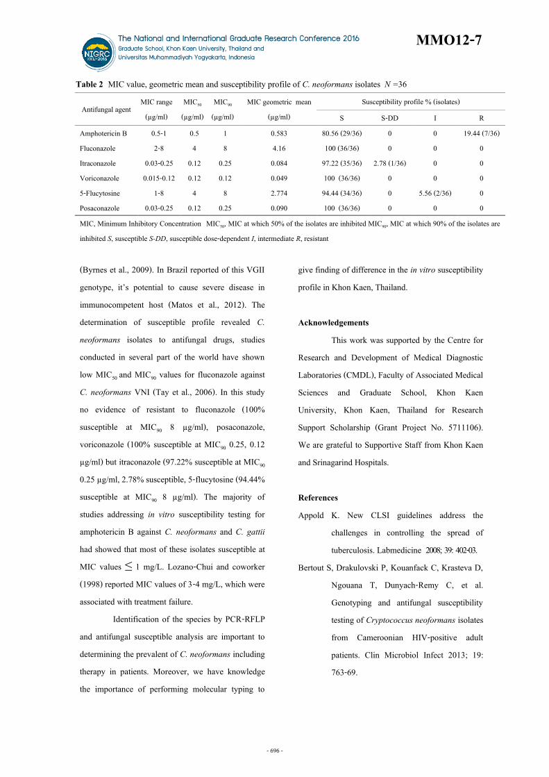
Figure 2 The PCR- RFLP profile of the URA5 gene
from Cryptococcus spp. Obtained double digested with Sau96I and Hhal. Lanes 1 and 16 are the molecular marker (DNA ladder 50 bp), lanes 2, 3, 4, 5 are genotype VNI, VNII, VNIII, VNIV, respectively. Lanes 6, 7, 8, 9 are the genotype VGI, VGII, VGIII and VGIV and lanes 10, 11, 12, 13, 14, 15 are C. neoformans clinical isolates from patients.
The in vitro susceptibility profile of C. neoformans 36 isolates to six antifungal agent drugs, MIC range, MIC50, MIC90 and MIC geometric mean (µg/ml) were showed in table 2. We found that the susceptible to fluconazole, voriconazole, and posaconazole were 100% inhibition by concentration of 8 µg/ml, 0.12 µg/ml and 0.25 µg/ml, respectively and the results of itraconazole and 5- flucytosine were presented 97.22 (35/36) and 94.44% (34/36) respectively. In addition, there are twenty-nine from thirty-six isolates were susceptible to amphotericin B but only 7 resistant isolates. Discussion and conclusion The molecular epidemiology has been studied in worldwide. The main etiology agents cryptococcosis are two species C. neoformans and C. gatti that present numerous differences geographical distribution, epidemiology and molecular character (Kaocharoen et al., 2013). Genotyping of pathogen species complex have been recognized VNI, VNII, VNB, VNIII and VNIV among C. neoformans isolates and VGI, VGII, VGIII and VGIV among C. gattii isolates from previous studies (Bertout et al., 2013; Ferreira-Paim et al., 2011). At that time, the molecular types VNI and VGII were founded the most common genotype worldwide. In this study, molecular type VNI (98.57%) being the most common molecular type and the rare molecular type VGII (1.43%) which only one case was founded. Previously study described C. gattii isolates belong to the VGII genotype (Trilles et al., 2008). This genotype was reported as the cryptococcosis outbreak that occurred on Vancouver Island (BC, Canada)
- 695 -
MMO12-7
(Byrnes et al., 2009). In Brazil reported of this VGII genotype, it’s potential to cause severe disease in immunocompetent host (Matos et al., 2012). The determination of susceptible profile revealed C. neoformans isolates to antifungal drugs, studies conducted in several part of the world have shown low MIC50 and MIC90 values for fluconazole against C. neoformans VNI (Tay et al., 2006). In this study no evidence of resistant to fluconazole (100% susceptible at MIC90 8 µg/ml), posaconazole, voriconazole (100% susceptible at MIC90 0.25, 0.12 µg/ml) but itraconazole (97.22% susceptible at MIC90 0.25 µg/ml, 2.78% susceptible, 5-flucytosine (94.44% susceptible at MIC90 8 µg/ml). The majority of studies addressing in vitro susceptibility testing for amphotericin B against C. neoformans and C. gattii had showed that most of these isolates susceptible at MIC values ≤ 1 mg/L. Lozano-Chui and coworker (1998) reported MIC values of 3-4 mg/L, which were associated with treatment failure. Identification of the species by PCR-RFLP and antifungal susceptible analysis are important to determining the prevalent of C. neoformans including therapy in patients. Moreover, we have knowledge the importance of performing molecular typing to
give finding of difference in the in vitro susceptibility profile in Khon Kaen, Thailand. Acknowledgements This work was supported by the Centre for Research and Development of Medical Diagnostic Laboratories (CMDL), Faculty of Associated Medical Sciences and Graduate School, Khon Kaen University, Khon Kaen, Thailand for Research Support Scholarship (Grant Project No. 5711106). We are grateful to Supportive Staff from Khon Kaen and Srinagarind Hospitals. References Appold K. New CLSI guidelines address the
challenges in controlling the spread of tuberculosis. Labmedicine 2008; 39: 402-03.
Bertout S, Drakulovski P, Kouanfack C, Krasteva D, Ngouana T, Dunyach-Remy C, et al. Genotyping and antifungal susceptibility testing of Cryptococcus neoformans isolates from Cameroonian HIV-positive adult patients. Clin Microbiol Infect 2013; 19: 763-69.
Table 2 MIC value, geometric mean and susceptibility profile of C. neoformans isolates N =36
Antifungal agent MIC range
(µg/ml) MIC50
(µg/ml) MIC90
(µg/ml) MIC geometric mean
(µg/ml) Susceptibility profile % (isolates)
S S-DD I R Amphotericin B 0.5-1 0.5 1 0.583 80.56 (29/36) 0 0 19.44 (7/36) Fluconazole 2-8 4 8 4.16 100 (36/36) 0 0 0 Itraconazole 0.03-0.25 0.12 0.25 0.084 97.22 (35/36) 2.78 (1/36) 0 0 Voriconazole 0.015-0.12 0.12 0.12 0.049 100 (36/36) 0 0 0 5-Flucytosine 1-8 4 8 2.774 94.44 (34/36) 0 5.56 (2/36) 0 Posaconazole 0.03-0.25 0.12 0.25 0.090 100 (36/36) 0 0 0 MIC, Minimum Inhibitory Concentration MIC50, MIC at which 50% of the isolates are inhibited MIC90, MIC at which 90% of the isolates are inhibited S, susceptible S-DD, susceptible dose-dependent I, intermediate R, resistant
- 696 -

MMO12-8
Boekhout T, Theelen B, Diaz M, Fell JW, Hop WC, Abeln EC, et al. Hybrid genotypes in the pathogenic yeast Cryptococcus neoformans. Microbiology 2001; 147: 891-907.
Bovers M, Hagen F, Kuramae EE, Boekhout T. Six monophyletic lineages identified within Cryptococcus neoformans and Cryptococcus gattii by multi-locus sequence typing. Fungal Genet Biol 2008; 45: 400-21.
Byrnes EJ, Bildfell RJ, Frank SA, Mitchell TG, Marr KA, Heitman J. Molecular evidence that the range of the vancouver Island outbreak of Cryptococcus gattii infection has expanded into the Pacific Northwest in the United States. J Infect Dis 2009; 199: 1081-86.
Currie BP, Freundlich LF, Casadevall A. Restriction fragment length polymorphism analysis of Cryptococcus neoformans isolates from environmental (pigeon excreta) and clinical sources in New York City. J Clin Microbiol 1994; 32: 1188-92.
Dromer F, Mathoulin S, Dupont B, Laporte A. Epidemiology of cryptococcosis in France: a 9-year survey (1985-1993). French Cryptococcosis Study Group. Clin Infect Dis 1996; 23: 82-90.
Ferreira-Paim K, Andrade-Silva L, Mora DJ, Pedrosa AL, Rodrigues V, Silva-Vergara ML. Genotyping of Cryptococcus neoformans isolated from captive birds in Uberaba, Minas Gerais, Brazil. Mycoses 2011; 54: 294-300.
Kaocharoen S, Ngamskulrungroj P, Firacative C, Trilles L, Piyabongkarn D, Banlunara W, et al. Molecular epidemiology reveals genetic diversity amongst isolates of the Cryptococcus neoformans/C. gattii species complex in Thailand. PLoS Negl Trop Dis 2013a; 7: e2297.
Latouche GN, Huynh M, Sorrell TC, Meyer W. PCR-restriction fragment length polymorphism analysis of the phospholipase B (PLB1) gene for subtyping of Cryptococcus neoformans isolates. Appl Environ Microbiol 2003; 69: 2080-86.
Manosuthi W, Sungkanuparph S, Thongyen S, Chumpathat N, Eampokalap B, Thawornwan U, et al. Antifungal susceptibilities of Cryptococcus neoformans cerebrospinal fluid isolates and clinical outcomes of cryptococcal meningitis in HIV-infected patients with/without fluconazole prophylaxis. J Med Assoc Thai 2006; 89: 795-802.
Matos CS, Andrade AD, Oliveira NS, Barros TF. Microbiological characteristics of clinical isolates of Cryptococcus spp. in Bahia, Brazil: molecular types and antifungal susceptibilities. Eur J Clin Microbiol Infect Dis 2012; 31: 1647-52.
Meyer W, Aanensen DM, Boekhout T, Cogliati M, Diaz MR, Esposto MC, et al. Consensus multi-locus sequence typing scheme for Cryptococcus neoformans and Cryptococcus gattii. Med Mycol 2009; 47: 61-70.
- 697 -

MMO12-9
Meyer W, Castaneda A, Jackson S, Huynh M, Castaneda E. Molecular typing of IberoAmerican Cryptococcus neoformans isolates. Emerg Infect Dis 2003; 9: 189-95.
Park JY, Shin JH, Uh Y, Kim EC, Kee SJ, Kim SH, et al. In Vitro amphotericin B susceptibility of korean bloodstream yeast isolates assessed by the CLSI broth microdilution method, Etest, and minimum fungicidal concentration test. Korean J Lab Med 2008; 28: 346-52.
Pfaller MA, Diekema DJ, Procop GW, Rinaldi MG. Multicenter comparison of the VITEK 2 yeast susceptibility test with the CLSI broth microdilution reference method for testing fluconazole against Candida spp. J Clin Microbiol 2007; 45: 796-802.
Pfaller MA, Diekema DJ, Procop GW, Wiederhold NP. Multicenter evaluation of the new Vitek 2 yeast susceptibility test using new CLSI clinical breakpoints for fluconazole. J Clin Microbiol 2014; 52: 2126-30.
Ros-Chumillas M, Egea-Cortines M, Lopez-Gomez A,Weiss J. Evaluation of a rapid DNA extraction method to detect yeast cells by PCR in orange juice. Food Control 2007; 18: 33-39.
Sukroongreung S, Nilakul C, Ruangsomboon O, Chuakul W, Eampokalap B. Serotypes of Cryptococcus neoformans isolated from patients prior to and during the AIDS era in Thailand. Mycopathologia 1996; 135: 75-8.
Tay ST, Haryanty TT, Ng KP, Rohani MY, Hamimah H. In vitro susceptibilities of Malaysian clinical isolates of Cryptococcus neoformans var. grubii and Cryptococcus gattii to five antifungal drugs. Mycoses 2006; 49: 324-30.
Trilles L, Lazera MD, Wanke B, Oliveira RV, Barbosa GG, Nishikawa MM, et al. Regional pattern of the molecular types of Cryptococcus neoformans and Cryptococcus gattii in Brazil. Mem Inst Oswaldo Cruz 2008; 103: 455-62.
Yamada Y, Makimura K, Mirhendi H, Ueda K, Nishiyama Y, Yamaguchi H, et al. Comparison of different methods for extraction of mitochondrial DNA from human pathogenic yeasts. Jpn J Infect Dis 2002; 55: 122-5.
- 698 -