Embed Size (px)
Citation preview
CASE REPORT
Pancreaticoduodenectomy for pancreas carcinoma occurringin the annular pancreas: report of a case
Hiromichi Kawaida1 • Hiroshi Kono1 • Mitsuaki Watanabe1 • Akira Maki1 •
Hidetake Amemiya1 • Masanori Matsuda1 • Hideki Fujii1 • Mitsuharu Fukasawa2 •
Ei Takahashi2 • Katsuhiro Sano3 • Tomohiro Inoue4
Received: 30 April 2015 / Accepted: 9 June 2015 / Published online: 8 July 2015
� The Author(s) 2015. This article is published with open access at Springerlink.com
Abstract The annular pancreas is a rare congenital
anomaly in which a ring of the pancreas parenchyma sur-
rounds the second part of the duodenum. Malignant tumors
are extremely rare in patients with an annular pancreas. A
64-year-old man presented with appetite loss and vomiting.
Abdominal contrast-enhanced computed tomography (CT)
indicated pancreas parenchyma surrounding the second
part of the duodenum, and a hypovascular area occupying
lesion in the annular pancreas. Subtotal stomach-preserving
pancreaticoduodenectomy was performed. Histopathology
showed pancreatic carcinoma occurring in the complete
annular pancreas.
Keywords Annular pancreas � Pancreas carcinoma �Pancreaticoduodenectomy
Introduction
An annular pancreas is a rare congenital anomaly in which
the second part of the duodenum is surrounded by a ring of
pancreas parenchyma. It was first described by Tiedemann
[1] in 1818 and was termed ‘‘annular pancreas’’ by Ecker in
1862 [2]. In adults, annular pancreas has been known to be
associated with peptic ulceration, duodenal obstruction,
pancreatitis, and obstructive jaundice. The presence of the
annular pancreas was reported in three of 20,000 autopsies
[3] and three of 24,519 surgical cases [4]. Malignant
tumors are extremely rare in patients with an annular
pancreas (Table 1). Herein, we report a case of pancreas
carcinoma occurring in the annular pancreas.
Clinical summary
A 64-year-old man was admitted to University of Yama-
nashi Hospital for complaint of abdominal pain and vom-
iting. On admission, physical examination revealed no
anemia and jaundice in the conjunctiva, and no tenderness
and no mass in the abdomen. Laboratory findings showed
elevated alkaline phosphatases, 898 U/l (100–310), aspar-
tate aminotransferase, 67 U/l (\30 U/l), alanine amino-
transferase, 144 U/l (\35U/l) and hemoglobin A1c, 7.7 %
(\6.2 %). Furthermore, carbohydrate antigen 19-9 was
elevated to 158 U/ml (\37 U/ml); however, carcinoem-
bryonic antigen was within the normal range.
Upper gastrointestinal radiology and esophagogastro-
duodenoscopy showed stenosis at the descending part of
the duodenum. Endoscopic ultrasonography showed a
hypoechogenic mass measuring 21 9 13 mm in the pan-
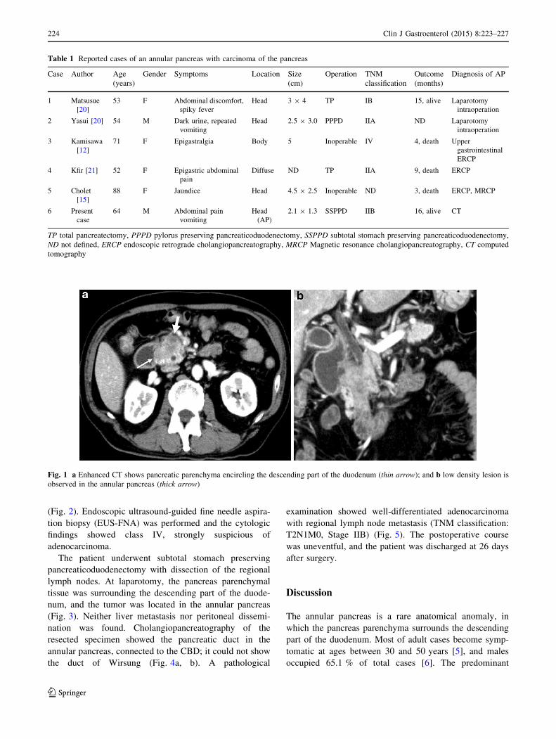
creas head. In addition, abdominal enhanced-CT revealed
the pancreatic parenchyma encircling the descending part
of the duodenum, and a hypovascular mass lesion was
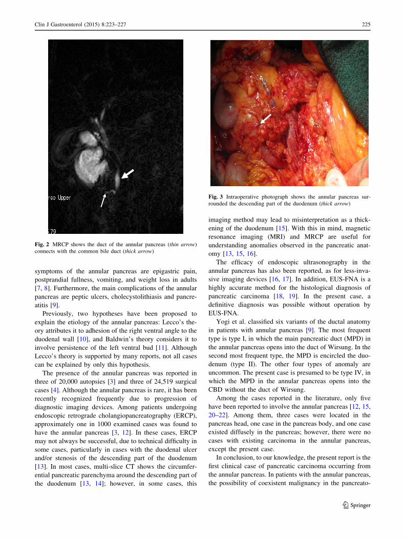
observed in the annular pancreas (Fig. 1a, b). Magnetic
resonance cholangiopancreatography (MRCP) showed the
pancreatic duct in the annular pancreas encircling the
duodenum connected with the common bile duct (CBD)
& Hiromichi Kawaida
1 First Department of Surgery, Faculty of Medicine, University
of Yamanashi, 1110 Shimokato, Chuo, Yamanashi 409-3898,
Japan
2 First Department of Internal Medicine, Faculty of Medicine,
University of Yamanashi, Chuo, Japan
3 Department of Radiology, Faculty of Medicine, University of
Yamanashi, Chuo, Japan
4 Department of Human Pathology, Faculty of Medicine,
University of Yamanashi, Chuo, Japan
123
Clin J Gastroenterol (2015) 8:223–227
DOI 10.1007/s12328-015-0579-6
(Fig. 2). Endoscopic ultrasound-guided fine needle aspira-
tion biopsy (EUS-FNA) was performed and the cytologic
findings showed class IV, strongly suspicious of
adenocarcinoma.
The patient underwent subtotal stomach preserving
pancreaticoduodenectomy with dissection of the regional
lymph nodes. At laparotomy, the pancreas parenchymal
tissue was surrounding the descending part of the duode-
num, and the tumor was located in the annular pancreas
(Fig. 3). Neither liver metastasis nor peritoneal dissemi-
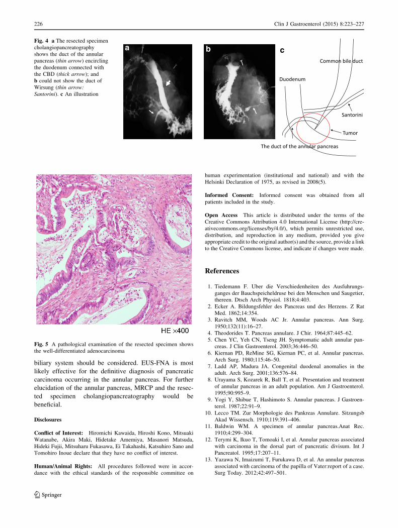
nation was found. Cholangiopancreatography of the
resected specimen showed the pancreatic duct in the
annular pancreas, connected to the CBD; it could not show
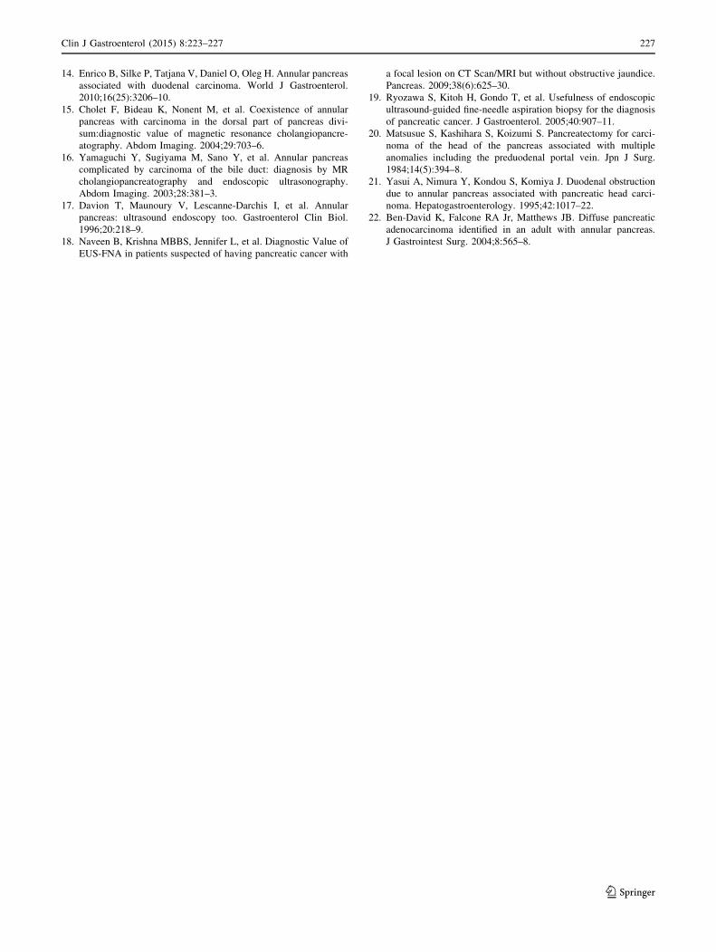
the duct of Wirsung (Fig. 4a, b). A pathological
examination showed well-differentiated adenocarcinoma
with regional lymph node metastasis (TNM classification:
T2N1M0, Stage IIB) (Fig. 5). The postoperative course
was uneventful, and the patient was discharged at 26 days
after surgery.
Discussion
The annular pancreas is a rare anatomical anomaly, in
which the pancreas parenchyma surrounds the descending
part of the duodenum. Most of adult cases become symp-
tomatic at ages between 30 and 50 years [5], and males
occupied 65.1 % of total cases [6]. The predominant
Table 1 Reported cases of an annular pancreas with carcinoma of the pancreas
Case Author Age
(years)
Gender Symptoms Location Size
(cm)
Operation TNM
classification
Outcome
(months)
Diagnosis of AP
1 Matsusue
[20]
53 F Abdominal discomfort,
spiky fever
Head 3 9 4 TP IB 15, alive Laparotomy
intraoperation
2 Yasui [20] 54 M Dark urine, repeated
vomiting
Head 2.5 9 3.0 PPPD IIA ND Laparotomy
intraoperation
3 Kamisawa
[12]
71 F Epigastralgia Body 5 Inoperable IV 4, death Upper
gastrointestinal
ERCP
4 Kfir [21] 52 F Epigastric abdominal
pain
Diffuse ND TP IIA 9, death ERCP
5 Cholet
[15]
88 F Jaundice Head 4.5 9 2.5 Inoperable ND 3, death ERCP, MRCP
6 Present
case
64 M Abdominal pain
vomiting
Head
(AP)
2.1 9 1.3 SSPPD IIB 16, alive CT
TP total pancreatectomy, PPPD pylorus preserving pancreaticoduodenectomy, SSPPD subtotal stomach preserving pancreaticoduodenectomy,
ND not defined, ERCP endoscopic retrograde cholangiopancreatography, MRCP Magnetic resonance cholangiopancreatography, CT computed
tomography
Fig. 1 a Enhanced CT shows pancreatic parenchyma encircling the descending part of the duodenum (thin arrow); and b low density lesion is
observed in the annular pancreas (thick arrow)
224 Clin J Gastroenterol (2015) 8:223–227
123
symptoms of the annular pancreas are epigastric pain,
postprandial fullness, vomiting, and weight loss in adults
[7, 8]. Furthermore, the main complications of the annular
pancreas are peptic ulcers, cholecystolithiasis and pancre-
atitis [9].
Previously, two hypotheses have been proposed to
explain the etiology of the annular pancreas: Lecco’s the-
ory attributes it to adhesion of the right ventral angle to the
duodenal wall [10], and Baldwin’s theory considers it to
involve persistence of the left ventral bud [11]. Although
Lecco’s theory is supported by many reports, not all cases
can be explained by only this hypothesis.
The presence of the annular pancreas was reported in
three of 20,000 autopsies [3] and three of 24,519 surgical
cases [4]. Although the annular pancreas is rare, it has been
recently recognized frequently due to progression of
diagnostic imaging devices. Among patients undergoing
endoscopic retrograde cholangiopancreatography (ERCP),
approximately one in 1000 examined cases was found to
have the annular pancreas [3, 12]. In these cases, ERCP
may not always be successful, due to technical difficulty in
some cases, particularly in cases with the duodenal ulcer
and/or stenosis of the descending part of the duodenum
[13]. In most cases, multi-slice CT shows the circumfer-
ential pancreatic parenchyma around the descending part of
the duodenum [13, 14]; however, in some cases, this
imaging method may lead to misinterpretation as a thick-
ening of the duodenum [15]. With this in mind, magnetic
resonance imaging (MRI) and MRCP are useful for
understanding anomalies observed in the pancreatic anat-
omy [13, 15, 16].
The efficacy of endoscopic ultrasonography in the
annular pancreas has also been reported, as for less-inva-
sive imaging devices [16, 17]. In addition, EUS-FNA is a
highly accurate method for the histological diagnosis of
pancreatic carcinoma [18, 19]. In the present case, a
definitive diagnosis was possible without operation by
EUS-FNA.
Yogi et al. classified six variants of the ductal anatomy
in patients with annular pancreas [9]. The most frequent
type is type I, in which the main pancreatic duct (MPD) in
the annular pancreas opens into the duct of Wirsung. In the
second most frequent type, the MPD is encircled the duo-
denum (type II). The other four types of anomaly are
uncommon. The present case is presumed to be type IV, in
which the MPD in the annular pancreas opens into the
CBD without the duct of Wirsung.
Among the cases reported in the literature, only five
have been reported to involve the annular pancreas [12, 15,
20–22]. Among them, three cases were located in the
pancreas head, one case in the pancreas body, and one case
existed diffusely in the pancreas; however, there were no
cases with existing carcinoma in the annular pancreas,
except the present case.
In conclusion, to our knowledge, the present report is the
first clinical case of pancreatic carcinoma occurring from
the annular pancreas. In patients with the annular pancreas,
the possibility of coexistent malignancy in the pancreato-
Fig. 2 MRCP shows the duct of the annular pancreas (thin arrow)
connects with the common bile duct (thick arrow)
Fig. 3 Intraoperative photograph shows the annular pancreas sur-
rounded the descending part of the duodenum (thick arrow)
Clin J Gastroenterol (2015) 8:223–227 225
123
biliary system should be considered. EUS-FNA is most
likely effective for the definitive diagnosis of pancreatic
carcinoma occurring in the annular pancreas. For further
elucidation of the annular pancreas, MRCP and the resec-
ted specimen cholangiopancreatography would be
beneficial.
Disclosures
Conflict of Interest: Hiromichi Kawaida, Hiroshi Kono, Mitsuaki
Watanabe, Akira Maki, Hidetake Amemiya, Masanori Matsuda,
Hideki Fujii, Mitsuharu Fukasawa, Ei Takahashi, Katsuhiro Sano and
Tomohiro Inoue declare that they have no conflict of interest.
Human/Animal Rights: All procedures followed were in accor-
dance with the ethical standards of the responsible committee on
human experimentation (institutional and national) and with the
Helsinki Declaration of 1975, as revised in 2008(5).
Informed Consent: Informed consent was obtained from all
patients included in the study.
Open Access This article is distributed under the terms of the
Creative Commons Attribution 4.0 International License (http://cre-
ativecommons.org/licenses/by/4.0/), which permits unrestricted use,
distribution, and reproduction in any medium, provided you give
appropriate credit to the original author(s) and the source, provide a link
to the Creative Commons license, and indicate if changes were made.
References
1. Tiedemann F. Uber die Verschiedenheiten des Ausfuhrungs-
ganges der Bauchspeicheldruse bei den Menschen und Saugetier,
thereen. Dtsch Arch Physiol. 1818;4:403.
2. Ecker A. Bildungsfehler des Pancreas und des Herzens. Z Rat
Med. 1862;14:354.
3. Ravitch MM, Woods AC Jr. Annular pancreas. Ann Surg.
1950;132(11):16–27.
4. Theodorides T. Pancreas annulare. J Chir. 1964;87:445–62.
5. Chen YC, Yeh CN, Tseng JH. Symptomatic adult annular pan-
creas. J Clin Gastroenterol. 2003;36:446–50.
6. Kiernan PD, ReMine SG, Kiernan PC, et al. Annular pancreas.
Arch Surg. 1980;115:46–50.
7. Ladd AP, Madura JA. Congenital duodenal anomalies in the
adult. Arch Surg. 2001;136:576–84.
8. Urayama S, Kozarek R, Ball T, et al. Presentation and treatment
of annular pancreas in an adult population. Am J Gastroenterol.
1995;90:995–9.
9. Yogi Y, Shibue T, Hashimoto S. Annular pancreas. J Gastroen-
terol. 1987;22:91–9.
10. Lecco TM. Zur Morphologie des Pankreas Annulare. Sitzungsb
Akad Wissensch. 1910;119:391–406.
11. Baldwin WM. A specimen of annular pancreas.Anat Rec.
1910;4:299–304.
12. Terymi K, Ikuo T, Tomoaki I, et al. Annular pancreas associated
with carcinoma in the dorsal part of pancreatic divisum. Int J
Pancreatol. 1995;17:207–11.
13. Yazawa N, Imaizumi T, Furukawa D, et al. An annular pancreas
associated with carcinoma of the papilla of Vater:report of a case.
Surg Today. 2012;42:497–501.
a b
Santorini
The duct of the annular pancreas
Common bile duct
Duodenum
Tumor
c Fig. 4 a The resected specimen
cholangiopancreatography
shows the duct of the annular
pancreas (thin arrow) encircling
the duodenum connected with
the CBD (thick arrow); and
b could not show the duct of
Wirsung (thin arrow:
Santorini). c An illustration
Fig. 5 A pathological examination of the resected specimen shows
the well-differentiated adenocarcinoma
226 Clin J Gastroenterol (2015) 8:223–227
123
14. Enrico B, Silke P, Tatjana V, Daniel O, Oleg H. Annular pancreas
associated with duodenal carcinoma. World J Gastroenterol.
2010;16(25):3206–10.
15. Cholet F, Bideau K, Nonent M, et al. Coexistence of annular
pancreas with carcinoma in the dorsal part of pancreas divi-
sum:diagnostic value of magnetic resonance cholangiopancre-
atography. Abdom Imaging. 2004;29:703–6.
16. Yamaguchi Y, Sugiyama M, Sano Y, et al. Annular pancreas
complicated by carcinoma of the bile duct: diagnosis by MR
cholangiopancreatography and endoscopic ultrasonography.
Abdom Imaging. 2003;28:381–3.
17. Davion T, Maunoury V, Lescanne-Darchis I, et al. Annular
pancreas: ultrasound endoscopy too. Gastroenterol Clin Biol.
1996;20:218–9.
18. Naveen B, Krishna MBBS, Jennifer L, et al. Diagnostic Value of
EUS-FNA in patients suspected of having pancreatic cancer with
a focal lesion on CT Scan/MRI but without obstructive jaundice.
Pancreas. 2009;38(6):625–30.
19. Ryozawa S, Kitoh H, Gondo T, et al. Usefulness of endoscopic
ultrasound-guided fine-needle aspiration biopsy for the diagnosis
of pancreatic cancer. J Gastroenterol. 2005;40:907–11.
20. Matsusue S, Kashihara S, Koizumi S. Pancreatectomy for carci-
noma of the head of the pancreas associated with multiple
anomalies including the preduodenal portal vein. Jpn J Surg.
1984;14(5):394–8.
21. Yasui A, Nimura Y, Kondou S, Komiya J. Duodenal obstruction
due to annular pancreas associated with pancreatic head carci-
noma. Hepatogastroenterology. 1995;42:1017–22.
22. Ben-David K, Falcone RA Jr, Matthews JB. Diffuse pancreatic
adenocarcinoma identified in an adult with annular pancreas.
J Gastrointest Surg. 2004;8:565–8.
Clin J Gastroenterol (2015) 8:223–227 227
123