Embed Size (px)
Citation preview
PowerLecture:PowerLecture:Chapter 5Chapter 5
The Skeletal SystemThe Skeletal System

Learning ObjectivesLearning Objectives
List the functions of bone.List the functions of bone. Identify human bones by name and Identify human bones by name and
location.location. Identify the locations and types of joints.Identify the locations and types of joints. Characterize several common disorders Characterize several common disorders
associated with the skeletal system. associated with the skeletal system.
Impacts/IssuesImpacts/Issues
Creaky JointsCreaky Joints

Creaky JointsCreaky Joints
Many people currently suffer, or may one Many people currently suffer, or may one day suffer, from osteoarthritis, in which the day suffer, from osteoarthritis, in which the joints between bones become stiff and joints between bones become stiff and painful due to the degeneration of the painful due to the degeneration of the cartilage lining.cartilage lining.
Conventional remedies range from pain Conventional remedies range from pain relievers to supplements to injections of relievers to supplements to injections of steroids.steroids.
More unconventional treatments employ More unconventional treatments employ combinations of various botanical extracts.combinations of various botanical extracts.

Creaky JointsCreaky Joints
Arthritis is a disorder of the Arthritis is a disorder of the skeletal systemskeletal system, the , the framework of bone, cartilage, framework of bone, cartilage, and ligaments on which the and ligaments on which the body is built. body is built.

How Would You Vote?How Would You Vote?To conduct an instant in-class survey using a classroom response To conduct an instant in-class survey using a classroom response system, access “JoinIn Clicker Content” from the PowerLecture main system, access “JoinIn Clicker Content” from the PowerLecture main menu. menu.
Should claims about “medicinal” exotic plant Should claims about “medicinal” exotic plant extracts have to be backed up by extracts have to be backed up by independent scientific testing?independent scientific testing? a. Yes, or companies could make any claim a. Yes, or companies could make any claim
about their product.about their product. b. No, so long as the extract does no harm, let b. No, so long as the extract does no harm, let
the buyer decide what to buy.the buyer decide what to buy.

Section 1Section 1
Bone—Mineralized Bone—Mineralized Connective TissueConnective Tissue

Bone – Mineralized Connective TissueBone – Mineralized Connective Tissue
BoneBone is a connective tissue with living cells is a connective tissue with living cells ((osteocytesosteocytes) and collagen fibers distributed ) and collagen fibers distributed through out a ground substance that is through out a ground substance that is hardened by calcium salts.hardened by calcium salts.
As bone develops, precursor cells called As bone develops, precursor cells called osteoblastsosteoblasts secrete collagen fibers and a secrete collagen fibers and a ground substance of proteins and ground substance of proteins and carbohydrates.carbohydrates.
Eventually, osteocytes reside within Eventually, osteocytes reside within lacunaelacunae in in the ground substance, which becomes the ground substance, which becomes mineralized by calcium deposits.mineralized by calcium deposits.

Bone – Mineralized Connective TissueBone – Mineralized Connective Tissue
Bones are surrounded by a sturdy membrane Bones are surrounded by a sturdy membrane called the called the periosteumperiosteum. .
There are two kinds of bone tissue.There are two kinds of bone tissue. Compact boneCompact bone tissue forms the bone’s shaft tissue forms the bone’s shaft
and the outer portion of its two ends.and the outer portion of its two ends.• Compact bone forms in thin, circular layers (Compact bone forms in thin, circular layers (osteonsosteons
or or Haversian systemsHaversian systems) with small canals at their ) with small canals at their centers, which contain blood vessels and nerves.centers, which contain blood vessels and nerves.
• Osteocytes in the lacunae communicate by way of Osteocytes in the lacunae communicate by way of canaliculi (little canals).canaliculi (little canals).
Spongy boneSpongy bone tissue is located inside the shaft tissue is located inside the shaft of long bones. of long bones.

Fig. 5.1, p. 88
blood vessel
space occupiedby living bone cell
osteon(Haversian system)
spongy bone tissue
compact bone tissue
outer layer of denseconnective tissueblood vessel
compact bone tissue
spongy bone tissue

Animation: Structure of Animation: Structure of the Human Thigh Bonethe Human Thigh Bone
CLICKTO PLAY

Bone – Mineralized Connective TissueBone – Mineralized Connective Tissue
A bone develops on a cartilage model.A bone develops on a cartilage model. Osteoblasts secrete material inside the shaft of Osteoblasts secrete material inside the shaft of
the cartilage model of long bones.the cartilage model of long bones. Calcium is deposited; cavities merge to form Calcium is deposited; cavities merge to form
the marrow cavity.the marrow cavity. Eventually osteoblasts become trapped within Eventually osteoblasts become trapped within
their own secretions and become osteocytes their own secretions and become osteocytes (mature bone cells).(mature bone cells).

In growing children, the In growing children, the epiphysesepiphyses (ends of (ends of bone) are separated from the shaft by an bone) are separated from the shaft by an epiphyseal plateepiphyseal plate (cartilage), which continues (cartilage), which continues to grow under the influence of growth hormone to grow under the influence of growth hormone until late adolescence.until late adolescence.
Bone – Mineralized Connective TissueBone – Mineralized Connective Tissue

Fig. 5.2, p. 89
Mature bone of adult
Remodeling and growth continue in newborn; secondary bone-forming centers appear at knobby ends of bone
When organs form in embryo, blood vessel invades model; osteoblasts start producing bone tissue; marrow cavity forms
Cartilage model of future bone in embryo
Forming bone collar
epiphysesStepped Art

Animation: How a Long Bone FormsAnimation: How a Long Bone Forms
CLICKTO PLAY

Bone – Mineralized Connective TissueBone – Mineralized Connective Tissue
Bone tissue is constantly “remodeled.”Bone tissue is constantly “remodeled.” Bone is renewed constantly as minerals are Bone is renewed constantly as minerals are
deposited by osteoblasts and withdrawn by deposited by osteoblasts and withdrawn by osteoclastsosteoclasts during the during the bone remodelingbone remodeling process.process.
• Before adulthood, bone turnover is especially Before adulthood, bone turnover is especially important in increasing the diameter of certain important in increasing the diameter of certain bones.bones.
• Bone turnover helps to maintain calcium levels for Bone turnover helps to maintain calcium levels for the entire body.the entire body.

Bone – Mineralized Connective TissueBone – Mineralized Connective Tissue
• A hormone called PTH causes bone cells to release A hormone called PTH causes bone cells to release enzymes that will dissolve bone tissue and release enzymes that will dissolve bone tissue and release calcium to the interstitial fluid and blood; calcitonin calcium to the interstitial fluid and blood; calcitonin stimulates the reverse.stimulates the reverse.
OsteoporosisOsteoporosis (decreased bone density) is (decreased bone density) is associated with decreases in osteoblast activity, associated with decreases in osteoblast activity, sex hormone production, exercise, and calcium sex hormone production, exercise, and calcium uptake.uptake.

Fig. 5.3, p. 89
a b

Video: Taller and TallerVideo: Taller and Taller
From ABC News, Human Biology in the Headlines, 2006 DVD.From ABC News, Human Biology in the Headlines, 2006 DVD.
CLICKTO PLAY

Section 2Section 2
The Skeleton: The The Skeleton: The Body’s Bony FrameworkBody’s Bony Framework

Bones are the main components of the Bones are the main components of the human skeletal system.human skeletal system.
There are four types of bones: long (arms), There are four types of bones: long (arms), short (ankle), flat (skull), and irregular short (ankle), flat (skull), and irregular (vertebrae).(vertebrae).
Bone marrowBone marrow fills the cavities of bones. fills the cavities of bones. • In long bones, In long bones, red marrowred marrow is confined to the ends; is confined to the ends;
yellow marrowyellow marrow fills the shaft portion. fills the shaft portion.• Irregular bones and flat bones are completely filled Irregular bones and flat bones are completely filled
with the red bone marrow responsible for blood cell with the red bone marrow responsible for blood cell formation.formation.
The Skeleton: The Skeleton: The Body’s Bony FrameworkThe Body’s Bony Framework
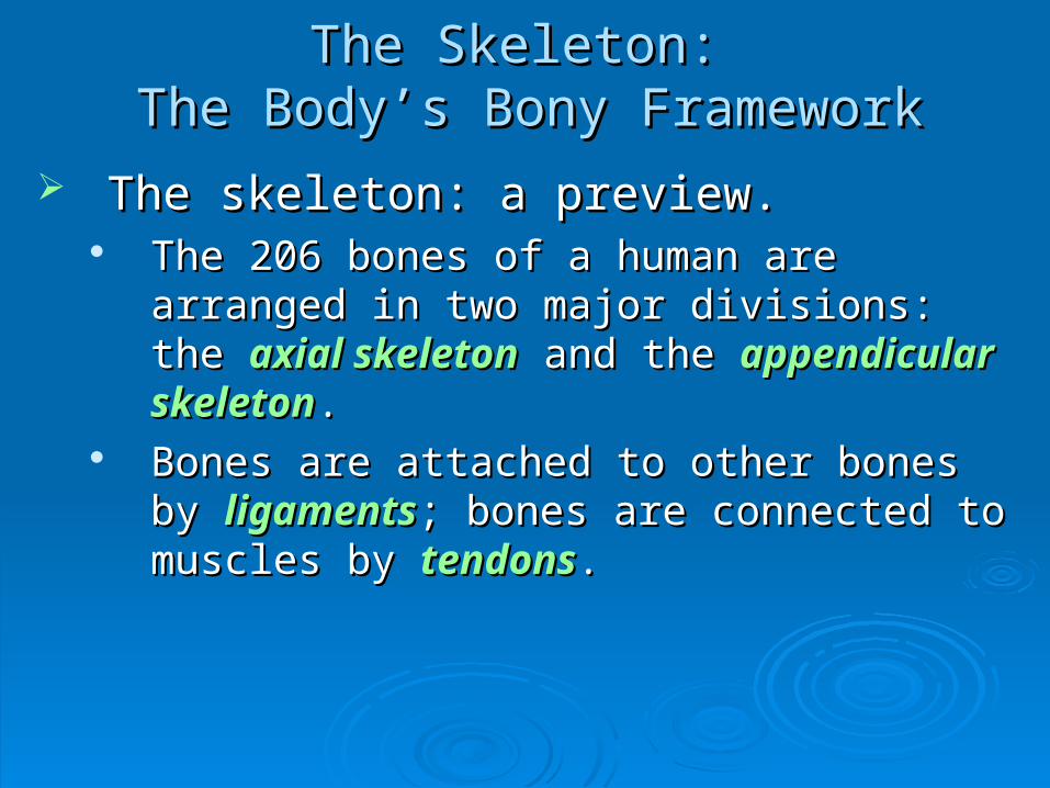
The skeleton: a preview.The skeleton: a preview. The 206 bones of a human are arranged in two The 206 bones of a human are arranged in two
major divisions: the major divisions: the axial skeletonaxial skeleton and the and the appendicular skeletonappendicular skeleton..
Bones are attached to other bones by Bones are attached to other bones by ligamentsligaments; bones are connected to muscles by ; bones are connected to muscles by tendonstendons..
The Skeleton: The Skeleton: The Body’s Bony FrameworkThe Body’s Bony Framework

Bone functions are vital in maintaining Bone functions are vital in maintaining homeostasis.homeostasis.
The bones are moved by muscles; thus the The bones are moved by muscles; thus the whole body is movable.whole body is movable.
The bones support and anchor muscles.The bones support and anchor muscles. Bones protect vital organs such as the brain Bones protect vital organs such as the brain
and lungs.and lungs. Bone tissue acts as a depository for calcium, Bone tissue acts as a depository for calcium,
phosphorus, and other ions.phosphorus, and other ions. Parts of some bones are sites of blood cell Parts of some bones are sites of blood cell
production.production.
The Skeleton: The Skeleton: The Body’s Bony FrameworkThe Body’s Bony Framework

Table 5.1, p. 90

Section 3Section 3
The Axial SkeletonThe Axial Skeleton

The Axial SkeletonThe Axial Skeleton
The skull protects the brain.The skull protects the brain. The The skullskull consists of more than two dozen consists of more than two dozen
bones.bones. The cranial vault, or The cranial vault, or brain casebrain case, is a grouping , is a grouping
of eight bones.of eight bones.• The The frontal bonefrontal bone makes up the forehead and makes up the forehead and
contains the contains the sinusessinuses..• Temporal bonesTemporal bones form the lower sides of the form the lower sides of the
cranium and surround the ear canals.cranium and surround the ear canals.• A A sphenoid bonesphenoid bone and an and an ethmoid boneethmoid bone form the form the
eye socket.eye socket.

frontal sinus
sphenoid sinus
ethmoid sinus
maxillary sinus
Fig. 5.6c, p. 93

The Axial SkeletonThe Axial Skeleton
• Parietal bonesParietal bones form a large part of the skull above form a large part of the skull above the temporal bones.the temporal bones.
• An An occipital boneoccipital bone forms the back of the skull and forms the back of the skull and encloses the encloses the foramen magnumforamen magnum, which is a , which is a passageway for the spinal cord.passageway for the spinal cord.

parietal bonefrontalbone
temporalbone
occipital bone
sphenoid bone
external auditory meatus (opening of the ear; part of the temporal bone)
ethmoid bone
lacrimal bone
zygomatic bone
maxilla
mandible
Fig. 5.6a, p. 92

The Axial SkeletonThe Axial Skeleton
Facial bones support and shape the face.Facial bones support and shape the face. A A mandiblemandible forms the lower jaw; two forms the lower jaw; two maxillarymaxillary
bonesbones form the upper jaw. form the upper jaw. Zygomatic bonesZygomatic bones form the cheekbones; form the cheekbones;
lacrimal boneslacrimal bones form the inner eye sockets. form the inner eye sockets. Palatine bonesPalatine bones make up make up
the nasal cavity; a the nasal cavity; a vomer vomer
bonebone forms the nasal forms the nasal
septum.septum.

hard palate
maxilla
palatine bone
vomer
temporal bone
parietal bone occipital bone
foramen magnum
jugular foramen
sphenoid bone
zygomatic bone
maxilla
Fig. 5.6b, p. 92

The Axial SkeletonThe Axial Skeleton
The vertebral column is the backbone.The vertebral column is the backbone. The The vertebral columnvertebral column, or backbone, extends , or backbone, extends
from the base of the skull to the hipbones.from the base of the skull to the hipbones. The spinal cord extends through a cavity The spinal cord extends through a cavity
formed by the formed by the vertebraevertebrae.. Humans have 33 vertebrae: 7 Humans have 33 vertebrae: 7 cervicalcervical, 12 , 12
thoracicthoracic, and 5 , and 5 lumbarlumbar, plus a , plus a sacrumsacrum formed formed of 5 fused vertebrae and a of 5 fused vertebrae and a coccyxcoccyx of 4 fused of 4 fused vertebrae.vertebrae.
Fibrocartilaginous Fibrocartilaginous intervertebral disksintervertebral disks serve serve as shock absorbers; they may slip (herniate) or as shock absorbers; they may slip (herniate) or rupture, leading to pain and immobility.rupture, leading to pain and immobility.

The Vertebral The Vertebral ColumnColumn
Figure 5.7Figure 5.7

intervertebraldisks
cervical vertebrae (7)
thoracic vertebrae (12)
lumbar vertebrae (5)
sacrum (5 fused)
coccyx (4 fused)Fig. 5.7, p. 93
1
32
4
567
1
2
3
4
5
6
7
8
9
10
11
12
1
2
3
4
5

The Axial SkeletonThe Axial Skeleton
The ribs and sternum support and help The ribs and sternum support and help protect internal organs.protect internal organs.
RibsRibs (12 pairs) are attached to the vertebrae (12 pairs) are attached to the vertebrae dorsally and serve as scaffolding for the upper dorsally and serve as scaffolding for the upper body torso.body torso.
Most of the ribs are attached to the Most of the ribs are attached to the sternumsternum ventrally.ventrally.
Animation: Axial SkeletonAnimation: Axial Skeleton
CLICKTO PLAY

Video: Painful PainkillersVideo: Painful Painkillers
From ABC News, Human Biology in the Headlines, 2006 DVD.From ABC News, Human Biology in the Headlines, 2006 DVD.
CLICKTO PLAY

Section 4Section 4
The Appendicular The Appendicular SkeletonSkeleton

The Appendicular SkeletonThe Appendicular Skeleton
The pectoral girdle and upper limbs provide The pectoral girdle and upper limbs provide flexibility.flexibility.
The The pectoral girdlepectoral girdle includes the bones of, and includes the bones of, and is attached to, the shoulder.is attached to, the shoulder.
• The The scapulascapula is a large, flat shoulder blade with a is a large, flat shoulder blade with a socket for the upper arm bone.socket for the upper arm bone.
• The The clavicleclavicle (collarbone) connects the scapula to (collarbone) connects the scapula to the sternum.the sternum.

The Appendicular SkeletonThe Appendicular Skeleton
Each upper limb includes some 30 separate Each upper limb includes some 30 separate bones.bones.
• The The humerushumerus is the bone of the upper arm. is the bone of the upper arm.• The The radiusradius and and ulnaulna extend from the hingelike joint extend from the hingelike joint
of the elbow to the wrist.of the elbow to the wrist.• The The carpalscarpals form the wrist; the form the wrist; the metacarpalsmetacarpals form form
the palm of the hand, and the the palm of the hand, and the phalangesphalanges the fingers. the fingers.

Fig. 5.8, p. 94
clavicle
humerus
ulna
radius
scapulasternum
carpals (8)metacarpals (5)phalanges (14)

The Appendicular SkeletonThe Appendicular Skeleton
The pelvic girdle and lower limbs support The pelvic girdle and lower limbs support body weight.body weight. The The pelvic girdlepelvic girdle includes the includes the pelvispelvis and the and the
legs.legs.• The pelvis is made up of coxal bones attaching to the The pelvis is made up of coxal bones attaching to the
sacrum in the back and forming the sacrum in the back and forming the pelvic archpelvic arch in the in the front.front.
• The pelvis is broader in females than males; this is The pelvis is broader in females than males; this is necessary for childbearing.necessary for childbearing.

The Appendicular SkeletonThe Appendicular Skeleton
The legs contain the body’s largest bones.The legs contain the body’s largest bones.• The The femurfemur is the longest bone, extending from the is the longest bone, extending from the
pelvis to the knee.pelvis to the knee.• The The tibiatibia and and fibulafibula form form
the lower leg; the kneecap the lower leg; the kneecap
bone is the bone is the patellapatella..
• TarsalTarsal bones compose the ankle, bones compose the ankle, metatarsalsmetatarsals the the foot, and phalanges the toes.foot, and phalanges the toes.

Fig. 5.4, p. 90
marrow cavity
compact bone tissue
spongy bonetissue
nutrient canal into and from marrow (for blood vessels and nerves)

Fig. 5.9, p. 95
pelvis
fibula
sacrum
femur
phalanges
patella
metatarsals
tarsals
tibia
pubic symphysis

Animation: Appendicular SkeletonAnimation: Appendicular Skeleton
CLICKTO PLAY

Skull bonescranial bones
facial bones
sternumRib cage
ribs
Vertebral column (backbone)vertebrae
intervertebral disks
Pectoral girdle and upperlimb bones
clavicle
scapula
humerusradius
ulna
carpalsmetacarpals
phalanges
pelvic girdlefemurpatella
tibiafibula
tarsals
phalangesmetatarsals
Pelvic girdle and lowerlimb bones
Fig. 5.5, p. 91
AXIAL SKELETON APPENDICULAR
SKELETON
ligament bridging a knee joint, here sliced down through the middle, side view.

Animation: The Human Skeleton SystemAnimation: The Human Skeleton System
CLICKTO PLAY

Section 5Section 5
Joints—Connections Joints—Connections Between BonesBetween Bones

Joints – Connections Between BonesJoints – Connections Between Bones
Synovial joints move freely.Synovial joints move freely. Synovial jointsSynovial joints are the most common type of are the most common type of
joint and move freely; they include the ball-and-joint and move freely; they include the ball-and-socket joints of the hips and the hingelike joints socket joints of the hips and the hingelike joints such as the knee.such as the knee.
These types of joints are stabilized by These types of joints are stabilized by ligaments.ligaments.
A capsule of dense connective tissue surrounds A capsule of dense connective tissue surrounds the bones of the joint and produces synovial the bones of the joint and produces synovial fluid that lubricates the joint.fluid that lubricates the joint.

Fig. 5.10a, p. 96
ligament
ligament (cut)
ligament
meniscus anterior cruciate ligament
posterior cruciate ligament
femur
fibulatibia

Fig. 5.10b, p. 96
quadriceps (straightens leg)
biceps femoris (bends leg)
femur
cartilage
ligament
fibula tibia
ligament (to knee cap)
knee cap (patella)
tendon (to thigh muscle)

© 2007 Thomson Higher Education
flexion at shoulder
extension at shoulder
flexion at knee
extension at knee
Fig. 5.11a (1), p. 97
© 2007 Thomson Higher Education
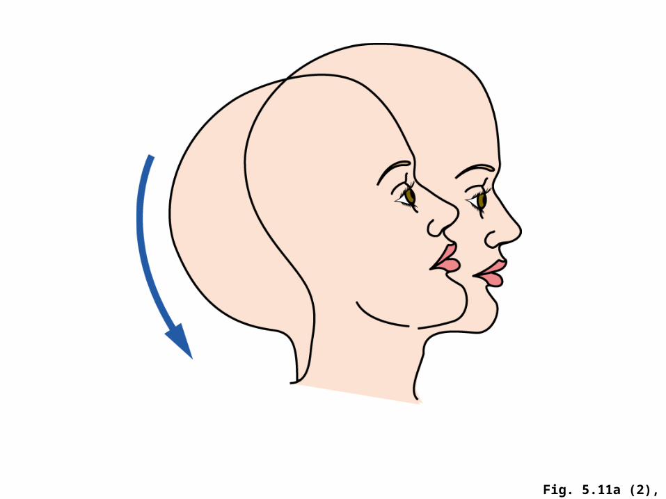
Fig. 5.11a (2), p. 97
hyperextension

© 2007 Thomson Higher Education © 2007 Thomson Higher Education
Fig. 5.11b, p. 97
circumduction rotation

© 2007 Thomson Higher Education
Fig. 5.11c, p. 97
abduction
abduction abduction
adduction
adduction
adduction

© 2007 Thomson Higher EducationFig. 5.11d, p. 97
supination
pronation

© 2007 Thomson Higher Education
gliding movement between carpals
Fig. 5.11e, p. 97

Joints – Connections Between BonesJoints – Connections Between Bones
Other joints move little or not at all.Other joints move little or not at all. Cartilaginous jointsCartilaginous joints (such as between the (such as between the
vertebrae) have no gap, but are held together vertebrae) have no gap, but are held together by cartilage and can move only a little.by cartilage and can move only a little.
Fibrous jointsFibrous joints also have no gap between the also have no gap between the bones and hardly move; flat cranial bones are bones and hardly move; flat cranial bones are an example.an example.

In-text Fig., p. 96
intervertebraldisks

Section 6Section 6
Disorders of the Disorders of the SkeletonSkeleton
Disorders of the SkeletonDisorders of the Skeleton
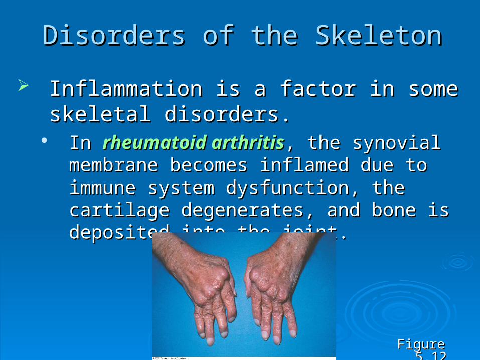
Inflammation is a factor in some skeletal Inflammation is a factor in some skeletal disorders.disorders.
In In rheumatoid arthritisrheumatoid arthritis, the synovial , the synovial membrane becomes inflamed due to immune membrane becomes inflamed due to immune system dysfunction, the cartilage degenerates, system dysfunction, the cartilage degenerates, and bone is deposited into the joint.and bone is deposited into the joint.
Figure 5.12Figure 5.12

Disorders of the SkeletonDisorders of the Skeleton
In In osteoarthritisosteoarthritis, the cartilage at the end of the , the cartilage at the end of the bone degenerates.bone degenerates.
Tendinitis Tendinitis is the inflammation of tendons and is the inflammation of tendons and synovial membranes around joints.synovial membranes around joints.
Carpal tunnel syndromeCarpal tunnel syndrome is the result of the is the result of the inflammation of the tendons in the space inflammation of the tendons in the space between a wrist ligament and the carpal bones, between a wrist ligament and the carpal bones, usually aggravated by chronic over use.usually aggravated by chronic over use.

Disorders of the SkeletonDisorders of the Skeleton
Joints also are vulnerable to strains, Joints also are vulnerable to strains, sprains, and dislocations.sprains, and dislocations.
A A strainstrain results from stretching or twisting a results from stretching or twisting a joint suddenly or too far.joint suddenly or too far.
A A sprainsprain is a tear of ligaments or tendons. is a tear of ligaments or tendons. A A dislocationdislocation causes two bones to no longer causes two bones to no longer
be in contact. be in contact.

Disorders of the SkeletonDisorders of the Skeleton
In factures, bones break.In factures, bones break. A A simple fracturesimple fracture is a crack in the bone; not is a crack in the bone; not
very serious.very serious. A A complete fracturecomplete fracture separates the bone into separates the bone into
two pieces, which must be quickly realigned for two pieces, which must be quickly realigned for proper healing. proper healing.
A A compound fracturecompound fracture is the most serious is the most serious because it means there are multiple breaks with because it means there are multiple breaks with the possibility of bone fragments penetrating the possibility of bone fragments penetrating the surrounding tissues.the surrounding tissues.

© 2007 Thomson Higher Education
simple compoundcomplete
Fig. 5.13, p. 98

Disorders of the SkeletonDisorders of the Skeleton
Other bone disorders include genetic Other bone disorders include genetic diseases, infections, and cancer.diseases, infections, and cancer.
Genetic diseases such as Genetic diseases such as osteogenesis osteogenesis imperfectaimperfecta can leave bones brittle and easily can leave bones brittle and easily broken.broken.
Figure 5.14Figure 5.14

Disorders of the SkeletonDisorders of the Skeleton
Bacterial and other Bacterial and other infections can spread infections can spread from the blood stream to from the blood stream to bone tissue or marrow.bone tissue or marrow.
OsteosarcomaOsteosarcoma, bone , bone cancer, usually occurs in cancer, usually occurs in long bones.long bones.
Figure 5.15Figure 5.15

Table 5.2, p. 100