Embed Size (px)
Citation preview
Hindawi Publishing CorporationCase Reports in DentistryVolume 2013, Article ID 249615, 4 pageshttp://dx.doi.org/10.1155/2013/249615
Case ReportRapidly Progressing Osteomyelitis of the Mandible
Yukiko Kusuyama,1 Ken Matsumoto,2 Shino Okada,1 Ken Wakabayashi,1
Noritami Takeuchi,1 and Yoshiaki Yura2
1 Department of Dentistry and Oral Surgery, Matsubara Tokushukai Hospital, 7-13-26 Amamihigashi, Matsubara-shi,Osaka 580-0032, Japan
2Department of Oral and Maxillofacial Surgery 𝐼𝐼, Osaka University, Graduate School of Dentistry, Osaka 565-0871, Japan
Correspondence should be addressed to Yukiko Kusuyama; [email protected]
Received 16 August 2013; Accepted 2 October 2013
Academic Editors: P. G. Arduino, M. B. D. Gaviao, and M. J. Wahl
Copyright © 2013 Yukiko Kusuyama et al. This is an open access article distributed under the Creative Commons AttributionLicense, which permits unrestricted use, distribution, and reproduction in any medium, provided the original work is properlycited.
Acute osteomyelitis exists as a refractory disease even now, which usually exhibits systemic symptoms such as fever or malaise andlocal redness or swelling. The present paper describes a case of acute osteomyelitis of the mandible that was rapidly progressingwithout typical symptoms. The patient had liver cirrhosis, which should be one of the systemic factors that affect immunesurveillance and metabolism. Actinomycotic druses and filaments were detected from the sequestrum. These were considered toplay a role in the rapid progression of osteomyelitis without typical symptoms. There has been no evidence of local recurrence 24months after surgery.
1. Introduction
Acute osteomyelitis of the jaws is not commonly seen inmodern oral and maxillofacial surgery practice. Generallyspeaking, this can be related to our society having becomemore health conscious, resulting in an increased awarenessof nutrition, as well as earlier and better access to healthcare than in the past [1, 2]. However, acute osteomyelitisexists as a refractory disease even now, which usually exhibitssystemic symptoms such as fever, malaise or high levels ofCRP and local redness, swelling, or pus discharge. It is knownthat osteomyelitis can be attributed to one or more of thepredisposing systemic diseases [3]. In immune-compromisedpatients, it is easy to expect that acute inflammatory reactionsare poor. Few case reports such as osteomyelitis of the jawswith poor acute inflammatory reactions and rapid progres-sion have been documented. The present paper describes acase of acute osteomyelitis of the mandible, with liver cirrho-sis, that was rapidly progressing without typical symptoms.
2. Case Report
A 77-year-old man was referred to our hospital for postex-traction hemorrhage and spontaneous pain in the socket of
the left mandibular first molar. The patient had a 1-monthhistory of spontaneous pain of the left mandibular firstmolar. At a nearby dental clinic, restorative treatment wasperformed. However, as the pain continued, the tooth wasfinally extracted on January 19, 2011. Next day he visited ourhospital.
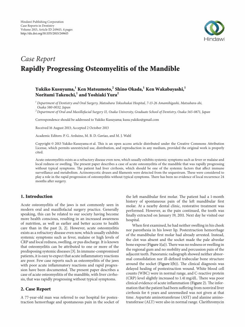
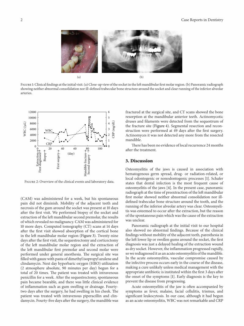
When first examined, he had neither swelling in his cheeknor paresthesia in his lower lip. Postextraction hemorrhageof the mandibular first molar had already arrested. Instead,the clot was absent and the socket made the pale alveolarbone expose (Figure 1(a)).Therewas no redness or swelling inthe regional gum and no mobility and percussion pain of theadjacent teeth. Panoramic radiograph showed neither abnor-mal consolidation nor ill-defined trabecular bone structurearound the socket (Figure 1(b)). The clinical diagnosis wasdelayed healing of postextraction wound. White blood cellcounts (WBC) were in normal range, and C-reactive protein(CRP) level slightly increased to 1.41mg/dL. There was poorclinical evidence of acute inflammation (Figure 2).The infor-mation that the patient had been suffering fromnonviral livercirrhosis for 6 years and unremedied was not given at thattime. Aspartate aminotransferase (AST) and alanine amino-transferase (ALT) were also in normal range. Clarithromycin
2 Case Reports in Dentistry
(a) (b)
Figure 1: Clinical findings at the initial visit. (a)Close-up viewof the socket in the leftmandibular firstmolar region. (b) Panoramic radiographshowing neither abnormal consolidation nor ill-defined trabecular bone structure around the socket and clear running of the inferior alveolararteries.
0
1
2
3
4
5
6
0
2000
4000
6000
8000
10000
12000
0 20 40 60 80 100
WBC
CRP
(day)
Firs
t dia
gnos
ed
Firs
t sur
gery
Man
dibl
e fra
ctur
eSe
cond
surg
ery
Biop
sy
Extr
actio
n
WBC
(/𝜇
L)
CRP
(mg/
dL)
Figure 2: Overview of the clinical events and laboratory data.
(CAM) was administered for a week, but his spontaneouspain did not diminish. Mobility of the adjacent teeth andnecrosis of the gum around the socket was present at 10 daysafter the first visit. We performed biopsy of the socket andextraction of the leftmandibular second premolar, the resultsof which revealed nomalignancy. CAMwas administered for10 more days. Computed tomography (CT) scans at 14 daysafter the first visit showed absorption of the cortical bonein the left mandibular molar region (Figure 3). Twenty-ninedays after the first visit, the sequestrectomy and corticectomyof the left mandibular molar region and the extraction ofthe left mandibular first premolar and second molar wereperformed under general anesthesia. The surgical site wasfilledwith gauze with pasta of dimethyl isopropyl azulene andclindamycin. Next day hyperbaric oxygen (HBO) utilization(2 atmosphere absolute, 90 minutes per day) begun for atotal of 20 times. The patient was treated with intravenouspenicillin for a week. After the sequestrectomy, spontaneouspain became bearable, and there was little clinical evidenceof inflammation such as gum swelling or drainage. Fourty-two days after the surgery, he had swelling in his cheek. Thepatient was treated with intravenous piperacillin and clin-damycin. Fourty-five days after the surgery, themandible was
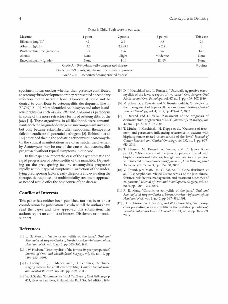
fractured at the surgical site, and CT scans showed the boneresorption at the mandibular anterior teeth. Actinomycoticdruses and filaments were detected from the sequestrum ofthe fracture site (Figure 4). Segmental resection and recon-struction were performed at 49 days after the first surgery.Actinomyces it was not detected any more from the resectedmandible.
There has been no evidence of local recurrence 24monthsafter the treatment.
3. Discussion
Osteomyelitis of the jaws is caused in association withhematogenous germ spread, drug- or radiation-related, orlocal odontogenic or nonodontogenic processes [1]. Schaferstates that dental infection is the most frequent cause ofosteomyelitis of the jaws [4]. In the present case, panoramicradiograph at the time of preextraction of the leftmandibularfirst molar showed neither abnormal consolidation nor ill-defined trabecular bone structure around the tooth, and therunning of the inferior alveolar artery was clear. Osteomyeli-tis was esteemed to occur after the extraction, but the reasonof the spontaneous pain which was the cause of the extractionwas unclear.
Panoramic radiograph at the initial visit to our hospitalalso showed no abnormal findings. Because of the clinicalfindings without mobility of the adjacent teeth, paresthesia inthe left lower lip or swollen gums around the socket, the firstdiagnosis was just a delayed healing of the extraction woundas dry socket. However, the inflammation progressed rapidly,sowe rediagnosed it as an acute osteomyelitis of themandible.In the acute osteomyelitis, vascular compromise caused bythe infective process occurs early in the course of the disease,making a cure unlikely unless medical management with theappropriate antibiotic is instituted within the first 3 days afterthe onset of the symptoms [1]. Early diagnosis is the key toprevent the disease from progressing.
Acute osteomyelitis of the jaw is often accompanied bysymptoms as fever, malaise, facial cellulitis, trismus, andsignificant leukocytosis. In our case, although it had begunas an acute osteomyelitis, WBC was not remarkable and CRP

Case Reports in Dentistry 3
(a) (b)
Figure 3: CT scans at 14 days after the initial visit showing remarkable absorption of the cortical bone in the left mandibular molar region.(a) Axial section. (b) Coronal section.
(a) (b)
Figure 4: Actinomycotic druses (A, H.E. stain, 200x) and filaments (B, Grocott stain, 400x) detected from the sequestrum of the fracturesite.
level increased only slightly (Figure 2), and there was neitherpus discharge nor swelling of the cheek until just beforethe fracture of themandible. Rapidly progressing osteomyeli-tis which was highly resistant to treatments without typicalsymptoms like this case is extremely rare [5]. Osteomyelitiswithout typical symptoms made the final diagnosis delayand might bring the inflammation to progress. Systemicfactors such as diabetes mellitus, agranulocytosis, leukemia,severe anemia, malnutrition, or alcohol abuse affect immunesurveillance and lead to impairing the osteomyelitis [1].The Cierny-Mader classification of long-bone osteomyelitisis based on the anatomy of bone infection and the phys-iology of the host [3]. Cierny described that not only theanatomic classification but also the condition of the host,regional vascularity, local milieu, and extent of necrosiswould influence the natural history of the disease. In thepresent case, the patient had liver cirrhosis. Liver cirrhosis
is one of the systemic factors in the classification that affectimmune surveillance and metabolism. This patient’s Child-Pugh score [6] was 8 points and the grade was B, significantfunctional compromise at the first surgery (Table 1). Child-Pugh grade can be used in patients with liver cirrhosis toassess the severity of the clinical condition [7]. Therefore,it was considered that impaired immunity introduced pooracute inflammatory reactions and the systemic compromiseplayed a role in the asymptomatic and rapid progression ofosteomyelitis.
Identification of responsible microorganisms can beextremely difficult. Simply swabbing a suspected area is notappropriate. The process of obtaining suitable material forculture is fraught with potential danger of contaminationfrom nearby oral site. In our case, actinomycotic druses andfilaments were detected from the sequestrum of the fracturesite, while they were not from the resected mandibular
4 Case Reports in Dentistry
Table 1: Child-Pugh score in our case.
Measure 1 point 2 points 3 points This caseBilirubin (mg/dL) <2 2-3 >3 2.1Albumin (g/dL) >3.5 2.8–3.5 <2.8 4Prothrombin time (seconds) 1–3 4–6 >6 14.6Ascites None Slight Moderate NoneEncephalopathy (grade) None I-II III-IV None
Grade A = 5-6 points; well-compensated disease 8 pointsGrade B = 7–9 points; significant functional compromise
Grade C = 10–15 points; decompensated disease
specimen. It was unclear whether their presence contributedto osteomyelitis development or they represented a secondaryinfection to the necrotic bone. However, it could not bedenied to contribute to osteomyelitis development like inBRONJ [8–10]. Marx identifiedActinomyces and other fastid-ious organisms such as Eikenella and Arachnia as pathogensin some of the more refractory forms of osteomyelitis of thejaws [11]. These organisms, in all likelihood, were contami-nants with the original odontogenicmicroorganism invasion,but only became established after suboptimal therapeuticsfailed to eradicate all potential pathogens [2]. Robinson et al.[12] described that in the pediatric actinomycotic osteomyeli-tis the clinical manifestations are often subtle. Involvementby Actinomyces may be one of the causes that osteomyelitisprogressed without typical symptoms in our case.
In this paper, we report the case of the asymptomatic andrapid progression of osteomyelitis of the mandible. Depend-ing on the predisposing factors, osteomyelitis progressesrapidly without typical symptoms. Correction of the under-lying predisposing factors, early diagnosis and evaluating thetherapeutic response of a multimodality treatment approachas needed would offer the best course of the disease.
Conflict of Interests
This paper has neither been published nor has been underconsideration for publication elsewhere. All the authors haveread the paper and have approved this submission. Theauthors report no conflict of interest. Disclosure or financialsupport.
References
[1] L. G. Mercuri, “Acute osteomyelitis of the jaws,” Oral andMaxillofacial Surgery Clinics of North America—Infections of theHead and Neck, vol. 3, no. 2, pp. 355–365, 1991.
[2] J. W. Hudson, “Osteomyelitis of the jaws: a 50-year perspective,”Journal of Oral and Maxillofacial Surgery, vol. 51, no. 12, pp.1294–1301, 1993.
[3] G. Cierny III, J. T. Mader, and J. J. Penninck, “A clinicalstaging system for adult osteomyelitis,” Clinical Orthopaedicsand Related Research, no. 414, pp. 7–24, 2003.
[4] W.G. Scafer, “Osteomyelitis,” inATextbook of Oral Pathology, p.453, Elsevier Saunders, Philadelphia, Pa, USA, 3rd edition, 1974.
[5] D. J. Krutchkoff and L. Runstad, “Unusually aggressive osteo-myelitis of the jaws. A report of two cases,” Oral Surgery OralMedicine and Oral Pathology, vol. 67, no. 5, pp. 499–507, 1989.
[6] M. Schwartz, S. Roayaie, andM. Konstadoulakis, “Strategies forthe management of hepatocellular carcinoma,” Nature ClinicalPractice Oncology, vol. 4, no. 7, pp. 424–432, 2007.
[7] F. Durand and D. Valla, “Assessment of the prognosis ofcirrhosis: child-pugh versus MELD,” Journal of Hepatology, vol.42, no. 1, pp. S100–S107, 2005.
[8] T. Mucke, J. Koschinski, H. Deppe et al., “Outcome of treat-ment and parameters influencing recurrence in patients withbisphosphonate-related osteonecrosis of the jaws,” Journal ofCancer Research and Clinical Oncology, vol. 137, no. 5, pp. 907–913, 2011.
[9] T. Hansen, M. Kunkel, A. Weber, and C. James Kirk-patrick, “Osteonecrosis of the jaws in patients treated withbisphosphonates—Histomorphologic analysis in comparisonwith infected osteoradionecrosis,” Journal of Oral Pathology andMedicine, vol. 35, no. 3, pp. 155–160, 2006.
[10] V. Thumbigere-Math, M. C. Sabino, R. Gopalakrishnan etal., “Bisphosphonate-related Osteonecrosis of the Jaw: clinicalfeatures, risk factors, management, and treatment outcomes of26 patients,” Journal of Oral and Maxillofacial Surgery, vol. 67,no. 9, pp. 1904–1913, 2009.
[11] R. E. Marx, “Chronic osteomyelitis of the jaws,” Oral andMaxillofacial Surgery Clinics of North America—Infections of theHead and Neck, vol. 3, no. 2, pp. 367–381, 1991.
[12] J. L. Robinson, W. L. Vaudry, and W. Dobrovolsky, “Actinomy-cosis presenting as osteomyelitis in the pediatric population,”Pediatric Infectious Disease Journal, vol. 24, no. 4, pp. 365–369,2005.

Submit your manuscripts athttp://www.hindawi.com
Hindawi Publishing Corporationhttp://www.hindawi.com Volume 2014
Oral OncologyJournal of
DentistryInternational Journal of
Hindawi Publishing Corporationhttp://www.hindawi.com Volume 2014
Hindawi Publishing Corporationhttp://www.hindawi.com Volume 2014
International Journal of
Biomaterials
Hindawi Publishing Corporationhttp://www.hindawi.com Volume 2014
BioMed Research International
Hindawi Publishing Corporationhttp://www.hindawi.com Volume 2014
Case Reports in Dentistry
Hindawi Publishing Corporationhttp://www.hindawi.com Volume 2014
Oral ImplantsJournal of
Hindawi Publishing Corporationhttp://www.hindawi.com Volume 2014
Anesthesiology Research and Practice
Hindawi Publishing Corporationhttp://www.hindawi.com Volume 2014
Radiology Research and Practice
Environmental and Public Health
Journal of
Hindawi Publishing Corporationhttp://www.hindawi.com Volume 2014
The Scientific World JournalHindawi Publishing Corporation http://www.hindawi.com Volume 2014
Hindawi Publishing Corporationhttp://www.hindawi.com Volume 2014
Dental SurgeryJournal of
Drug DeliveryJournal of
Hindawi Publishing Corporationhttp://www.hindawi.com Volume 2014
Hindawi Publishing Corporationhttp://www.hindawi.com Volume 2014
Oral DiseasesJournal of
Hindawi Publishing Corporationhttp://www.hindawi.com Volume 2014
Computational and Mathematical Methods in Medicine
ScientificaHindawi Publishing Corporationhttp://www.hindawi.com Volume 2014
PainResearch and TreatmentHindawi Publishing Corporationhttp://www.hindawi.com Volume 2014
Preventive MedicineAdvances in
Hindawi Publishing Corporationhttp://www.hindawi.com Volume 2014
EndocrinologyInternational Journal of
Hindawi Publishing Corporationhttp://www.hindawi.com Volume 2014
Hindawi Publishing Corporationhttp://www.hindawi.com Volume 2014
OrthopedicsAdvances in