Embed Size (px)
Citation preview

Lithuanian Journal of Physics, Vol. 48, No. 3, pp. 265–273 (2008) doi:10.3952/lithjphys.48308
RESISTANCE OF AIRBORNE FUNGAL PROPAGULES TOULTRAVIOLET IRRADIATION: LABORATORY STUDY
V. Ulevicius a, D. Peciulyte b, K. Plauškaite a, and N. Špirkauskaite a
a Institute of Physics, Savanoriu 231, LT-02300 Vilnius, LithuaniaE-mail: [email protected]
b Institute of Botany, Žaliuju ežeru 49, LT-08406 Vilnius, Lithuania
Received 30 July 2008; accepted 19 September 2008
The influence of ultraviolet (UV) radiation on fungi Aspergillus niger Tiegh. isolate OG168, Paecilomyces puntonii (Vuill.)Nann. isolate OG68, and Penicillium expansum Link isolate PO88 was studied under laboratory conditions. A test system wasdeveloped for this study. An aerosol chamber provided a dust-free space of 1.5 m3. The source of ultraviolet rays was an UVlamp (DPT 220, 240–320 nm, 15 W). Fungal propagules were injected into the UV exposed chamber space from an externalbioaerosol generator. Aerosols from the aerosol chamber after irradiation to UV were sampled into an impinger AGI-30 andmeasured with the optical aerosol spectrometer LAS-15M (Institute of Physics, Lithuania). The changes in fungi survivalcaused by exposure to UV radiation were evaluated by determining their relative recovery. The laboratory study indicatedthat the fungal propagules responded to UV radiation distinctively. P. puntonii propagules were injured without possibility torepair. On the contrary, P. expansum propagules repaired after a long enough exposure to UV radiation, but this ability waslimited. The stressed A. niger propagules recovered after the 80 min exposure to UV radiation and the relative recovery reacheda plateau. The mutagenic effects of UV light on tested fungi have shown that frequent occurrence of different morphologicalmutants was detected after the 30 min exposure of conidia. The mean geometrical diameter of fungal propagules exposed toUV irradiation in the aerosol chamber was in the range of 2.5 to 2.8 µm.
Keywords: aerosol chamber, fungal propagules, relative recovery, mutation, UV radiation
PACS: 92.60.Mt, 92.20.Bk, 87.19.xg
1. Introduction
Thousands of microorganisms (fungi, bacteria, vi-ruses, etc.) as one of components of ambient particulatematter constantly exist in the shape of aerosol particlesin all size ranges and forms in outdoor and indoor air[1–5]. In recent years with an increase of the atmo-sphere pollution, attention of researchers to investiga-tions of the survival of the microorganisms as objectsinhabiting an increasingly changing polluted environ-ment has raised [1, 6–8]. Solar radiation, especiallyultraviolet (UV) light, is a specific biophysical param-eter playing an important role in the atmospheric mi-crobial structure life [6, 7]. The effects of the light aredependent on microorganism species [1, 6], their phys-iological state [8, 9], light intensity duration [10, 11],wavelength [10, 12], etc.
As stratospheric ozone depletion progresses, moreharmful solar UVB (i. e. 290–320 nm) radiation whichis responsible for inactivation of microorganisms [4, 9]will penetrate the atmosphere to the Earth’s surface andwill induce variability of atmospheric microbial popu-
lations. Solar radiation appears to be a factor whichcould determine the atmospheric microbial structureand is limiting the upper boundary of the biosphere onthe planet [3].
It is known that high doses of UV radiation areharmful to all living organisms in the following order:UVC > UVB > UVA. The ecological significance ofUV radiation may be even greater than that of ionizingradiation in the environment. UV rays are absorbed bya substance whose maximum absorption is at a wave-length of 200–300 nm. The main cellular target sub-stance of UV for microbial death is known to be theDNR which has the maximum absorption at 265 nm[1]. In the case of UVC (below 290 nm) and UVB ra-diation, the cause is a direct damage to nucleic acidsand proteins that can lead to genetic mutation or celldeath [8]. The mechanism of the UVA (320–400 nm)damage of cells is less understood, but it probably in-volves the generation of reactive oxygen molecules thatcan damage many different components of cells, in-cluding nucleic acids and proteins. Furthermore, low
c© Lithuanian Physical Society, 2008c© Lithuanian Academy of Sciences, 2008 ISSN 1648-8504

266 V. Ulevicius et al. / Lithuanian J. Phys. 48, 265–273 (2008)
doses of UVA radiation can induce physiological re-sponses in microorganisms, probably by activating spe-cific genes [6]. The wide application of UVC radi-ation for disinfection of the indoor air and surfacesis also an important factor determining genetic varia-tions of microorganisms. Many of the physiologicaland pathological effects of UVA radiation can be pro-voked by violet-blue light. This is most likely due toa common photochemical transduction process involv-ing flavinoids and carotenoids. The mechanism associ-ated with gene activation is unclear, and it is uncertainwhether low doses of UVC and UVB radiation can in-duce similar responses. The percentage of the UVB islow (<1% of total energy) [9] but it has a highly ac-tive component of the solar spectrum which has the po-tential to cause wide-ranging effects, and it is assumedthat shorter wavelengths have a major role in regulat-ing both the size and the balance of atmospheric mi-crobial populations [3, 6, 10]. UV light has been shownto be lethal and mutagenic in a variety of organisms,including plants [13, 14], bacteria [4, 5, 15, 16], yeast[17], and fungi [11, 12, 18–20]. There is a wide varietyof microorganisms (germs) which cause contaminationand vary in their structure and sensitivity to UV radi-ation. Airborne fungal propagules are highly adaptedfor survival because the air is their natural dispersalmedium and they have various protective mechanisms.It is well documented that the main cellular target sub-stance of UV for bacteria death is DNR and cell walls,in yeasts it is DNR, cell walls, and cell nuclei, and infungi the main target is DNR, cell walls + pigments,and cell nuclei. Thus, fungal propagules dispersed inthe air are less vulnerable to UV radiation damage.
Fungal propagules make up the main part of the mi-croorganisms in the ambient aerosol particle [1, 6, 12]and participate in processes occurring in other compo-nents, e. g. chemical. Therefore, changes in the cellprocesses, morphological structure, or in possible lifecycle of the fungi recovered after UV radiation will bereflected in the chemical and physical composition ofthe aerosols and will directly influence the atmosphericprocesses. To our knowledge, only a few investigationson the effect of UV radiation on survival of fungi andbacteria in the air have been performed using aerosoltechnology [16, 22]. Most of the investigations wereperformed by the method when a monolayer of fun-gal propagules on the static flat surface was irradiated[2, 7, 8, 11, 20].
The aim of this study was the laboratory evaluationof the UV radiation induced changes in survivability ofairborne fungal propagules by establishing conditions
close to natural. The present study was carried out toassess the UV irradiation effect on aerosols of the fun-gal propagules, to compare resistance differences dueto fungal morphology, to determine the lethality andthe increase in mutation frequency.
2. Materials and methods
2.1. Culture preparation
When choosing fungi for the laboratory study, wetook into consideration several criteria, i. e.: the fun-gal propagules must be most prevalent in the out-door environment, easily aerosolized, able to grow faston the laboratory media. The test fungi, Aspergillusniger Tiegh. (strain OG168), Paecilomyces puntonii(Vuill.) Nann. (strain OG68), and Penicillium expan-sum Link (strain PO88), were selected as representa-tives of species which are found in the ambient airand are known as microorganisms robust to environ-mental factors. Aspergillus and Penicillium are amongthe most common genera of fungi, isolated from theLithuanian outdoor environment [21]. The fungi wereincubated at 25 ◦C and stored at 4 ◦C on the malt ex-tract agar (MEA, Oxoid). P. expansum conidia are sub-globose to ellipsoidal, with a physical diameter of 3.3–3.5 × 2.5–3.0 µm, greenish, smooth-walled. A. nigerconidia are globose to subglobose, 3.5–5 µm in phys-ical diameter, brown, ornamented with irregular warts,spines, and ridges. Fungus P. puntonii was selected be-cause it has been reported as a causative agent of al-lergic alveolitis, humidifier disease, and as a sensitivemicroorganism [9]. P. puntonii was chosen as speciesof Paecilomyces genus with more stable morphology.Conidia of P. puntonii are ellipsoidal to cylindrical,with a physical diameter of 1.7–2.0 × 3.5–4.0 µm,smooth-walled.
The fungi were grown on MEA at 25 ◦C for 14 days.Conidia of the 14-day culture of the fungi served as thematerial for irradiation. The suspensions of conidia fil-tered through cotton and sterile gauze in the ceramicfilter were used in the experiment when they were irra-diated as suspensions.
2.2. The method of irradiation in the aerosol chamber
The test system was developed to determine changesin survivability of airborne fungal propagules exposedto UV irradiation under conditions close to the outdoorenvironment and well-mixed air (Fig. 1). An aerosolchamber provided a dust-free space of 1.5 m3. The

V. Ulevicius et al. / Lithuanian J. Phys. 48, 265–273 (2008) 267
Fig. 1. Schematic representation of the experimental set-up.
chamber frame was made of strong aluminum tubes,which were covered with a double layer of the Teflonfilm. The two inlets of the chamber were sealed withHEPA filters. Air was pumped through the chamberproviding an aerosol free space in the chamber. Thetop of the chamber was constructed so that it could beopened to place instruments inside the chamber. Thebottom was covered with a plastic sheet wrapped intothe Teflon film. An UV lamp (DPT 220, 240–320 nm,15 W), a fan, relative humidity and temperature sen-sors were mounted at the bottom of the chamber. Asmall opening in the aerosol chamber generated fun-gal propagules from an external bioaerosol generatorinto the UV exposed space. Other small openings wereused to extract aerosols into an impinger AGI–30 andan optical aerosol spectrometer LAS–15M (Institute ofPhysics, Lithuania), and to supply power for the instru-ments inside the aerosol chamber.
The agar-tube disperser, one of the three bioaerosolgenerators developed at the University of Cincinnati(USA) [22, 23], was built for aerosolization of the fun-gal propagules. The external body of the disperser con-sisted of a cylindrical tube (0.3 m long and 0.03 m in-ner diameter). Agar in a half-cylindrical container withmicrobial growth on it was placed inside the bioaerosolgenerator tube. The air flow was directed through thetube resulting in the release of propagules from the mi-crobial growth. For this study one orifice was added atthe entrance of the dispersion tube to increase the airturbulence in the tube and thus enhance the release ofpropagules. Test fungi were incubated on MEA in thehalf-cylindrical containers. A piece of fungus culture(14-days old) was transferred with an inoculating looponto the medium surface trying to spread conidia on
it. Inoculated containers were incubated for 7 days at25 ◦C in the dark. Fungal propagules were dispersedinto UV exposed space of the aerosol chamber directlyfrom the grown biomass.
Fungal propagules from the UV radiation space werecollected into the impinger containing 20 ml sterilequarter ringer solution. Sampling with the AGI-30 im-pinger was performed at the flow rate of 10 l/m for5 min after every 15 min exposure to UV radiation.Samples were diluted in succession in the sterile quar-ter ringer solution and a viable count was performed intriplicate on MEA in plate media. The plates were in-cubated for 2, 3, and 7 days at 25 ◦C in the dark priorto counting the number of colony-forming units (num-ber of viable fungal propagules). When the UV dosewas sufficiently high to ensure the 100% level of mi-crobial inactivation, no colonies were observed on theMEA dishes. When the colonies were counted after 2,3, and 7 days, all the dishes were left for prolonged in-cubation and were counted again to ensure no bias dueto microbial injury and further recovery.
The deposition velocity of fungal spores in thechamber was estimated using the methodology from[16]. The average deposition velocity was 0.06±0.03 m/h.
In parallel, the optical aerosol spectrometer wasused for the determination of size distribution of fun-gal propagules and their total concentration. The rela-tive recovery (%) of fungi in aerosol samples removedfrom the aerosol chamber was estimated as a ratio ofthe number of colonies grown on MEA obtained withthe impinger to the total number of the fungus propag-ules measured with the optical aerosol spectrometer.
268 V. Ulevicius et al. / Lithuanian J. Phys. 48, 265–273 (2008)
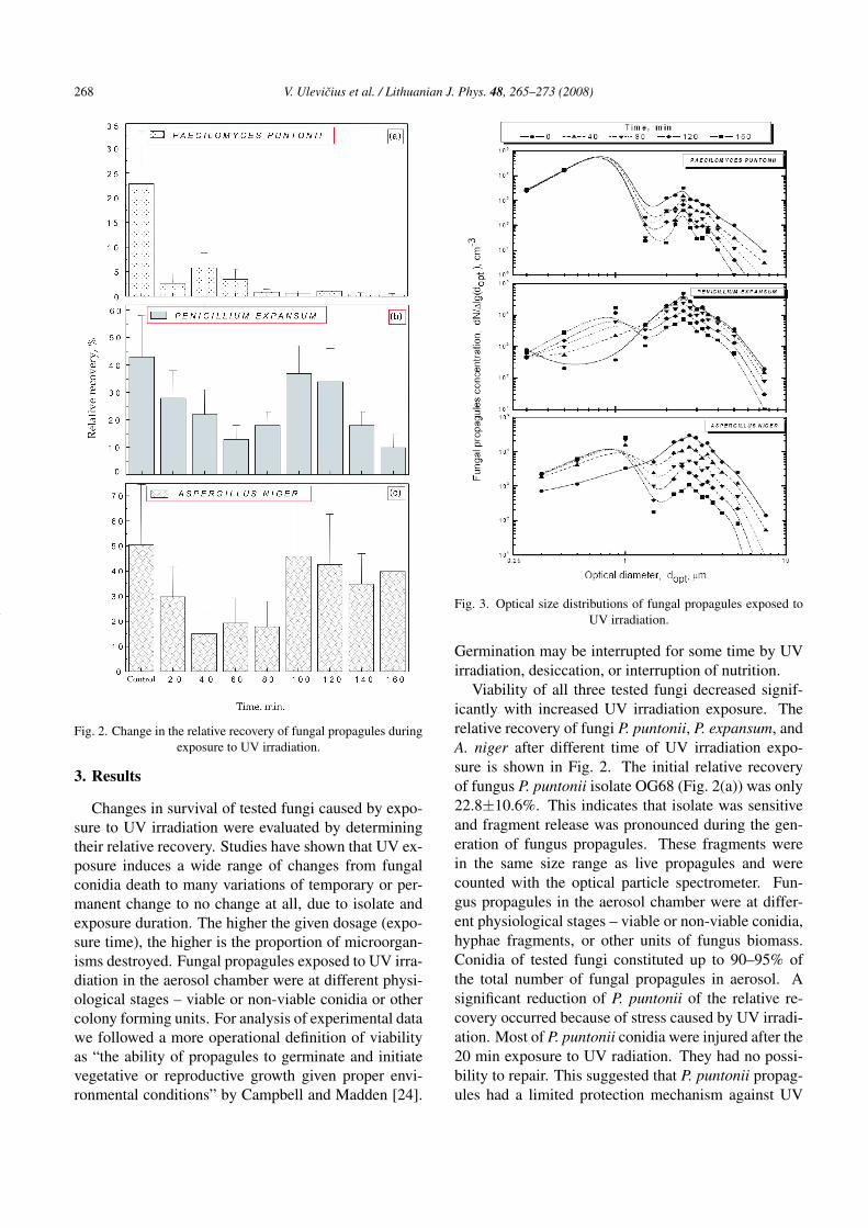
Fig. 2. Change in the relative recovery of fungal propagules duringexposure to UV irradiation.
3. Results
Changes in survival of tested fungi caused by expo-sure to UV irradiation were evaluated by determiningtheir relative recovery. Studies have shown that UV ex-posure induces a wide range of changes from fungalconidia death to many variations of temporary or per-manent change to no change at all, due to isolate andexposure duration. The higher the given dosage (expo-sure time), the higher is the proportion of microorgan-isms destroyed. Fungal propagules exposed to UV irra-diation in the aerosol chamber were at different physi-ological stages – viable or non-viable conidia or othercolony forming units. For analysis of experimental datawe followed a more operational definition of viabilityas “the ability of propagules to germinate and initiatevegetative or reproductive growth given proper envi-ronmental conditions” by Campbell and Madden [24].
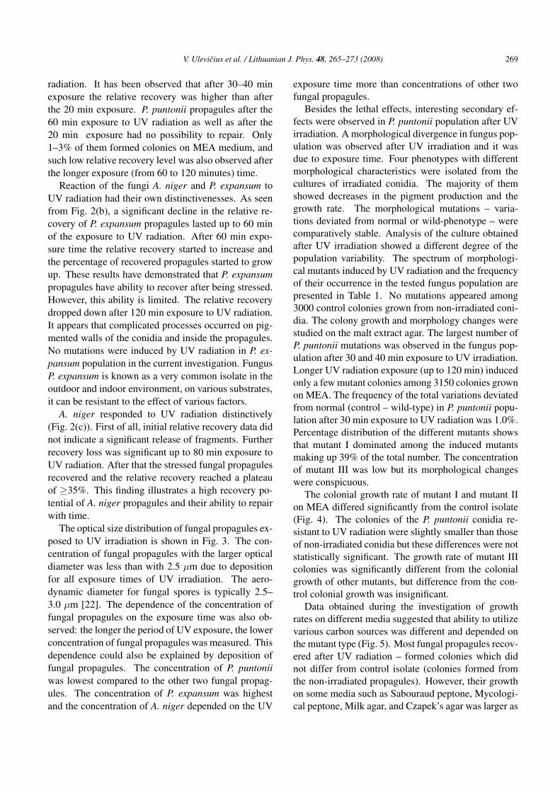
Fig. 3. Optical size distributions of fungal propagules exposed toUV irradiation.
Germination may be interrupted for some time by UVirradiation, desiccation, or interruption of nutrition.
Viability of all three tested fungi decreased signif-icantly with increased UV irradiation exposure. Therelative recovery of fungi P. puntonii, P. expansum, andA. niger after different time of UV irradiation expo-sure is shown in Fig. 2. The initial relative recoveryof fungus P. puntonii isolate OG68 (Fig. 2(a)) was only22.8±10.6%. This indicates that isolate was sensitiveand fragment release was pronounced during the gen-eration of fungus propagules. These fragments werein the same size range as live propagules and werecounted with the optical particle spectrometer. Fun-gus propagules in the aerosol chamber were at differ-ent physiological stages – viable or non-viable conidia,hyphae fragments, or other units of fungus biomass.Conidia of tested fungi constituted up to 90–95% ofthe total number of fungal propagules in aerosol. Asignificant reduction of P. puntonii of the relative re-covery occurred because of stress caused by UV irradi-ation. Most of P. puntonii conidia were injured after the20 min exposure to UV radiation. They had no possi-bility to repair. This suggested that P. puntonii propag-ules had a limited protection mechanism against UV
V. Ulevicius et al. / Lithuanian J. Phys. 48, 265–273 (2008) 269
radiation. It has been observed that after 30–40 minexposure the relative recovery was higher than afterthe 20 min exposure. P. puntonii propagules after the60 min exposure to UV radiation as well as after the20 min exposure had no possibility to repair. Only1–3% of them formed colonies on MEA medium, andsuch low relative recovery level was also observed afterthe longer exposure (from 60 to 120 minutes) time.
Reaction of the fungi A. niger and P. expansum toUV radiation had their own distinctivenesses. As seenfrom Fig. 2(b), a significant decline in the relative re-covery of P. expansum propagules lasted up to 60 minof the exposure to UV radiation. After 60 min expo-sure time the relative recovery started to increase andthe percentage of recovered propagules started to growup. These results have demonstrated that P. expansumpropagules have ability to recover after being stressed.However, this ability is limited. The relative recoverydropped down after 120 min exposure to UV radiation.It appears that complicated processes occurred on pig-mented walls of the conidia and inside the propagules.No mutations were induced by UV radiation in P. ex-pansum population in the current investigation. FungusP. expansum is known as a very common isolate in theoutdoor and indoor environment, on various substrates,it can be resistant to the effect of various factors.
A. niger responded to UV radiation distinctively(Fig. 2(c)). First of all, initial relative recovery data didnot indicate a significant release of fragments. Furtherrecovery loss was significant up to 80 min exposure toUV radiation. After that the stressed fungal propagulesrecovered and the relative recovery reached a plateauof ≥35%. This finding illustrates a high recovery po-tential of A. niger propagules and their ability to repairwith time.
The optical size distribution of fungal propagules ex-posed to UV irradiation is shown in Fig. 3. The con-centration of fungal propagules with the larger opticaldiameter was less than with 2.5 µm due to depositionfor all exposure times of UV irradiation. The aero-dynamic diameter for fungal spores is typically 2.5–3.0 µm [22]. The dependence of the concentration offungal propagules on the exposure time was also ob-served: the longer the period of UV exposure, the lowerconcentration of fungal propagules was measured. Thisdependence could also be explained by deposition offungal propagules. The concentration of P. puntoniiwas lowest compared to the other two fungal propag-ules. The concentration of P. expansum was highestand the concentration of A. niger depended on the UV
exposure time more than concentrations of other twofungal propagules.
Besides the lethal effects, interesting secondary ef-fects were observed in P. puntonii population after UVirradiation. A morphological divergence in fungus pop-ulation was observed after UV irradiation and it wasdue to exposure time. Four phenotypes with differentmorphological characteristics were isolated from thecultures of irradiated conidia. The majority of themshowed decreases in the pigment production and thegrowth rate. The morphological mutations – varia-tions deviated from normal or wild-phenotype – werecomparatively stable. Analysis of the culture obtainedafter UV irradiation showed a different degree of thepopulation variability. The spectrum of morphologi-cal mutants induced by UV radiation and the frequencyof their occurrence in the tested fungus population arepresented in Table 1. No mutations appeared among3000 control colonies grown from non-irradiated coni-dia. The colony growth and morphology changes werestudied on the malt extract agar. The largest number ofP. puntonii mutations was observed in the fungus pop-ulation after 30 and 40 min exposure to UV irradiation.Longer UV radiation exposure (up to 120 min) inducedonly a few mutant colonies among 3150 colonies grownon MEA. The frequency of the total variations deviatedfrom normal (control – wild-type) in P. puntonii popu-lation after 30 min exposure to UV radiation was 1.0%.Percentage distribution of the different mutants showsthat mutant I dominated among the induced mutantsmaking up 39% of the total number. The concentrationof mutant III was low but its morphological changeswere conspicuous.
The colonial growth rate of mutant I and mutant IIon MEA differed significantly from the control isolate(Fig. 4). The colonies of the P. puntonii conidia re-sistant to UV radiation were slightly smaller than thoseof non-irradiated conidia but these differences were notstatistically significant. The growth rate of mutant IIIcolonies was significantly different from the colonialgrowth of other mutants, but difference from the con-trol colonial growth was insignificant.
Data obtained during the investigation of growthrates on different media suggested that ability to utilizevarious carbon sources was different and depended onthe mutant type (Fig. 5). Most fungal propagules recov-ered after UV radiation – formed colonies which didnot differ from control isolate (colonies formed fromthe non-irradiated propagules). However, their growthon some media such as Sabouraud peptone, Mycologi-cal peptone, Milk agar, and Czapek’s agar was larger as

270 V. Ulevicius et al. / Lithuanian J. Phys. 48, 265–273 (2008)
Fig. 4. Colony radial growth rate (evaluated as the colony diameter after 7 days of growth at 25 ◦C on MEA) of P. puntonii and its mutantsobtained after 30 min exposure to UV irradiation in the aerosol chamber.
Table 1. The morphological characteristics of the control isolate P. puntonii and UV-induced mutant strainsgrown on MEA at 25 ◦C temperature in the dark.
Morphological Colony Colony Appearance of the coloniesFungus mutants grown diameter Intensity
(% of total rate after Colour Reverse ofnumber) (mm/h) 7 days (cm) sporulation
Wild isolate – 3.3–3.6 5.5–6.0 Tan White to pale Abundantyellowish
Control isolate afterexposure to UV – 2.7–3.0 4.5–5.1 Tan Yellow Abundantirradiation (not mutated)
Mutant I 39 5.1–5.7 8.5–9.5 White Colourless Limited
Mutant II 31 0.7–0.9 1.2–1.5 Light-grey Colourless Absent or limited
Light-brown, sometimes Sparse butMutant III 6 3.9–4.3 6.5–7.2 with white / brown Colourless less than in
mycelium segments control isolate
Mutant IV 24 2.6–3.0 4.3–5.0 Light-brown with Green Abundantgreen shadows
compared with control isolate. UV radiation stimulatedthe growth rate of P. puntonii on these media. Mutantcultures had uniform morphology of colonies on all thegrowth media, but the growth rate differed dependingon the medium composition.
Mutant I also utilized starch better than control iso-late. The growth rate of mutant II was very low (from0.027 to 0.181 mm/h) on all the investigated media(Fig. 5). Mutant III grew better on malt extract andmalt extract + yeast extract medium than control iso-
late. Mutant IV utilized all carbon substrates similarlyas control isolate.
Each mutant type was studied in the period of 5–8 continuous generations. The populations of mutantI and mutant II were genetically stable – morpholog-ical changes did not revert to the control isolate. Mu-tant III and mutant IV were genetically unstable and re-verted to control isolate after 5th or 4th passage, respec-tively. The population of mutant III after each new pas-sage split into 3 types of colonies: subdivided colonies,

V. Ulevicius et al. / Lithuanian J. Phys. 48, 265–273 (2008) 271
Fig. 5. Growth rate of P. puntonii control isolate, isolate after UV radiation and four mutants at 25 ◦C on different media: malt extractagar (MEA), malt extract + yeast extract (MEA+ye), malt extract + starch + yeast extract (MEA+st+ye), Sabouraud peptone agar (SPA),
mycological peptone (MP), milk agar (MA), Czapek’s agar + carboxymethyl cellulose (CA+CMC), Czapek’s agar (CA).
colonies consisting of very thin mycelium, and thosewith velvety appearance.
4. Discussion and conclusions
Irradiation of airborne organisms has been the pri-mary focus of many studies, but only a few investiga-tions have been performed on the recovery and phys-iological changes of microorganisms some time afterUV radiation. The destruction of microorganisms byUV radiation is an exponential process. The higher thegiven dosage, the higher the proportion of destroyedmicroorganisms. Consequently, the dose necessary todestroy 99% of fungal propagules is double the value todestroy 90% of fungal propagules. It follows thereforethat the dosage required to kill 99.9% is three times thevalue to destroy 90% and the dosage required to kill99.99% is four times the value to destroy 90%. Vari-ations in ultra-violet light sensitivity can be due to thecell size, structure of cell wall or membrane, pigmenta-tion, or the existence and capacity of repair systems.
Apart from the capacity for repair mechanisms, tol-erance to UV radiation may be related to pigmentation,which for microorganisms is believed to play a protec-tive function against injurious solar radiation [25]. Forexample, a cell wall is often pigmented with melanin,making the fungal propagules less vulnerable to UVradiation damage [26]. Mason et al. [27] suggestedthat in some organisms melanin may act as a biolog-ical electron exchange polymer. Due to this prop-erty and its free radical state, melanin has the abil-
ity to protect the cell against the reducing and oxidiz-ing conditions, which might otherwise set reactive freeradicals capable of disrupting metabolism. In manycases, it has been suggested that pigments in fungi arethe main protective mechanisms against UV radiation[25, 28]. Therefore, exposure to UV radiation may nothave a lethal effect on fungal propagules; however, itmay cause changes in their metabolic activity. Af-ter deposition and growth, fungi may develop differ-ent forms, which are adapted for better survival underunfavourable conditions. This means that some of fun-gal species may become very resistant to mechanical,chemical, biological attack and may have significant in-fluence on ecosystems.
The laboratory study of the survival of airborne fun-gal propagules exposed to UV irradiation in the aerosolchamber under conditions close to the outdoor environ-ment has indicated that UV exposure induces a widerange of changes from fungal conidia death to manyvariations of temporary or permanent changes to nochanges at all due to isolate and exposure duration. In-vestigations have shown that the growth rate of differ-ent mutant types depend on the chemical compositionof media. The fungal propagules responded to UV ra-diation distinctively. P. puntonii propagules were in-jured without the possibility to repair. On the contrary,P. expansum propagules repaired after a long enoughexposure to UV radiation. But this ability was lim-ited. The stressed A. niger propagules recovered after80 min exposure to UV radiation and the relative re-covery reached a plateau. The mutagenic effects of UVlight on tested fungi were quite frequent: occurrence

272 V. Ulevicius et al. / Lithuanian J. Phys. 48, 265–273 (2008)
of different morphological mutants was detected after30 min exposure of conidia. The mean geometricaldiameter of fungal propagules exposed to UV irradi-ation in the aerosol chamber was in the range of 2.5 to2.8 µm.
References
[1] T. Yanagita, Natural Microbial Communities, Ecologi-cal and Physiological Features (Japan Scientific Soci-eties Press, Tokyo and Springer Verlag, Berlin, 1990).
[2] J. Rotem, B. Wooding, and D.E. Aulor, The role ofsolar radiation, especially ultraviolet, in the mortalityof fungal spores, Phytopathol. 75, 510–514 (1985).
[3] Y. Tong and B. Lighthart, Solar radiation has a lethaleffect on natural populations of culturable outdooratmospheric bacteria, Atmos. Environ. 31, 897–900(1996).
[4] G.J. Herd, N.G. Muller, and J. Frick, Major role ofUltraviolet-B in controlling bacterioplankton growth inthe surface layer of the ocean, Nature 361, 717–719(1993).
[5] G. Ko, M.W. First, and H.A. Burge, Influence of rel-ative humidity on particle size and UV sensitivity ofSerratia marcescens and Mycobacterium bovis BCGaerosols, Tuber. Lung Dis. 80, 217–228 (2000).
[6] P.E. Hockberger, A history of ultraviolet photobiologyfor humans, animals and microorganisms, Photochem.Photobiol. 76, 561–579 (2002).
[7] E.C. Polard, S. Person, M. Rader, and D.J. Fluke, Re-lation of ultraviolet light mutagenesis to a radiation-damage inducible system in Escherichia coli, Radiat.Res. 72, 519–532 (1989).
[8] T. Schwarz, UV light affects cells membrane and cyto-plasmic targets, J. Photochem. Photobiol. B 44, 91–96(1998).
[9] R.P. Sinha, M. Klisch, A. Gröniger, and D.P. Häder,Mycosporine-like amino acids in the marine red algaGracilaria cornea – effects of UV and heat, Environ.Exp. Bot. 43, 33–43 (2000).
[10] P.G. Ayres, T.S. Gunasekera, S. Rasanayagam, andN.D. Paul, Effects of UV-B radiation on foliarsaprophytes and pathogens, in: Fungi and Envi-ronmental Change, eds. J.C. Frankland, N. Magan,G.M. Gadd (Cambridge University Press, Cambridge,1996), pp. 32–50.
[11] D. Peciulyte and V. Ulevicius, Effect of ultraviolet ir-radiation on the germination, growth and variabilityof Paecilomyces puntonii (Vuill.) Nonnizzi, Biol. 3–4,68–74 (2000).
[12] D. Peciulyte and V. Ulevicius, Fungal response to ul-traviolet irradiation, Ecol. 3, 7–9 (1999).
[13] W. Bilger, T. Johnson, and U. Schreiber, UV-excitedchlorophyll fluorescence as a tool for the assessment
of UV-protection by the epidermis of plants, J. Exp.Bot. 52, 2007–2014 (2001).
[14] T.M. Robson, V.A. Pancotto, S.D. Flint, C.L. Ballaré,O.E. Sala, A.L. Scopel, and M.M. Caldwell, Six yearsof solar UV-B manipulations affected growth of Sphag-num and vascular plants in a Tierra del Fuego peatland,New Phytologist 160, 379–389 (2003).
[15] A. Muela, J.M. Garcia-Bringas, I.I. Arana, andI.I. Barcina, The effect of simulated solar radiation onescherichia coli: The relative roles of UV-B, UV-A,and photosynthetically active radiation, Microb. Ecol.39, 65–71 (2000).
[16] J. Peccia, H.M. Werth, S. Miller, and M. Hernandez,Effects of relative humidity on the ultraviolet inducedinactivation of airborne bacteria, Aerosol Sci. Technol.35, 728–740 (2001).
[17] M.L. Smith and A.J. Fornacwe, p-53-mediater protec-tive responses to UV irradiation, Proc. Natl. Acad. Sci.USA 94, 12255–12257 (1997).
[18] D.E. Aylor and S. Sanogo, Germinability of Venturiainaequalis conidia exposed to sunlight, Phytopathol.87, 628–633 (1997).
[19] E. Levetin, R. Shaughnessy, C.A. Rogers, andR. Scheir. Effectiveness of germicidal UV radia-tion for reducing fungal contamination within air-handling units, Appl. Environ. Microbiol. 67, 3712–3715 (2001).
[20] J. Rotem and H.J. Aust, The effect of UV and solar ra-diation and temperature on survival of fungal propag-ules, J. Phytopathol. 133, 76–84 (1991).
[21] K.H. Domsch, W. Gams, and T.H. Anderson, Com-pendium of Soil Fungi (Academic Press, London,1980).
[22] T. Reponen, K. Willeke, V. Ulevicius, S. Grinshpun,and J. Donnelly, Techniques for dispersion of microor-ganisms into air, Aerosol Sci. Technol. 27, 405–421(1996).
[23] V. Ulevicius, K. Willeke, S. Grinshpun, J. Donnelly,X. Lin, and G. Mainelis, Aerosol generation by bub-bling liquid: Characteristics and generator develop-ment, Aerosol Sci. Technol. 26, 175–190 (1996).
[24] C.L. Campbell and L.V. Madden, Introduction to Plantdisease Epidemiology (John Wiley & Sons, New York,1990).
[25] C.M. Ignoffo and C. Garcia, Influence of conidialcolour on inactivation of several entomogenous fungiby simulated sunlight, Environ. Entomol. 21, 913–917(1992).
[26] N.N. Zhdanova and A.I. Vasilevskaja, Melanin Con-taining Fungi in the Extremal Conditions (NaukovaDumka, Kiev, 1988) [in Russian].
[27] H.S. Mason, H.E. Ingram, and B. Allen, The free radi-cal property of melanins, Arch. Biochem. Biophys. 86,230–255 (1960).

V. Ulevicius et al. / Lithuanian J. Phys. 48, 265–273 (2008) 273
[28] A. Asthana and R.W. Tuveson, Effects of UV and pho-totoxins on selected fungal pathogens of citrus, Int.
J. Plant Sci. 153, 442–452 (1992).
MIKROMICETU AEROZOLIO ATSPARUMAS ULTRAVIOLETINEI SPINDULIUOTEI:KAMERINIAI TYRIMAI
V. Ulevicius a, D. Peciulyte b, K. Plauškaite a, N. Špirkauskaite a
a Fizikos institutas, Vilnius, Lietuvab Botanikos institutas, Vilnius, Lietuva
SantraukaTirtas ultravioletines (UV) spinduliuotes poveikis Aspergillus
niger Tiegh (OG168), Paecilomyces puntonii (Vuill.) Nann.(OG68) ir Penicillium expansum Link (PO88) mikromicetu pra-dams, panaudojus aerozolio technologija. Tyrimai atlikti sukon-struotoje eksperimentineje 1,5 m3 aerozolio kameroje. Kamerabuvo užpildoma mikromicetu pradais, generuojamais bioaerozoliugeneratoriumi. Jie buvo veikiami skirtingos trukmes (iki 160 min)ultravioletine spinduliuote. Bandiniai iš kameros rinkti i steriluvandeni, kur 15 min intervalais optiniu aerozolio spektrometruLAS-15m (Fizikos institutas) matuota mikromicetu pradu koncen-tracija ir dydžiu pasiskirstymas. Mikromicetams auginti buvo nau-dota agarizuota alaus misa. Mikromicetai auginti 7 dienas tam-soje, 25 ◦C temperaturoje. Mikromicetu pradu gyvybingumas buvo
vertinamas pagal koncentraciju, išmatuotu aerozolio spektrometruvandens terpeje, santyki. Nustatyta, kad P. puntonii pradu pažai-dos negrižtamos: dauguma ju žuvo paveikus UV spinduliuote20 min. P. expansum pradu gyvybingumas pradžioje sumažejo, betpo 60 min poveikio UV spinduliuote pradejo dideti, o po 120 min– vel mažeti. Šiu mikromicetu pradu apsauginiai mechanizmainuo UV spinduliuotes buvo riboti. Po 80 min UV spinduliuotespoveikio A. niger pradams ju gyvybingumas pradejo dideti ir veliaumažai kito. A. niger pradu apsauginiai mechanizmai nuo UV spin-duliuotes buvo išvystyti geriausiai, lyginant juos su P. expansumir P. puntonii. Daugiausia mikromicetu pradu mutaciju nustatytapaveikus UV spinduliuote 30 min P. puntonii. Eksperimentinejeaerozolio kameroje veikiamu UV spinduliuote mikromicetu praduvidutinis geometrinis skersmuo buvo nuo 2,2 iki 2,8 µm.