Embed Size (px)
Citation preview
Stomatal immunity against fungal invasion comprisesnot only chitin-induced stomatal closure but alsochitosan-induced guard cell deathWenxiu Yea,b,c,1, Shintaro Munemasab, Tomonori Shinyad, Wei Wua, Tao Maa, Jiang Lua, Toshinori Kinoshitac,e,Hanae Kakuf
, Naoto Shibuyaf, and Yoshiyuki Muratab,1
aSchool of Agriculture and Biology, Shanghai Jiao Tong University, Minhang, 200240 Shanghai, China; bGraduate School of Environmental and Life Science,Okayama University, Tsushima-Naka, 700-8530 Okayama, Japan; cInstitute of Transformative Bio-Molecules, Nagoya University, Chikusa, 464-8602 Nagoya,Japan; dInstitute of Plant Science and Resources, Okayama University, Kurashiki, 710-0046 Okayama, Japan; eGraduate School of Science, Nagoya University,Chikusa, 464-8602 Nagoya, Japan; and fDepartment of Life Sciences, School of Agriculture, Meiji University, Kawasaki, 214-8571 Kanagawa, Japan
Edited by Natasha V. Raikhel, Center for Plant Cell Biology, Riverside, CA, and approved July 14, 2020 (received for review December 21, 2019)
Many pathogenic fungi exploit stomata as invasion routes, causingdestructive diseases of major cereal crops. Intensive interaction isexpected to occur between guard cells and fungi. In the presentstudy, we took advantage of well-conserved molecules derivedfrom the fungal cell wall, chitin oligosaccharide (CTOS), and chitosanoligosaccharide (CSOS) to study how guard cells respond to fungalinvasion. In Arabidopsis, CTOS induced stomatal closure through asignaling mediated by its receptor CERK1, Ca2+, and a major S-typeanion channel, SLAC1. CSOS, which is converted from CTOS by chitindeacetylases from invading fungi, did not induce stomatal closure,suggesting that this conversion is a fungal strategy to evade stoma-tal closure. At higher concentrations, CSOS but not CTOS inducedguard cell death in a manner dependent on Ca2+ but not CERK1.These results suggest that stomatal immunity against fungal inva-sion comprises not only CTOS-induced stomatal closure but alsoCSOS-induced guard cell death.
Ca2+ signaling | chitin oligosaccharide | chitosan oligosaccharide | fungalresistance | stomatal immunity
Stomata, surrounded by pairs of guard cells, regulate gas ex-change between plants and the environment, thus being criti-
cal for plant growth. On the other hand, a variety of bacteria,oomycetes, and fungi exploit stomatal openings as major invasionroutes (1, 2). To prevent microbe invasion, plants can recognizethe so-called microbe-associated molecular patterns (MAMPs)that are highly conserved in the whole class of microbes, such asflagellin for bacteria and chitin oligosaccharides (CTOSs) forfungi, leading to stomatal immunity or stomatal defense, includingstomatal closure and inhibition of stomatal opening (3–5). Recentstudies have focused on bacterium–guard cell interaction andfound that pathogenic bacteria secrete phytotoxins and proteins,termed as effectors, to suppress bacterial MAMP-induced sto-matal immunity (3–5). Although many pathogenic fungi penetratethrough stomata, posing major threats to crop production andconsequently human nutrition, the fungus–guard cell interactionmechanism is much less understood.Among the fungi penetrating through stomata, rust fungi are
notorious for causing many destructive diseases of major cerealcrops, such as wheat, maize, and barley (6). In an interaction be-tween a wheat leaf rust fungus and Arabidopsis, it has been observedthat penetration of the rust fungi induced a transient stomatal clo-sure, followed by the hypersensitive response of guard cells causingguard cell death that eventually prevented the fungal infection (7).Although the details of the molecular mechanism remain unknown,these results suggest that guard cells actively respond to rust fungalinvasion even in the form of suicide.Chitin, an insoluble polymer of β-1,4-linked N-acetylglucosamine,
is a major component of the fungal cell wall. During fungal invasion,plant chitinases (Enzyme Commission [EC] 3.2.1.14) hydrolyze chi-tin, releasing CTOS that triggers plant immune responses, mediated
by a receptor complex formed by lysin motif (LysM)-type receptor-like kinases, lysin motif receptor kinase 5 (LYK5), and Chitin Elic-itor Receptor Kinase 1 (CERK1), in Arabidopsis (8, 9). To suppressplant immune responses, many fungi secrete chitin de-N-acetylases(EC 3.5.1.41) to convert chitin to chitosan, a poor substrate ofchitinases, avoiding cell wall degradation, and CTOS to chitosanoligosaccharide (CSOS) (10, 11). Fully deacetylated CSOS does notbind to the CTOS receptor or induce immune responses, includingproduction of reactive oxygen species (ROS) and activation ofmitogen-associated protein kinases (MAPKs) in Arabidopsis (12,13). Thus, conversion of CTOS to CSOS is thought to dampenplant immune responses against fungal invasion.Recent studies have observed CTOS-induced stomatal closure
in Arabidopsis (14), but the molecular mechanism remainslargely unknown. It is known that rust fungi secrete chitindeacetylases when they contact with guard cells intimately (10,15). These results led us to hypothesize that conversion of CTOSto CSOS is a strategy for rust fungi to evade CTOS-inducedstomatal immunity. However, guard cell responses to CSOSremain largely unclarified.
Significance
Fungal disease is a major threat to agriculture and consequentlyhuman nutrition. Many pathogenic fungi penetrate throughstomata. This study reveals that chitin oligosaccharide (CTOS)from fungal cell wall induces stomatal closure through its re-ceptor CERK1, Ca2+, and S-type anion channel SLAC1, whichwould prevent fungal penetration. It is also shown that con-version of CTOS to chitosan oligosaccharide (CSOS) is a possiblefungal strategy to circumvent stomatal immunity. At higherconcentration, guard cells perceive CSOS, resulting in guard celldeath, which potentially contributes to plant defense haltingfungal infection through stomata. The finding of active guardcell death represents a conceptual advance in understanding ofstomatal immunity and would guide future research on guardcell–microbe interaction and crop protection.
Author contributions: W.Y., S.M., T.S., N.S., and Y.M. designed research; W.Y., S.M., W.W.,and T.M. performed research; W.Y., J.L., T.K., H.K., N.S., and Y.M. contributed new re-agents/analytic tools; W.Y., S.M., W.W., T.M., and Y.M. analyzed data; and W.Y., N.S., andY.M. wrote the paper.
The authors declare no competing interest.
This article is a PNAS Direct Submission.
This open access article is distributed under Creative Commons Attribution-NonCommercial-NoDerivatives License 4.0 (CC BY-NC-ND).1To whom correspondence may be addressed. Email: [email protected] or [email protected].
This article contains supporting information online at https://www.pnas.org/lookup/suppl/doi:10.1073/pnas.1922319117/-/DCSupplemental.
First published August 10, 2020.
20932–20942 | PNAS | August 25, 2020 | vol. 117 | no. 34 www.pnas.org/cgi/doi/10.1073/pnas.1922319117
Dow
nloa
ded
by g
uest
on
Dec
embe
r 1,
202
1
Cytosolic Ca2+ is a critical second messenger in stomatalmovement (16–18). The influx of Ca2+ from the apoplast is me-diated by Ca2+-permeable cation channels (ICa channels) that areactivated by plasma membrane hyperpolarization (19–22). Eleva-tion of free cytosolic Ca2+ concentration ([Ca2+]cyt) is critical forS-type anion channel activation in guard cells (23–25). Furtherstudies show that Ca2+-dependent protein kinase 6 (CPK6) and aCa2+-independent protein kinase, Open Stomata 1 (OST1), areimportant for stomatal closure and activation of S-type anionchannels in guard cells (25–33). In addition to stomatal move-ment, Ca2+ is also an important second messenger in signalingleading to plant cell death (34, 35).In this study, we investigated CTOS signaling in guard cells
and guard cell responses to CSOS in Arabidopsis to clarify themolecular basis for the interaction between guard cells and fungi.
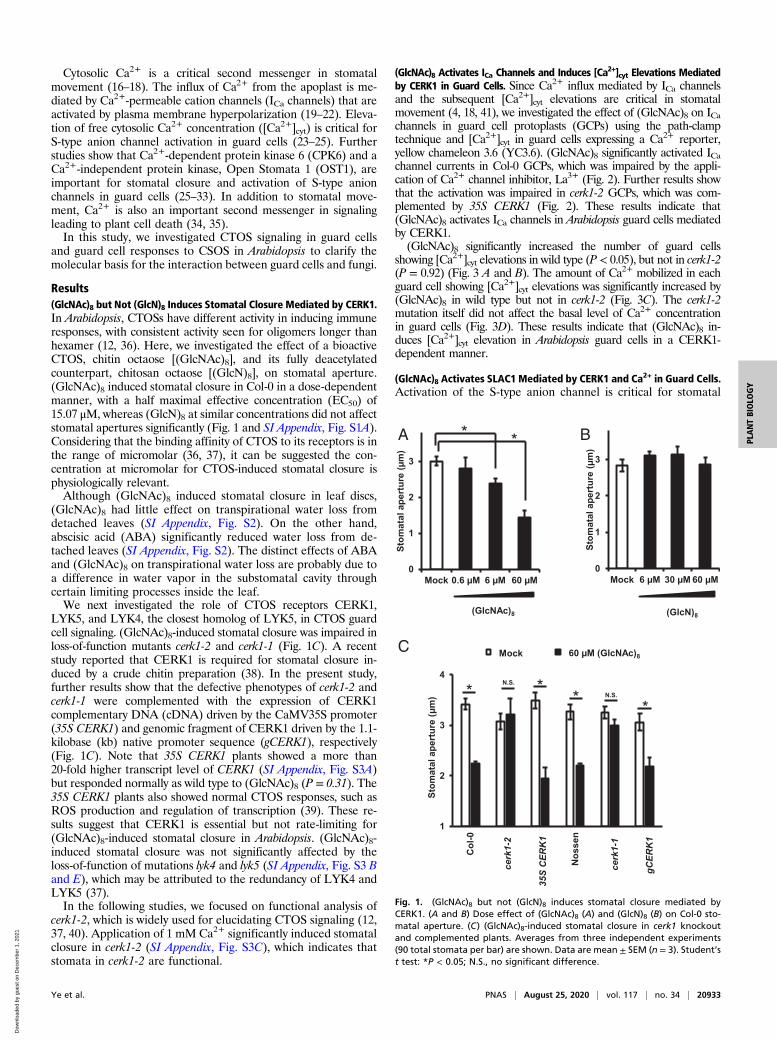
Results(GlcNAc)8 but Not (GlcN)8 Induces Stomatal Closure Mediated by CERK1.In Arabidopsis, CTOSs have different activity in inducing immuneresponses, with consistent activity seen for oligomers longer thanhexamer (12, 36). Here, we investigated the effect of a bioactiveCTOS, chitin octaose [(GlcNAc)8], and its fully deacetylatedcounterpart, chitosan octaose [(GlcN)8], on stomatal aperture.(GlcNAc)8 induced stomatal closure in Col-0 in a dose-dependentmanner, with a half maximal effective concentration (EC50) of15.07 μM, whereas (GlcN)8 at similar concentrations did not affectstomatal apertures significantly (Fig. 1 and SI Appendix, Fig. S1A).Considering that the binding affinity of CTOS to its receptors is inthe range of micromolar (36, 37), it can be suggested the con-centration at micromolar for CTOS-induced stomatal closure isphysiologically relevant.Although (GlcNAc)8 induced stomatal closure in leaf discs,
(GlcNAc)8 had little effect on transpirational water loss fromdetached leaves (SI Appendix, Fig. S2). On the other hand,abscisic acid (ABA) significantly reduced water loss from de-tached leaves (SI Appendix, Fig. S2). The distinct effects of ABAand (GlcNAc)8 on transpirational water loss are probably due toa difference in water vapor in the substomatal cavity throughcertain limiting processes inside the leaf.We next investigated the role of CTOS receptors CERK1,
LYK5, and LYK4, the closest homolog of LYK5, in CTOS guardcell signaling. (GlcNAc)8-induced stomatal closure was impaired inloss-of-function mutants cerk1-2 and cerk1-1 (Fig. 1C). A recentstudy reported that CERK1 is required for stomatal closure in-duced by a crude chitin preparation (38). In the present study,further results show that the defective phenotypes of cerk1-2 andcerk1-1 were complemented with the expression of CERK1complementary DNA (cDNA) driven by the CaMV35S promoter(35S CERK1) and genomic fragment of CERK1 driven by the 1.1-kilobase (kb) native promoter sequence (gCERK1), respectively(Fig. 1C). Note that 35S CERK1 plants showed a more than20-fold higher transcript level of CERK1 (SI Appendix, Fig. S3A)but responded normally as wild type to (GlcNAc)8 (P = 0.31). The35S CERK1 plants also showed normal CTOS responses, such asROS production and regulation of transcription (39). These re-sults suggest that CERK1 is essential but not rate-limiting for(GlcNAc)8-induced stomatal closure in Arabidopsis. (GlcNAc)8-induced stomatal closure was not significantly affected by theloss-of-function of mutations lyk4 and lyk5 (SI Appendix, Fig. S3 Band E), which may be attributed to the redundancy of LYK4 andLYK5 (37).In the following studies, we focused on functional analysis of
cerk1-2, which is widely used for elucidating CTOS signaling (12,37, 40). Application of 1 mM Ca2+ significantly induced stomatalclosure in cerk1-2 (SI Appendix, Fig. S3C), which indicates thatstomata in cerk1-2 are functional.
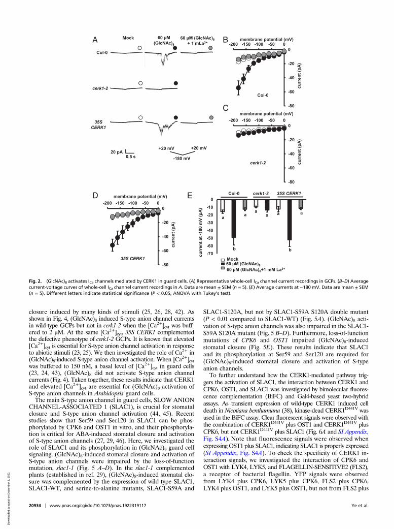
(GlcNAc)8 Activates ICa Channels and Induces [Ca2+]cyt Elevations Mediatedby CERK1 in Guard Cells. Since Ca2+ influx mediated by ICa channelsand the subsequent [Ca2+]cyt elevations are critical in stomatalmovement (4, 18, 41), we investigated the effect of (GlcNAc)8 on ICachannels in guard cell protoplasts (GCPs) using the path-clamptechnique and [Ca2+]cyt in guard cells expressing a Ca2+ reporter,yellow chameleon 3.6 (YC3.6). (GlcNAc)8 significantly activated ICachannel currents in Col-0 GCPs, which was impaired by the appli-cation of Ca2+ channel inhibitor, La3+ (Fig. 2). Further results showthat the activation was impaired in cerk1-2 GCPs, which was com-plemented by 35S CERK1 (Fig. 2). These results indicate that(GlcNAc)8 activates ICa channels in Arabidopsis guard cells mediatedby CERK1.(GlcNAc)8 significantly increased the number of guard cells
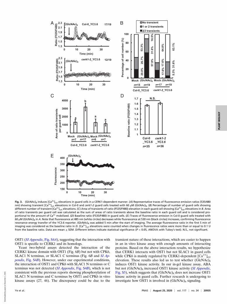
showing [Ca2+]cyt elevations in wild type (P < 0.05), but not in cerk1-2(P = 0.92) (Fig. 3 A and B). The amount of Ca2+ mobilized in eachguard cell showing [Ca2+]cyt elevations was significantly increased by(GlcNAc)8 in wild type but not in cerk1-2 (Fig. 3C). The cerk1-2mutation itself did not affect the basal level of Ca2+ concentrationin guard cells (Fig. 3D). These results indicate that (GlcNAc)8 in-duces [Ca2+]cyt elevation in Arabidopsis guard cells in a CERK1-dependent manner.
(GlcNAc)8 Activates SLAC1 Mediated by CERK1 and Ca2+ in Guard Cells.Activation of the S-type anion channel is critical for stomatal
0
1
2
3
Mock 0.6 μM 6 μM 60 μM
)mμ( erutrep a lata
motS
(GlcNAc)8
* *
0
1
2
3
Mock 6 μM 30 μM 60 μMSt
omat
al a
pert
ure
(μm
)
(GlcN)8
A B
1
2
3
4
Col
-0
Nos
sen
Control (GlcNAc)8gC
ERK
1
cerk
1-1
* ***
Stom
atal
ape
rtur
e (μ
m)
35S
CER
K1
60 μM (GlcNAc)8
cerk
1-2
Mock
N.S.
N.S.
C
Fig. 1. (GlcNAc)8 but not (GlcN)8 induces stomatal closure mediated byCERK1. (A and B) Dose effect of (GlcNAc)8 (A) and (GlcN)8 (B) on Col-0 sto-matal aperture. (C) (GlcNAc)8-induced stomatal closure in cerk1 knockoutand complemented plants. Averages from three independent experiments(90 total stomata per bar) are shown. Data are mean ± SEM (n = 3). Student’st test: *P < 0.05; N.S., no significant difference.
Ye et al. PNAS | August 25, 2020 | vol. 117 | no. 34 | 20933
PLANTBIOLO
GY
Dow
nloa
ded
by g
uest
on
Dec
embe
r 1,
202
1
closure induced by many kinds of stimuli (25, 26, 28, 42). Asshown in Fig. 4, (GlcNAc)8 induced S-type anion channel currentsin wild-type GCPs but not in cerk1-2 when the [Ca2+]cyt was buff-ered to 2 μM. At the same [Ca2+]cyt, 35S CERK1 complementedthe defective phenotype of cerk1-2 GCPs. It is known that elevated[Ca2+]cyt is essential for S-type anion channel activation in responseto abiotic stimuli (23, 25). We then investigated the role of Ca2+ in(GlcNAc)8-induced S-type anion channel activation. When [Ca2+]cytwas buffered to 150 nM, a basal level of [Ca2+]cyt in guard cells(23, 24, 43), (GlcNAc)8 did not activate S-type anion channelcurrents (Fig. 4). Taken together, these results indicate that CERK1and elevated [Ca2+]cyt are essential for (GlcNAc)8 activation ofS-type anion channels in Arabidopsis guard cells.The main S-type anion channel in guard cells, SLOW ANION
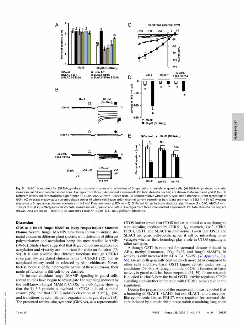
CHANNEL-ASSOCIATED 1 (SLAC1), is crucial for stomatalclosure and S-type anion channel activation (44, 45). Recentstudies show that Ser59 and Ser120 in SLAC1 can be phos-phorylated by CPK6 and OST1 in vitro, and their phosphoryla-tion is critical for ABA-induced stomatal closure and activationof S-type anion channels (27, 29, 46). Here, we investigated therole of SLAC1 and its phosphorylation in (GlcNAc)8 guard cellsignaling. (GlcNAc)8-induced stomatal closure and activation ofS-type anion channels were impaired by the loss-of-functionmutation, slac1-1 (Fig. 5 A–D). In the slac1-1 complementedplants (established in ref. 29), (GlcNAc)8-induced stomatal clo-sure was complemented by the expression of wild-type SLAC1,SLAC1-WT, and serine-to-alanine mutants, SLAC1-S59A and
SLAC1-S120A, but not by SLAC1-S59A S120A double mutant(P < 0.01 compared to SLAC1-WT) (Fig. 5A). (GlcNAc)8 acti-vation of S-type anion channels was also impaired in the SLAC1-S59A S120A mutant (Fig. 5 B–D). Furthermore, loss-of-functionmutations of CPK6 and OST1 impaired (GlcNAc)8-inducedstomatal closure (Fig. 5E). These results indicate that SLAC1and its phosphorylation at Ser59 and Ser120 are required for(GlcNAc)8-induced stomatal closure and activation of S-typeanion channels.To further understand how the CERK1-mediated pathway trig-
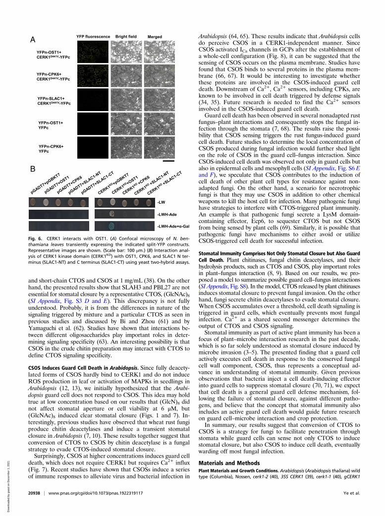
gers the activation of SLAC1, the interaction between CERK1 andCPK6, OST1, and SLAC1 was investigated by bimolecular fluores-cence complementation (BiFC) and Gal4-based yeast two-hybridassays. As transient expression of wild-type CERK1 induced celldeath in Nicotiana benthamiana (38), kinase-dead CERK1D441V wasused in the BiFC assay. Clear fluorescent signals were observed withthe combination of CERK1D441V plus OST1 and CERK1D441V plusCPK6, but not CERK1D441V plus SLAC1 (Fig. 6A and SI Appendix,Fig. S4A). Note that fluorescence signals were observed whenexpressing OST1 plus SLAC1, indicating SLAC1 is properly expressed(SI Appendix, Fig. S4A). To check the specificity of CERK1 in-teraction signals, we investigated the interaction of CPK6 andOST1 with LYK4, LYK5, and FLAGELLIN-SENSITIVE2 (FLS2),a receptor of bacterial flagellin. YFP signals were observedfrom LYK4 plus CPK6, LYK5 plus CPK6, FLS2 plus CPK6,LYK4 plus OST1, and LYK5 plus OST1, but not from FLS2 plus
A B
E
-80
-60
-40
-20
0-200 -150 -100 -50 0
curr
ent (
pA)
membrane potential (mV)
Col-0
-80
-60
-40
-20
0-200 -150 -100 -50 0
curr
ent (
pA)
membrane potential (mV)
35S CERK1
-80
-60
-40
-20
0-200 -150 -100 -50 0
curr
ent (
pA)
membrane potential (mV)
cerk1-2
C
D
cerk1-2
Col-0
35SCERK1
Mock 60 μM(GlcNAc)8
60 μM (GlcNAc)8+ 1 mLa3+
0.5 s20 pA
+20 mV +20 mV
-180 mV
-70-60-50-40-30-20-10
0Col-0 cerk1-2 35S CERK1
curr
ent a
t -18
0 m
V(p
A)
a aa
a aa
b b
Mock60 μM (GlcNAc)8
a a
60 μM (GlcNAc)8+1 mM La3+
Fig. 2. (GlcNAc)8 activates ICa channels mediated by CERK1 in guard cells. (A) Representative whole-cell ICa channel current recordings in GCPs. (B–D) Averagecurrent-voltage curves of whole-cell ICa channel current recordings in A. Data are mean ± SEM (n = 5). (E) Average currents at −180 mV. Data are mean ± SEM(n = 5). Different letters indicate statistical significance (P < 0.05, ANOVA with Tukey’s test).
20934 | www.pnas.org/cgi/doi/10.1073/pnas.1922319117 Ye et al.
Dow
nloa
ded
by g
uest
on
Dec
embe
r 1,
202
1
OST1 (SI Appendix, Fig. S4A), suggesting that the interaction withOST1 is specific to CERK1 and its homologs.Yeast two-hybrid assays detected the interaction of the
CERK1 kinase domain with OST1 (Fig. 6B) but not with CPK6,SLAC1 N terminus, or SLAC1 C terminus (Fig. 6B and SI Ap-pendix, Fig. S4B). However, under our experimental conditions,the interaction of OST1 and CPK6 with SLAC1 N terminus or Cterminus was not detected (SI Appendix, Fig. S4B), which is notconsistent with the previous reports showing phosphorylation ofSLAC1 N terminus and C terminus by OST1 and CPK6 in vitrokinase assays (27, 46). The discrepancy could be due to the
transient nature of these interactions, which are easier to happenin an in vitro kinase assay with enough amounts of interactingproteins. Based on the above interaction results, we hypothesizethat CERK1 interacts with OST1 but not SLAC1 in guard cellswhile CPK6 is mainly regulated by CERK1-dependent [Ca2+]cytelevation. These results also led us to test whether (GlcNAc)8induces OST1 kinase activity. In our in-gel kinase assay, ABAbut not (GlcNAc)8 increased OST1 kinase activity (SI Appendix,Fig. S5), which suggests that (GlcNAc)8 does not increase OST1kinase activity in guard cells. Further research is undergoing toinvestigate how OST1 is involved in (GlcNAc)8 signaling.
1.31.51.71.92.12.3
0 10 20 30
Rat
io (F
535/
F480
)Time (min)
(GlcNAc)8 Col-0_YC3.6 17/19A
1.31.51.71.92.12.3
0 10 20 30
Rat
io (F
535/
F480
)
Time (min)
(GlcNAc)8cerk1-2_YC3.6 12/19
0
20
40
60
80
100
Col-0 cpk6-2
Perc
enta
ge o
f cel
l num
ber (
%)
No transient1 or 2 transients≧3 transients
(GlcNAc)8Mock Mock (GlcNAc)8
n=15 n=19 n=17 n=19
53.3
%26
.7%
20.0
%
10.5
%31
.6%
57.9
%
64.8
%17
.6%
17.6
%
63.1
%31
.6%
5.3%
Col-0_YC3.6 cerk1-2_YC3.6
0
1000
2000
3000
4000
mock GN8Mock (GlcNAc)8 Mock (GlcNAc)8n=7 n=17 n=6 n=7Ar
eaof
ratio
tran
sien
tspe
r gua
rd c
ell
a
b
aa
Col-0_YC3.6 cerk1-2_YC3.6
1
1.1
1.2
1.3
1.4
1.5
1.6
1.7
Col-0 cerk1cerk1-2_YC3.6
n=35 n=36
Bas
elin
e ra
tio (F
535/
F480
)
Col-0_YC3.6
N.S.
B
C D
150
200
250
300
350
400
0 10 20 30
ecnecseroulF
Time (min)
(GlcNAc)8E
Fig. 3. (GlcNAc)8 induces [Ca2+]cyt elevations in guard cells in a CERK1-dependent manner. (A) Representative traces of fluorescence emission ratios (535/480
nm) showing transient [Ca2+]cyt elevations in Col-0 and cerk1-2 guard cells treated with 60 μM (GlcNAc)8. (B) Percentage of number of guard cells showingdifferent number of transient [Ca2+]cyt elevations. (C) Area of transients of ratio (F535/F480) elevation in each guard cell showing [Ca2+]cyt elevations in B. Areaof ratio transients per guard cell was calculated as the sum of areas of ratio transients above the baseline ratio in each guard cell and is considered pro-portional to the amount of Ca2+ mobilized. (D) Baseline ratio (F535/F480) in guard cells. (E) Traces of fluorescence emission in Col-0 guard cells treated with60 μM (GlcNAc)8 in A. Note that fluorescence at 485 nm (white circles) decreases while fluorescence at 530 nm (black circles) increases, confirming fluorescenceresonance energy transfer of the YC3.6 reporter. (GlcNAc)8 was added 5 min after the start of imaging. The average fluorescence ratio in the first 5 min ofimaging was considered as the baseline ratio in D. [Ca2+]cyt elevations were counted when changes in fluorescence ratios were more than or equal to 0.1 Ufrom the baseline ratio. Data are mean ± SEM. Different letters indicate statistical significance (P < 0.05, ANOVA with Tukey’s test). N.S., not significant.
Ye et al. PNAS | August 25, 2020 | vol. 117 | no. 34 | 20935
PLANTBIOLO
GY
Dow
nloa
ded
by g
uest
on
Dec
embe
r 1,
202
1
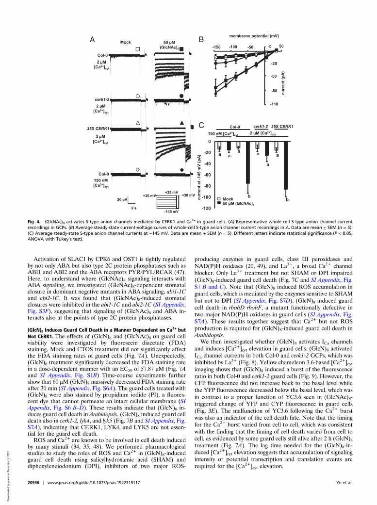
Activation of SLAC1 by CPK6 and OST1 is tightly regulatedby not only ABA but also type 2C protein phosphatases such asABI1 and ABI2 and the ABA receptors PYR/PYL/RCAR (47).Here, to understand where (GlcNAc)8 signaling interacts withABA signaling, we investigated (GlcNAc)8-dependent stomatalclosure in dominant negative mutants in ABA signaling, abi1-1Cand abi2-1C. It was found that (GlcNAc)8-induced stomatalclosures were inhibited in the abi1-1C and abi2-1C (SI Appendix,Fig. S3F), suggesting that signaling of (GlcNAc)8 and ABA in-teracts also at the points of type 2C protein phosphatases.
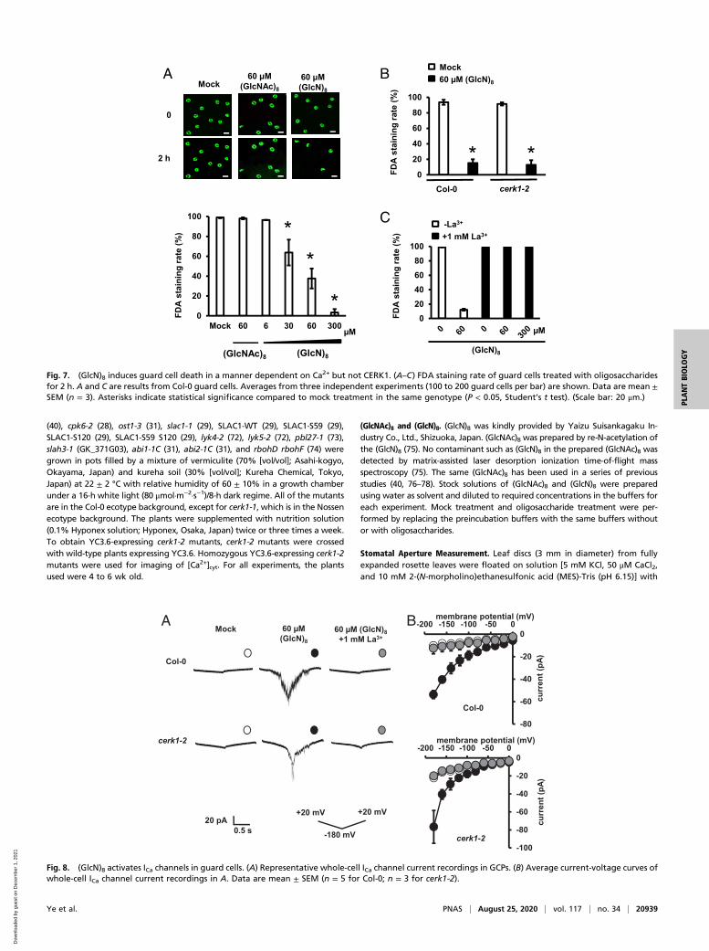
(GlcN)8 Induces Guard Cell Death in a Manner Dependent on Ca2+ butNot CERK1. The effects of (GlcN)8 and (GlcNAc)8 on guard cellviability were investigated by fluorescein diacetate (FDA)staining. Mock and CTOS treatment did not significantly affectthe FDA staining rates of guard cells (Fig. 7A). Unexpectedly,(GlcN)8 treatment significantly decreased the FDA staining ratein a dose-dependent manner with an EC50 of 57.87 μM (Fig. 7Aand SI Appendix, Fig. S1B) Time-course experiments furthershow that 60 μM (GlcN)8 massively decreased FDA staining rateafter 30 min (SI Appendix, Fig. S6A). The guard cells treated with(GlcN)8 were also stained by propidium iodide (PI), a fluores-cent dye that cannot permeate an intact cellular membrane (SIAppendix, Fig. S6 B–D). These results indicate that (GlcN)8 in-duces guard cell death in Arabidopsis. (GlcN)8 induced guard celldeath also in cerk1-2, lyk4, and lyk5 (Fig. 7B and SI Appendix, Fig.S7A), indicating that CERK1, LYK4, and LYK5 are not essen-tial for the guard cell death.ROS and Ca2+ are known to be involved in cell death induced
by many stimuli (34, 35, 48). We performed pharmacologicalstudies to study the roles of ROS and Ca2+ in (GlcN)8-inducedguard cell death using salicylhydroxamic acid (SHAM) anddiphenyleneiodonium (DPI), inhibitors of two major ROS-
producing enzymes in guard cells, class III peroxidases andNAD(P)H oxidases (20, 49), and La3+, a broad Ca2+ channelblocker. Only La3+ treatment but not SHAM or DPI impaired(GlcN)8-induced guard cell death (Fig. 7C and SI Appendix, Fig.S7 B and C). Note that (GlcN)8 induced ROS accumulation inguard cells, which is mediated by the enzymes sensitive to SHAMbut not to DPI (SI Appendix, Fig. S7D). (GlcN)8 induced guardcell death in rbohD rbohF, a mutant functionally defective intwo major NAD(P)H oxidases in guard cells (SI Appendix, Fig.S7A). These results together suggest that Ca2+ but not ROSproduction is required for (GlcN)8-induced guard cell death inArabidopsis.We then investigated whether (GlcN)8 activates ICa channels
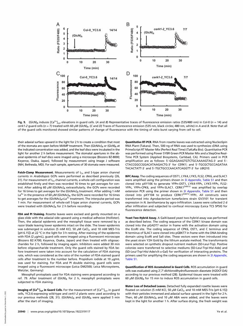
and induces [Ca2+]cyt elevation in guard cells. (GlcN)8 activatedICa channel currents in both Col-0 and cerk1-2 GCPs, which wasinhibited by La3+ (Fig. 8). Yellow chameleon 3.6-based [Ca2+]cytimaging shows that (GlcN)8 induced a burst of the fluorescenceratio in both Col-0 and cerk1-2 guard cells (Fig. 9). However, theCFP fluorescence did not increase back to the basal level whilethe YFP fluorescence decreased below the basal level, which wasin contrast to a proper function of YC3.6 seen in (GlcNAc)8-triggered change of YFP and CYP fluorescence in guard cells(Fig. 3E). The malfunction of YC3.6 following the Ca2+ burstwas also an indicator of the cell death fate. Note that the timingfor the Ca2+ burst varied from cell to cell, which was consistentwith the finding that the timing of cell death varied from cell tocell, as evidenced by some guard cells still alive after 2 h (GlcN)8treatment (Fig. 7A). The lag time needed for the (GlcN)8-in-duced [Ca2+]cyt elevation suggests that accumulation of signalingintensity or potential transcription and translation events arerequired for the [Ca2+]cyt elevation.
-150 -100 -50 0 50
curr
ent (
pA)
membrane potential (mV)
-20
-50
-80
-110
0-50-100-150 50
-120
-100
-80
-60
-40
-20
0150 nM [Ca2+]cyt 2 μM [Ca2+]cyt
Col-0 cerk1-2 35S CERK1
curr
ent a
t -14
5 m
V(p
A )
a aa a
a
a
bbMock
60 μM (GlcNAc)8
B
C
20 pA
2 s
+30 mV+35 mV
-145 mV
+30 mV
cerk1-2
Col-0
35S CERK1
Col-0150 nM[Ca2+]cyt
Mock 60 μM (GlcNAc)8
A
2 μM [Ca2+]cyt
2 μM [Ca2+]cyt
2 μM [Ca2+]cyt
Fig. 4. (GlcNAc)8 activates S-type anion channels mediated by CERK1 and Ca2+ in guard cells. (A) Representative whole-cell S-type anion channel currentrecordings in GCPs. (B) Average steady-state current-voltage curves of whole-cell S-type anion channel current recordings in A. Data are mean ± SEM (n = 5).(C) Average steady-state S-type anion channel currents at −145 mV. Data are mean ± SEM (n = 5). Different letters indicate statistical significance (P < 0.05,ANOVA with Tukey’s test).
20936 | www.pnas.org/cgi/doi/10.1073/pnas.1922319117 Ye et al.
Dow
nloa
ded
by g
uest
on
Dec
embe
r 1,
202
1
DiscussionCTOS as a Model Fungal MAMP to Study Fungus-Induced StomatalClosure. Several fungal MAMPs have been shown to induce sto-matal closure in different plant species, with chitosans of differentpolymerization and acetylation being the most studied MAMPs(50–52). Studies have suggested that degree of polymerization andacetylation and viscosity are important for chitosan function (53,54). It is also possible that chitosan functions through CERK1since partially acetylated chitosan binds to CERK1 (12), and itsacetylated moiety could be released by plant chitinases. Never-theless, because of the heterogenic nature of these chitosans, theirmode of function is difficult to be clarified.To further elucidate fungal MAMP signaling in guard cells,
recent studies have begun to investigate the signaling induced bythe well-known fungal MAMP, CTOS, in Arabidopsis, showingthat the 14-3-3 protein is involved in CTOS-induced stomatalclosure (55) and that CTOS induces elevation of [Ca2+]cyt (56)and transitions in actin filament organization in guard cells (14).The presented results using synthetic (GlcNAc)8 as a representative
CTOS further reveal that CTOS induces stomatal closure through acore signaling mediated by CERK1, ICa channels, Ca2+, CPK6,PP2Cs, OST1, and SLAC1 in Arabidopsis. Given that OST1 andSLAC1 are guard cell-specific genes, it will be interesting to in-vestigate whether their homologs play a role in CTOS signaling inother cell types.Although OST1 is required for stomatal closure induced by
ABA, methyl jasmonate, CO2, flg22, and fungal MAMPs, itsactivity is only increased by ABA (31, 57–59) (SI Appendix, Fig.S5). Guard cells generally contain much more ABA compared toother cells and have basal OST1 kinase activity under restingconditions (59, 60). Although a model of OST1 function at basalactivity in guard cells has been proposed (31, 59), future researchis needed to clarify how the basal OST1 activity regulates CTOSsignaling and whether interaction with CERK1 plays a role in theregulation.During the preparation of the manuscript, it was reported that
a homolog of SLAC1, SLAH3, but not SLAC1, and a receptor-like cytoplasmic kinase, PBL27, were required for stomatal clo-sure induced by a crude chitin preparation containing long-chain
-150 -100 -50 0 50
curr
ent (
pA)
membrane potential (mV)
-20
-50
-80
-110
0-50-100-150 50
-140
C
-150
-100
-50
0
Col-0 slac1-1SLAC1-
S59A S120A
curr
ent a
t -14
5 m
V( p
A)
aaa
c
a
b
Mock60 μM (GlcNAc)8
0
1
2
3
4
Mock GN8Col-0 slac1-1SLAC1-WT SLAC1-S59ASLAC1-S120A SLAC1-S59A S120A
slac1-1
( erutrep a latamotS
μm)
60 μM(GlcNAc)8
a a a a aa
bcc c
bc
ab
a
D
E
20 pA
2 s
+30 mV
+35 mV
-145 mV
+30 mV
slac1-1
Col-0
SLAC1-S59A S120A
Mock 60 μM (GlcNAc)8
0
1
2
3
Col-0
Control GN8
*
cpk6-2 ost1-3
Stom
atal
ape
rtur
e ( μ
m)
60 μM (GlcNAc)8
N.S.N.S.
B
A
Fig. 5. SLAC1 is required for (GlcNAc)8-induced stomatal closure and activation of S-type anion channels in guard cells. (A) (GlcNAc)8-induced stomatalclosure in slac1-1 and complemented lines. Averages from three independent experiments (90 total stomata per bar) are shown. Data are mean ± SEM (n = 3).Different letters indicate statistical significance (P < 0.05, ANOVA with Tukey’s test). (B) Representative whole-cell S-type anion channel current recordings inGCPs. (C) Average steady-state current-voltage curves of whole-cell S-type anion channel current recordings in A. Data are mean ± SEM (n = 5). (D) Averagesteady-state S-type anion channel currents at −145 mV. Data are mean ± SEM (n = 5). Different letters indicate statistical significance (P < 0.05, ANOVA withTukey’s test). (E) (GlcNAc)8-induced stomatal closure in Col-0, cpk6-2, and ost1-3. Averages from three independent experiments (90 total stomata per bar) areshown. Data are mean ± SEM (n = 3). Student’s t test: *P < 0.05. N.S., no significant difference.
Ye et al. PNAS | August 25, 2020 | vol. 117 | no. 34 | 20937
PLANTBIOLO
GY
Dow
nloa
ded
by g
uest
on
Dec
embe
r 1,
202
1
and short-chain CTOS and CSOS at 1 mg/mL (38). On the otherhand, the presented results show that SLAH3 and PBL27 are notessential for stomatal closure by a representative CTOS, (GlcNAc)8(SI Appendix, Fig. S3 D and E). This discrepancy is not fullyunderstood. Probably, it is from the differences in nature of thesignaling triggered by mixture and a particular CTOS as seen inprevious studies and discussed by Bi and Zhou (61) and byYamaguchi et al. (62). Studies have shown that interactions be-tween different oligosaccharides play important roles in deter-mining signaling specificity (63). An interesting possibility is thatCSOS in the crude chitin preparation may interact with CTOS todefine CTOS signaling specificity.
CSOS Induces Guard Cell Death in Arabidopsis. Since fully deacety-lated forms of CSOS hardly bind to CERK1 and do not induceROS production in leaf or activation of MAPKs in seedlings inArabidopsis (12, 13), we initially hypothesized that the Arabi-dopsis guard cell does not respond to CSOS. This idea may holdtrue at low concentration based on our results that (GlcN)8 didnot affect stomatal aperture or cell viability at 6 μM, but(GlcNAc)8 induced clear stomatal closure (Figs. 1 and 7). In-terestingly, previous studies have observed that wheat rust fungiproduce chitin deacetylases and induce a transient stomatalclosure in Arabidopsis (7, 10). These results together suggest thatconversion of CTOS to CSOS by chitin deacetylase is a fungalstrategy to evade CTOS-induced stomatal closure.Surprisingly, CSOS at higher concentrations induces guard cell
death, which does not require CERK1 but requires Ca2+ influx(Fig. 7). Recent studies have shown that CSOSs induce a seriesof immune responses to alleviate virus and bacterial infection in
Arabidopsis (64, 65). These results indicate that Arabidopsis cellsdo perceive CSOS in a CERK1-independent manner. SinceCSOS activated ICa channels in GCPs after the establishment ofa whole-cell configuration (Fig. 8), it can be suggested that thesensing of CSOS occurs on the plasma membrane. Studies havefound that CSOS binds to several proteins in the plasma mem-brane (66, 67). It would be interesting to investigate whetherthese proteins are involved in the CSOS-induced guard celldeath. Downstream of Ca2+, Ca2+ sensors, including CPKs, areknown to be involved in cell death triggered by defense signals(34, 35). Future research is needed to find the Ca2+ sensorsinvolved in the CSOS-induced guard cell death.Guard cell death has been observed in several nonadapted rust
fungus–plant interactions and consequently stops the fungal in-fection through the stomata (7, 68). The results raise the possi-bility that CSOS sensing triggers the rust fungus-induced guardcell death. Future studies to determine the local concentration ofCSOS produced during fungal infection would further shed lighton the role of CSOS in the guard cell–fungus interaction. SinceCSOS-induced cell death was observed not only in guard cells butalso in epidermal cells and mesophyll cells (SI Appendix, Fig. S6 Eand F), we speculate that CSOS contributes to the induction ofcell death of other plant cell types for resistance against non-adapted fungi. On the other hand, a scenario for necrotrophicfungi is that they may use CSOS in addition to other chemicalweapons to kill the host cell for infection. Many pathogenic fungihave strategies to interfere with CTOS-triggered plant immunity.An example is that pathogenic fungi secrete a LysM domain-containing effector, Ecp6, to sequester CTOS but not CSOSfrom being sensed by plant cells (69). Similarly, it is possible thatpathogenic fungi have mechanisms to either avoid or utilizeCSOS-triggered cell death for successful infection.
Stomatal Immunity Comprises Not Only Stomatal Closure but Also GuardCell Death. Plant chitinases, fungal chitin deacetylases, and theirhydrolysis products, such as CTOS and CSOS, play important rolesin plant–fungus interaction (8, 9). Based on our results, we pro-posed a model to summarize possible guard cell–fungus interactions(SI Appendix, Fig. S8). In the model, CTOS released by plant chitinasesinduces stomatal closure to prevent fungal invasion. On the otherhand, fungi secrete chitin deacetylases to evade stomatal closure.When CSOS accumulates over a threshold, cell death signaling istriggered in guard cells, which eventually prevents most fungalinfection. Ca2+ as a shared second messenger determines theoutput of CTOS and CSOS signaling.Stomatal immunity as part of active plant immunity has been a
focus of plant–microbe interaction research in the past decade,which is so far solely understood as stomatal closure induced bymicrobe invasion (3–5). The presented finding that a guard cellactively executes cell death in response to the conserved fungalcell wall component, CSOS, thus represents a conceptual ad-vance in understanding of stomatal immunity. Given previousobservations that bacteria inject a cell death-inducing effectorinto guard cells to suppress stomatal closure (70, 71), we expectthat cell death is a general guard cell defense mechanism, fol-lowing the failure of stomatal closure, against different patho-gens, and believe that the concept that stomatal immunity alsoincludes an active guard cell death would guide future researchon guard cell–microbe interaction and crop protection.In summary, our results suggest that conversion of CTOS to
CSOS is a strategy for fungi to facilitate penetration throughstomata while guard cells can sense not only CTOS to inducestomatal closure, but also CSOS to induce cell death, eventuallywarding off most fungal infection.
Materials and MethodsPlant Materials and Growth Conditions. Arabidopsis (Arabidopsis thaliana) wildtype (Columbia), Nossen, cerk1-2 (40), 35S CERK1 (39), cerk1-1 (40), gCERK1
YFP fluorescence Bright field Merged
YFPn-OST1+CERK1D441V-YFPc
YFPn-CPK6+CERK1D441V-YFPc
YFPn-SLAC1+CERK1D441V-YFPc
YFPn-OST1+YFPc
YFPn-CPK6+YFPc
A
B
-LW
-LWH-Ade
-LWH-Ade+α-Gal
Fig. 6. CERK1 interacts with OST1. (A) Confocal microscopy of N. ben-thamiana leaves transiently expressing the indicated split-YFP constructs.Representative images are shown. (Scale bar: 100 μm.) (B) Interaction anal-ysis of CERK1 kinase domain (CERK1KD) with OST1, CPK6, and SLAC1 N ter-minus (SLAC1-NT) and C terminus (SLAC1-CT) using yeast two-hybrid assays.
20938 | www.pnas.org/cgi/doi/10.1073/pnas.1922319117 Ye et al.
Dow
nloa
ded
by g
uest
on
Dec
embe
r 1,
202
1
(40), cpk6-2 (28), ost1-3 (31), slac1-1 (29), SLAC1-WT (29), SLAC1-S59 (29),SLAC1-S120 (29), SLAC1-S59 S120 (29), lyk4-2 (72), lyk5-2 (72), pbl27-1 (73),slah3-1 (GK_371G03), abi1-1C (31), abi2-1C (31), and rbohD rbohF (74) weregrown in pots filled by a mixture of vermiculite (70% [vol/vol]; Asahi-kogyo,Okayama, Japan) and kureha soil (30% [vol/vol]; Kureha Chemical, Tokyo,Japan) at 22 ± 2 °C with relative humidity of 60 ± 10% in a growth chamberunder a 16-h white light (80 μmol·m−2·s−1)/8-h dark regime. All of the mutantsare in the Col-0 ecotype background, except for cerk1-1, which is in the Nossenecotype background. The plants were supplemented with nutrition solution(0.1% Hyponex solution; Hyponex, Osaka, Japan) twice or three times a week.To obtain YC3.6-expressing cerk1-2 mutants, cerk1-2 mutants were crossedwith wild-type plants expressing YC3.6. Homozygous YC3.6-expressing cerk1-2mutants were used for imaging of [Ca2+]cyt. For all experiments, the plantsused were 4 to 6 wk old.
(GlcNAc)8 and (GlcN)8. (GlcN)8 was kindly provided by Yaizu Suisankagaku In-dustry Co., Ltd., Shizuoka, Japan. (GlcNAc)8 was prepared by re-N-acetylation ofthe (GlcN)8 (75). No contaminant such as (GlcN)8 in the prepared (GlcNAc)8 wasdetected by matrix-assisted laser desorption ionization time-of-flight massspectroscopy (75). The same (GlcNAc)8 has been used in a series of previousstudies (40, 76–78). Stock solutions of (GlcNAc)8 and (GlcN)8 were preparedusing water as solvent and diluted to required concentrations in the buffers foreach experiment. Mock treatment and oligosaccharide treatment were per-formed by replacing the preincubation buffers with the same buffers withoutor with oligosaccharides.
Stomatal Aperture Measurement. Leaf discs (3 mm in diameter) from fullyexpanded rosette leaves were floated on solution [5 mM KCl, 50 μM CaCl2,and 10 mM 2-(N-morpholino)ethanesulfonic acid (MES)-Tris (pH 6.15)] with
0
20
40
60
80
100
Mock 60 6 30 60 300
FDA
stai
ning
rate
(%)
(GlcN)8(GlcNAc)8
**
*μM
0
2 h
Mock60 μM
(GlcNAc)8
60 μM(GlcN)8
A
C
020406080
100
FDA
stai
ning
rate
(%)
-La3+
+1 mM La3+
μM
(GlcN)8
B
0
20
40
60
80
100
FDA
stai
ning
rate
(%)
cerk1-2Col-0
Mock60 μM (GlcN)8
* *
Fig. 7. (GlcN)8 induces guard cell death in a manner dependent on Ca2+ but not CERK1. (A–C) FDA staining rate of guard cells treated with oligosaccharidesfor 2 h. A and C are results from Col-0 guard cells. Averages from three independent experiments (100 to 200 guard cells per bar) are shown. Data are mean ±SEM (n = 3). Asterisks indicate statistical significance compared to mock treatment in the same genotype (P < 0.05, Student’s t test). (Scale bar: 20 μm.)
-80
-60
-40
-20
0-200 -150 -100 -50 0
curr
ent (
pA)
membrane potential (mV)
Col-0
-100
-80
-60
-40
-20
0-200 -150 -100 -50 0
curr
ent (
pA)
membrane potential (mV)
cerk1-2
cerk1-2
Col-0
Mock 60 μM (GlcN)8
60 μM (GlcN)8+1 mM La3+
0.5 s20 pA
+20 mV +20 mV
-180 mV
A B
Fig. 8. (GlcN)8 activates ICa channels in guard cells. (A) Representative whole-cell ICa channel current recordings in GCPs. (B) Average current-voltage curves ofwhole-cell ICa channel current recordings in A. Data are mean ± SEM (n = 5 for Col-0; n = 3 for cerk1-2).
Ye et al. PNAS | August 25, 2020 | vol. 117 | no. 34 | 20939
PLANTBIOLO
GY
Dow
nloa
ded
by g
uest
on
Dec
embe
r 1,
202
1
their adaxial surface upward in the light for 2 h to create a condition that mostof the stomata are open before MAMP treatment. Then (GlcNAc)8 or (GlcN)8 atthe indicated concentration was added, and the leaf discs were incubated in thelight for another 2 h before measurement. The stomatal apertures in the ab-axial epidermis of leaf discs were imaged using a microscope (Biozero BZ-8000;Keyence, Osaka, Japan), followed by measurement using Image J software(NIH, Bethesda, MD). For each sample, apertures of 30 stomata were measured.
Patch-Clamp Measurement. Measurements of ICa and S-type anion channelcurrents in Arabidopsis GCPs were performed as described previously (26,31). For measurement of ICa channel currents, a whole-cell configuration wasestablished firstly and then was recorded 16 times to get averages for con-trol. After adding 60 μM (GlcNAc)8 extracellularly, the GCPs were recordedfor 16 times to get averages for the (GlcNAc)8 treatment. After adding 1 mMLa3+ in the presence of 60 μM (GlcNAc)8, the GCPs were recorded for 16 timesto get averages for the (GlcNAc)8+La
3+ treatment. The interpulse period was1 min. For measurement of whole-cell S-type anion channel currents, GCPswere treated with (GlcNAc)8 for 2 h before recordings.
FDA and PI Staining. Rosette leaves were excised and gently mounted on aglass slide with the adaxial side upward using a medical adhesive (Hollister).Then, the adaxial epidermis and the mesophyll tissue were removed by arazor blade leaving lower epidermis intact on the slide. The lower epidermiswas submerged in solution (5 mM KCl, 50 μM CaCl2, and 10 mM MES-Tris[pH 6.15]) at 22 °C in the light for 3 h resting. After staining of the epidermiswith FDA (2 μg/mL), guard cells were imaged using a fluorescent microscope(Biozero BZ-X700; Keyence, Osaka, Japan) and then treated with oligosac-charides for 2 h, followed by imaging again. Inhibitors were added 30 minbefore oligosaccharide treatment. Only the guard cells stained by FDA be-fore treatment were taken into account for the calculation of FDA stainingrate, which was considered as the ratio of the number of FDA-stained guardcells after treatment to the number before. Propidium iodide at 10 μg/mLwas used for staining. For FDA and PI double staining, guard cells wereimaged using a fluorescent microscope (Leica DM2500; Leica Microsystems,Wetzlar, Germany)
Mesophyll protoplasts used for FDA staining were prepared according toref. 79. After treatment of (GlcN)8 for 2 h, mesophyll protoplasts weresubjected to FDA staining.
Imaging of [Ca2+]cyt in Guard Cells. For the measurement of [Ca2+]cyt in guardcells, YC3.6-expressing wild-type and cerk1-2 plants were used according toour previous methods (28, 31). (GlcNAc)8 and (GlcN)8 were applied 5 minafter the start of imaging.
Quantitative RT-PCR. RNA from rosette leaves was extracted using NucleoSpinRNA Plant (Takara). Then, 500 ng of RNA was used to synthesize cDNA usingPrimeScript RT Master Mix (Perfect Real Time) (TaKaRa Bio). Quantitative PCRwas performed using Power SYBR Green PCRMaster Mix and a StepOne Real-Time PCR System (Applied Biosystems, Carlsbad, CA). Primers used in PCRamplification are as follows: 5′-GGAGAAGTGTCTGCAAAAGTAG-3′ and 5′-CTACCGGCCGGACATAAGACTG-3‘ for CERK1; and 5′-TGCGCTGCCAGATAATACACTATT-3′ and 5′-TGCTGCCCAACATCAGGTT-3′ for UBQ10.
BiFC Assay. The coding sequences of OST1, LYK4, LYK5, FLS2, CPK6, and SLAC1were amplified using the primers shown in SI Appendix, Table S1 and thencloned into pXY106 to generate YFPn-OST1, LYK4-YFPc, LYK5-YFPc, FLS2-YFPc, YFPn-CPK6, and YFPn-SLAC1. CERK1D441V was amplified by overlapextension PCR using the primer shown in SI Appendix, Table S1 and thencloned into pXY104 to produce CERK1D441V-YFPc. All constructs weretransformed into Agrobacterium tumefaciens strain GV3101 for transientexpression in N. benthamiana by agro-infiltration. Leaves were collected 2 dafter infiltration and subjected to confocal microscopy (Leica TCS SP5II) forfluorescence detection.
Yeast Two-Hybrid Assay. A Gal4-based yeast two-hybrid assay was performedas described below. The coding sequence of the CERK1 kinase domain wascloned into the pGADT7 vector in frame with the activation domain usingthe EcoRI site. The coding sequence of CPK6, OST1, and C terminus andN terminus of SLAC1 were cloned into pGBKT7 in frame with the DNA-bindingdomain using EcoRI and SalI sites. These vectors were then introduced intothe yeast strain Y2H Gold by the lithium acetate method. The transformantswere selected on synthetic dropout nutrient medium (SD-Leu/-Trp). Positivecolonies were transferred to selective mediums (SD-Leu/-Trp/-His/-Ade) and(SD-Leu/-Trp/-His/-Ade/+X-α-Gal) for verification of interacting proteins. Theprimers used for amplifying the coding sequences are shown in SI Appendix,Table S1.
Quantification of ROS Accumulated in Guard Cells. ROS accumulation in guardcells was evaluated using 2’,7’-dichlorodihydrofluorescein diacetate (H2DCF-DA)according to our previous method (28). Epidermal tissues were treated with60 μM (GlcN)8 for 15 min to induce ROS accumulation in guard cells.
Water Loss of Detached Leaves. Detached fully expanded rosette leaves werefloated on solution (5 mM KCl, 50 μM CaCl2, and 10 mM MES-Tris [pH 6.15])with their petioles immersed and adaxial surface upward in the light for 2 h.Then, 60 μM (GlcNAc)8 and 10 μM ABA were added, and the leaves werekept in the light for another 1 h. After surface drying, the fresh weight was
0123456
0 10 20 30)084F/535F( oita
R
Time (min)
Col-0_YC3.6
(GlcN)8
14/14
0
1
2
3
4
5
0 10 20 30
Rat
io (F
535/
F480
)
Time (min)
(GlcN)8
cerk1-2_YC3.6 7/7
0
20
40
60
80
100
0 10 20 30
ecnecseroulF
Time (min)
Col-0_YC3.6 14/14
0
20
40
60
80
100
0 10 20 30
Fluo
resc
ence
Time (min)
cerk1-2_YC3.6 7/7
A B
C D
Fig. 9. (GlcN)8 induces [Ca2+]cyt elevations in guard cells. (A and B) Representative traces of fluorescence emission ratios (535/480 nm) in Col-0 (n = 14) andcerk1-2 guard cells (n = 7) treated with 60 μM (GlcN)8. (C and D) Traces of fluorescence emission (535 nm, black circles; 480 nm, white) in A and B. Note that allof the guard cells monitored showed similar patterns of change of fluorescence with the timing of ratio burst varying from cell to cell.
20940 | www.pnas.org/cgi/doi/10.1073/pnas.1922319117 Ye et al.
Dow
nloa
ded
by g
uest
on
Dec
embe
r 1,
202
1
determined at indicated time points up to 2 h. Relative water loss wasrepresented by the weight of the leaves at various time points divided by theoriginal weight.
In-Gel Kinase Assay. Ten- to 14-d-old seedlings were treated with 10 μM ABAand 60 μM (GlcNAc)8 for 30 min and then flash frozen in liquid nitrogen forprotein extraction. An in-gel kinase assay using protein extracted from theseedlings was performed as described previously (29). The radioactivity ofthe gel was visualized using Imaging Plate BAS-IP MS 2325 (FUJIFILM, Tokyo,Japan) and BAS imaging analyzer FLA-7000 (FUJIFILM).
Statistical Analysis. Student’s t test and ANOVA with Tukey’s test were used toassess the significance of differences between datasets. The response of [Ca2+]cytwas assessed by χ2 test. Differences were considered significant for P < 0.05.
Accession Numbers. Arabidopsis Genome Initiative numbers for the genes dis-cussed in this article are as follows: ABI1 (AT4G26080), ABI2 (AT5G57050), CERK1(AT3G21630), CPK6 (AT2G17290), FLS2 (AT5G46330), LYK4 (AT2G23770), LYK5(AT2G33580), OST1 (AT4G33950), PBL27 (AT5G18610), RBOHD (AT5G47910),RBOHF (AT1G64060), SLAC1 (At1g12480), and SLAH3 (AT5G24030).
Data Availability. All study data are included in the article and SI Appendix.
ACKNOWLEDGMENTS. We thank Dr. Yoshitake Desaki (Meiji University) forpreparing materials; Dr. Julian I. Schroeder (University of California SanDiego) for providing seeds of slac1-1, SLAC1-WT, SLAC1-S59A, SLAC1-S120A,and SLAC1-S59A S120A; Dr. Takashi Hirayama (Institute of Plant Science andResources, Okayama University) for providing abi1-1C and abi2-1C seeds;and Dr. Yuichiro Takahashi (Okayama University) and Dr. Kazuhiro Toyoda(Okayama University) for the use of the BAS imaging analyzer FLA-7000 andimaging plates. This work was supported by the National Natural ScienceFoundation of China (31901984 to W.Y.); the Start-up Fund from ShanghaiJiao Tong University (WF220515002 to W.Y.); Grants-in-Aid for Japan Societyfor the Promotion of Science Fellows (267977 and 17F17091 to W.Y.) fromthe Japan Society for the Promotion of Science, National Key R & D Programof China (100300 to J.L.); the Shanghai Science and Technology Commission(18391900400 to J.L.); the China Agriculture Research System (CARS-29-yc-2to J.L.); Grants-in-Aid for Scientific Research from the Ministry of Education,Culture, Sports, Science and Technology, Japan (15H05956 to T.K.) and theAdvanced Low Carbon Technology Research and Development Programfrom the Japan Science and Technology Agency (to T.K.); Grants-in-Aid forScientific Research on Innovative Areas (15H01240 to H.K.); and the Ministryof Education, Culture, Sports, Science and Technology (MEXT)-SupportedProgram for the Strategic Research Foundation at Private Universities2014–2018 (S1411023 to H.K.) from the Ministry of Education, Culture,Sports, Science and Technology, Japan.
1. M. Melotto, W. Underwood, S. Y. He, Role of stomata in plant innate immunity andfoliar bacterial diseases. Annu. Rev. Phytopathol. 46, 101–122 (2008).
2. M. K. Grimmer, M. John Foulkes, N. D. Paveley, Foliar pathogenesis and plant waterrelations: A review. J. Exp. Bot. 63, 4321–4331 (2012).
3. D. Arnaud, I. Hwang, A sophisticated network of signaling pathways regulates sto-matal defenses to bacterial pathogens. Mol. Plant 8, 566–581 (2015).
4. W. Ye, Y. Murata, Microbe associated molecular pattern signaling in guard cells.Front. Plant Sci. 7, 583 (2016).
5. M. Melotto, L. Zhang, P. R. Oblessuc, S. Y. He, Stomatal defense a decade later. PlantPhysiol. 174, 561–571 (2017).
6. J. Zhao, M. Wang, X. Chen, Z. Kang, Role of alternate hosts in epidemiology andpathogen variation of cereal rusts. Annu. Rev. Phytopathol. 54, 207–228 (2016).
7. R. Shafiei, C. Hang, J. G. Kang, G. J. Loake, Identification of loci controlling non-hostdisease resistance in Arabidopsis against the leaf rust pathogen Puccinia triticina.Mol.Plant Pathol. 8, 773–784 (2007).
8. A. Sánchez-Vallet, J. R. Mesters, B. P. H. J. Thomma, P. de Wit, The battle for chitinrecognition in plant-microbe interactions. FEMS Microbiol. Rev. 39, 171–183 (2015).
9. T. Shinya, T. Nakagawa, H. Kaku, N. Shibuya, Chitin-mediated plant-fungal interac-tions: Catching, hiding and handshaking. Curr. Opin. Plant Biol. 26, 64–71 (2015).
10. N. E. El Gueddari, U. Rauchhaus, B. M. Moerschbacher, H. B. Deising, Developmentallyregulated conversion of surface-exposed chitin to chitosan in cell walls of plantpathogenic fungi. New Phytol. 156, 103–112 (2002).
11. S. Cord-Landwehr, R. L. Melcher, S. Kolkenbrock, B. M. Moerschbacher, A chitin de-acetylase from the endophytic fungus Pestalotiopsis sp. efficiently inactivates theelicitor activity of chitin oligomers in rice cells. Sci. Rep. 6, 38018 (2016).
12. E. K. Petutschnig, A. M. E. Jones, L. Serazetdinova, U. Lipka, V. Lipka, The lysin motifreceptor-like kinase (LysM-RLK) CERK1 is a major chitin-binding protein in Arabidopsisthaliana and subject to chitin-induced phosphorylation. J. Biol. Chem. 285, 28902–28911(2010).
13. E. Gubaeva et al., “Slipped sandwich” model for chitin and chitosan perception inarabidopsis. Mol. Plant Microbe Interact. 31, 1145–1153 (2018).
14. M. Shimono et al., Quantitative evaluation of stomatal cytoskeletal patterns duringthe activation of immune signaling in Arabidopsis thaliana. PLoS One 11, e0159291(2016).
15. K. Mendgen, M. Hahn, H. Deising, Morphogenesis and mechanisms of penetration byplant pathogenic fungi. Annu. Rev. Phytopathol. 34, 367–386 (1996).
16. K. Shimazaki, M. Doi, S. M. Assmann, T. Kinoshita, Light regulation of stomatalmovement. Annu. Rev. Plant Biol. 58, 219–247 (2007).
17. K. E. Hubbard, R. S. Siegel, G. Valerio, B. Brandt, J. I. Schroeder, Abscisic acid and CO2
signalling via calcium sensitivity priming in guard cells, new CDPK mutant phenotypesand a method for improved resolution of stomatal stimulus-response analyses. Ann.Bot. 109, 5–17 (2012).
18. Y. Murata, I. C. Mori, S. Munemasa, Diverse stomatal signaling and the signal inte-gration mechanism. Annu. Rev. Plant Biol. 66, 369–392 (2015).
19. D. W. A. Hamilton, A. Hills, B. Kohler, M. R. Blatt, Ca2+ channels at the plasmamembrane of stomatal guard cells are activated by hyperpolarization and abscisicacid. Proc. Natl. Acad. Sci. U.S.A. 97, 4967–4972 (2000).
20. Z. M. Pei et al., Calcium channels activated by hydrogen peroxide mediate abscisicacid signalling in guard cells. Nature 406, 731–734 (2000).
21. D. W. A. Hamilton, A. Hills, M. R. Blatt, Extracellular Ba2+ and voltage interact to gateCa2+ channels at the plasma membrane of stomatal guard cells. FEBS Lett. 491,99–103 (2001).
22. Y. Wang, Z. H. Chen, B. Zhang, A. Hills, M. R. Blatt, PYR/PYL/RCAR abscisic acid re-ceptors regulate K+ and Cl- channels through reactive oxygen species-mediated ac-tivation of Ca2+ channels at the plasma membrane of intact Arabidopsis guard cells.Plant Physiol. 163, 566–577 (2013).
23. R. S. Siegel et al., Calcium elevation-dependent and attenuated resting calcium-dependent abscisic acid induction of stomatal closure and abscisic acid-induced en-hancement of calcium sensitivities of S-type anion and inward-rectifying K channels inArabidopsis guard cells. Plant J. 59, 207–220 (2009).
24. Z. H. Chen, A. Hills, C. K. Lim, M. R. Blatt, Dynamic regulation of guard cell anionchannels by cytosolic free Ca2+ concentration and protein phosphorylation. Plant J.61, 816–825 (2010).
25. S. Xue et al., Central functions of bicarbonate in S-type anion channel activation andOST1 protein kinase in CO2 signal transduction in guard cell. EMBO J. 30, 1645–1658(2011).
26. I. C. Mori et al., CDPKs CPK6 and CPK3 function in ABA regulation of guard cell S-typeanion- and Ca(2+)-permeable channels and stomatal closure. PLoS Biol. 4, e327 (2006).
27. D. Geiger et al., Activity of guard cell anion channel SLAC1 is controlled by drought-stress signaling kinase-phosphatase pair. Proc. Natl. Acad. Sci. U.S.A. 106, 21425–21430(2009).
28. W. Ye et al., Calcium-dependent protein kinase CPK6 positively functions in inductionby yeast elicitor of stomatal closure and inhibition by yeast elicitor of light-inducedstomatal opening in Arabidopsis. Plant Physiol. 163, 591–599 (2013).
29. B. Brandt et al., Calcium specificity signaling mechanisms in abscisic acid signaltransduction in Arabidopsis guard cells. eLife 4, e3599 (2015).
30. A. Guzel Deger et al., Guard cell SLAC1-type anion channels mediate flagellin-inducedstomatal closure. New Phytol. 208, 162–173 (2015).
31. W. Ye et al., Open stomata 1 kinase is essential for yeast elicitor-induced stomatalclosure in arabidopsis. Plant Cell Physiol. 56, 1239–1248 (2015).
32. E. Merilo et al., Stomatal VPD response: There is more to the story than ABA. PlantPhysiol. 176, 851–864 (2018).
33. F. Pantin, M. R. Blatt, Stomatal response to humidity: Blurring the boundary betweenactive and passive movement. Plant Physiol. 176, 485–488 (2018).
34. R. Ali et al., Death don’t have no mercy and neither does calcium: Arabidopsis CYCLICNUCLEOTIDE GATED CHANNEL2 and innate immunity. Plant Cell 19, 1081–1095(2007).
35. X. Gao et al., Bifurcation of Arabidopsis NLR immune signaling via Ca2+-dependentprotein kinases. PLoS Pathog. 9, e1003127 (2013).
36. T. Liu et al., Chitin-induced dimerization activates a plant immune receptor. Science336, 1160–1164 (2012).
37. Y. Cao et al., The kinase LYK5 is a major chitin receptor in Arabidopsis and forms achitin-induced complex with related kinase CERK1. eLife 3, e3766 (2014).
38. Y. Liu et al., Anion channel SLAH3 is a regulatory target of chitin receptor-associatedkinase PBL27 in microbial stomatal closure. eLife 8, e44474 (2019).
39. M. Suzuki et al., Autophosphorylation of specific threonine and tyrosine residues inarabidopsis CERK1 is essential for the activation of chitin-induced immune signaling.Plant Cell Physiol. 57, 2312–2322 (2016).
40. A. Miya et al., CERK1, a LysM receptor kinase, is essential for chitin elicitor signaling inArabidopsis. Proc. Natl. Acad. Sci. U.S.A. 104, 19613–19618 (2007).
41. M. Jezek, M. R. Blatt, The membrane transport system of the guard cell and its in-tegration for stomatal dynamics. Plant Physiol. 174, 487–519 (2017).
42. D. Hua et al., A plasma membrane receptor kinase, GHR1, mediates abscisic acid- andhydrogen peroxide-regulated stomatal movement in Arabidopsis. Plant Cell 24,2546–2561 (2012).
43. A. Grabov, M. R. Blatt, Membrane voltage initiates Ca2+ waves and potentiates Ca2+increases with abscisic acid in stomatal guard cells. Proc. Natl. Acad. Sci. U.S.A. 95,4778–4783 (1998).
44. J. Negi et al., CO2 regulator SLAC1 and its homologues are essential for anion ho-meostasis in plant cells. Nature 452, 483–486 (2008).
45. T. Vahisalu et al., SLAC1 is required for plant guard cell S-type anion channel functionin stomatal signalling. Nature 452, 487–491 (2008).
Ye et al. PNAS | August 25, 2020 | vol. 117 | no. 34 | 20941
PLANTBIOLO
GY
Dow
nloa
ded
by g
uest
on
Dec
embe
r 1,
202
1
46. B. Brandt et al., Reconstitution of abscisic acid activation of SLAC1 anion channel byCPK6 and OST1 kinases and branched ABI1 PP2C phosphatase action. Proc. Natl. Acad.Sci. U.S.A. 109, 10593–10598 (2012).
47. S. Munemasa et al., Mechanisms of abscisic acid-mediated control of stomatal aper-ture. Curr. Opin. Plant Biol. 28, 154–162 (2015).
48. R. Mittler, ROS are good. Trends Plant Sci. 22, 11–19 (2017).49. A. R. Khokon et al., Involvement of extracellular oxidative burst in salicylic acid-
induced stomatal closure in Arabidopsis. Plant Cell Environ. 34, 434–443 (2011).50. N. Srivastava, V. K. Gonugunta, M. R. Puli, A. S. Raghavendra, Nitric oxide production
occurs downstream of reactive oxygen species in guard cells during stomatal closureinduced by chitosan in abaxial epidermis of Pisum sativum. Planta 229, 757–765(2009).
51. S. Koers, A. Guzel-Deger, I. Marten, M. R. G. Roelfsema, Barley mildew and its elicitorchitosan promote closed stomata by stimulating guard-cell S-type anion channels.Plant J. 68, 670–680 (2011).
52. M. A. Salam et al., MAP kinases, MPK9 and MPK12, regulate chitosan-induced sto-matal closure. Biosci. Biotechnol. Biochem. 76, 1785–1787 (2012).
53. H. Kauss, W. Jeblick, A. Domard, The degrees of polymerization and N-acetylation ofchitosan determine its ability to elicit callose formation in suspension cells and pro-toplasts of Catharanthus roseus. Planta 178, 385–392 (1989).
54. M. Iriti, F. Faoro, Chitosan as a MAMP, searching for a PRR. Plant Signal. Behav. 4,66–68 (2009).
55. R. Lozano-Durán, G. Bourdais, S. Y. He, S. Robatzek, The bacterial effector HopM1suppresses PAMP-triggered oxidative burst and stomatal immunity. New Phytol. 202,259–269 (2014).
56. N. F. Keinath et al., Live cell imaging with R-GECO1 sheds light on flg22- and chitin-induced transient [Ca(2+)]cyt patterns in arabidopsis. Mol. Plant 8, 1188–1200 (2015).
57. J. L. Montillet et al., An abscisic acid-independent oxylipin pathway controls stomatalclosure and immune defense in Arabidopsis. PLoS Biol. 11, e1001513 (2013).
58. Y. Yin et al., Involvement of OST1 protein kinase and PYR/PYL/RCAR receptors inmethyl jasmonate-induced stomatal closure in arabidopsis guard cells. Plant CellPhysiol. 57, 1779–1790 (2016).
59. L. Zhang et al., FRET kinase sensor development reveals SnRK2/OST1 activation byABA but not by MeJA and high CO2 during stomatal closure. eLife 9, e56351 (2020).
60. R. Waadt et al., FRET-based reporters for the direct visualization of abscisic acidconcentration changes and distribution in Arabidopsis. eLife 3, e01739 (2014).
61. G. Bi, J. Zhou, Future studies are needed to determine specificity of PBL27 andMAPKKK5 in immune signaling. Plant Cell supplemental comment (2019). www.plantcell.org/content/plantcell/suppl/2019/09/24/tpc.17.00981.DC2/TPC17.00981Reply.pdf. Accessed 24 September 2019.
62. K. Yamaguchi et al, Variable dependencies of PBL27 and MAPKKK5 in chitin-inducedMAPK activation in Arabidopsis: Types of chitin and plant growth conditions matter.Plant Cell supplemental comment (2019). www.plantcell.org/content/plantcell/suppl/2019/09/24/tpc.17.00981.DC2/TPC17.00981Comment.pdf. Accessed 24 September 2019.
63. F. Feng et al., A combination of chitooligosaccharide and lipochitooligosacchariderecognition promotes arbuscular mycorrhizal associations in Medicago truncatula.Nat. Commun. 10, 5047 (2019).
64. X. Jia, Q. Meng, H. Zeng, W. Wang, H. Yin, Chitosan oligosaccharide induces resis-
tance to Tobacco mosaic virus in Arabidopsis via the salicylic acid-mediated signalling
pathway. Sci. Rep. 6, 26144 (2016).65. X. Jia, H. Zeng, W. Wang, F. Zhang, H. Yin, Chitosan oligosaccharide induces resistance
to Pseudomonas syringae pv. tomato DC3000 in Arabidopsis thaliana by activating
both salicylic acid- and jasmonic acid-mediated pathways. Mol. Plant Microbe Inter-
act. 31, 1271–1279 (2018).66. H. Chen, L. Xu, Isolation and characterization of a novel chitosan-binding protein
from non-heading Chinese cabbage leaves. J. Integr. Plant Biol. 47, 452–456 (2005).67. D. Liu et al., Identification of chitosan oligosaccharides binding proteins from the
plasma membrane of wheat leaf cell. Int. J. Biol. Macromol. 111, 1083–1090 (2018).68. U. S. Gill, S. R. Uppalapati, J. Nakashima, K. S. Mysore, Characterization of Brachy-
podium distachyon as a nonhost model against switchgrass rust pathogen Puccinia
emaculata. BMC Plant Biol. 15, 113 (2015).69. R. de Jonge et al., Conserved fungal LysM effector Ecp6 prevents chitin-triggered
immunity in plants. Science 329, 953–955 (2010).70. D. Lee, G. Bourdais, G. Yu, S. Robatzek, G. Coaker, Phosphorylation of the plant im-
mune regulator RPM1-INTERACTING PROTEIN4 enhances plant plasma membrane
H+-ATPase activity and inhibits flagellin-triggered immune responses in arabidopsis.
Plant Cell 27, 2042–2056 (2015).71. Z. Zhou et al., An arabidopsis plasma membrane proton ATPase modulates JA sig-
naling and is exploited by the Pseudomonas syringae effector protein AvrB for sto-
matal invasion. Plant Cell 27, 2032–2041 (2015).72. Y. Desaki et al., OsCERK1 plays a crucial role in the lipopolysaccharide-induced im-
mune response of rice. New Phytol. 217, 1042–1049 (2018).73. T. Shinya et al., Selective regulation of the chitin-induced defense response by the
Arabidopsis receptor-like cytoplasmic kinase PBL27. Plant J. 79, 56–66 (2014).74. M. M. Islam et al., Reactive carbonyl species function as signal mediators downstream
of H2O2 production and regulate [Ca2+]cyt elevation in ABA signal pathway in arabi-
dopsis guard cells. Plant Cell Physiol. 60, 1146–1159 (2019).75. Y. Ito, H. Kaku, N. Shibuya, Identification of a high-affinity binding protein for
N-acetylchitooligosaccharide elicitor in the plasma membrane of suspension-cultured
rice cells by affinity labeling. Plant J. 12, 347–356 (1997).76. H. Kaku et al., Plant cells recognize chitin fragments for defense signaling through a
plasma membrane receptor. Proc. Natl. Acad. Sci. U.S.A. 103, 11086–11091 (2006).77. M. Hayafune et al., Chitin-induced activation of immune signaling by the rice re-
ceptor CEBiP relies on a unique sandwich-type dimerization. Proc. Natl. Acad. Sci.
U.S.A. 111, E404–E413 (2014).78. T. Shinya et al., Functional characterization of CEBiP and CERK1 homologs in arabi-
dopsis and rice reveals the presence of different chitin receptor systems in plants.
Plant Cell Physiol. 53, 1696–1706 (2012).79. K. Ueno, T. Kinoshita, S. Inoue, T. Emi, K. Shimazaki, Biochemical characterization of
plasma membrane H+-ATPase activation in guard cell protoplasts of Arabidopsis
thaliana in response to blue light. Plant Cell Physiol. 46, 955–963 (2005).
20942 | www.pnas.org/cgi/doi/10.1073/pnas.1922319117 Ye et al.
Dow
nloa
ded
by g
uest
on
Dec
embe
r 1,
202
1