Embed Size (px)
Citation preview
Struktura proteina srži hepatitis B virusa i njegoveinterakcije s proteinima virusne ovojnice
Pritišanac, Iva
Undergraduate thesis / Završni rad
2011
Degree Grantor / Ustanova koja je dodijelila akademski / stručni stupanj: University of Zagreb, Faculty of Science / Sveučilište u Zagrebu, Prirodoslovno-matematički fakultet
Permanent link / Trajna poveznica: https://urn.nsk.hr/urn:nbn:hr:217:079010
Rights / Prava: In copyright
Download date / Datum preuzimanja: 2022-01-08
Repository / Repozitorij:
Repository of Faculty of Science - University of Zagreb
SVEUČILIŠTE U ZAGREBU
PRIRODOSLOVNO - MATEMATIČKI FAKULTET
BIOLOŠKI ODSJEK
STRUKTURA PROTEINA SRŽI HEPATITIS B VIRUSA I
NJEGOVE INTERAKCIJE S PROTEINIMA VIRUSNE OVOJNICE
STRUCTURE OF THE HEPATITIS B VIRUS CORE PROTEIN
AND ITS INTERACTIONS WITH THE ENVELOPE PROTEINS
SEMINARSKI RAD
Iva Pritišanac
Preddiplomski studij molekularne biologije
(Undergraduate Study Program of Molecular Biology)
Mentor : Prof. dr. sc. Mirna Ćurković-Perica
Zagreb, 2011.
UNIVERSITY OF ZAGREB
FACULTY OF SCIENCE
DEPARTMENT OF BIOLOGY
STRUCTURE OF THE HEPATITIS B VIRUS CORE PROTEIN
AND ITS INTERACTIONS WITH THE ENVELOPE PROTEINS
FINAL SEMINAR
Iva Pritišanac
Undergraduate Study Program of Molecular Biology
Mentor : Prof. dr. sc. Mirna Ćurković-Perica
Zagreb, 2011.
1
Contents:
1. THE INTRODUCTION....................................................................................................... 2
2. THE HBV CORE PROTEIN (HBc)-PRIMARY STRUCTURE
AND FUNCTIONALLY IMPORTANT REGIONS.......................................................... 3
2.1. The HBV core protein C-terminal region ........................................................................... 5
3. THE HBV CORE PROTEIN MONOMER FOLD.............................................................. 6
3.1. The structure of the HBV core protein (HBc) fold determined by the protein X-ray
protein crystallography........................................................................................................ 8
4. THE DIMER OF THE TWO HBV CORE PROTEIN MONOMERS-
THE MAIN CORE STRUCTURAL UNIT ....................................................................... 10
4.1. The HBV core protein monomer structural features responsible for the compact dimer
formation ........................................................................................................................... 12
5. THE INTERACTIONS OF THE HBV CORE PROTEIN WITH
THE VIRAL NUCLEIC ACID AND THE PRE-S1 REGION OF
THE S ENVELOPE PROTEIN ......................................................................................... 18
5.1. The structural differences between the HBV RNA containing core particles and HBV
DNA containing core particles…………………………………………………………………..
5.2. The model for the induction of the conformational changes in the HBV core protein uponthe reverse transcription of the viral RNA pre-genome to viral partially ds DNAgenome………………………………………………………………………………………
5.3. Interactions of the mature HBV core protein with the pre-S1 of the S envelope protein….
6. CONCLUSION .................................................................................................................... 19
7. REFERENCES..................................................................................................................... 21
8. SUMMARY ......................................................................................................................... 22
9. SAŽETAK..........................................................................................................................
2
1. The introduction:
The hepatitis B virus is a small, human, hepatotropic, enveloped virus, the prototype member
of the Orthohepadnavirus genus, Hepadnaviridae family. It was discovered and first
described around forty years ago (Blumberg et al. 1960, Dane et al. 1970) and still represents
severe danger to the human health as it is highly infective and in addition increases the risk of
liver cancer and liver cirrhosis development in chronic carriers, estimated to be 350 million
people worldwide (Perz et al. 2006). Per year, approximately 1 million people worldwide die
due to the HBV infection and subsequent liver cancer development (Patient et al. 2009).
These findings account for the current research focus set on the understanding of the structure
and intracellular morphogenesis of the HBV in order to detect the effective targets for new
antiviral strategies.
The HBV infective particle, also known as Dane particle, is spherical virion, 42 nm in
diameter, its icosahedral core of either T=4 (34 nm diameter) or T=3 (30 nm diameter)
symmetry. The core is composed of the 22kD HBV core protein (HBc), 183-185 amino acid
residues long, its exact length determined by the viral genotype, amino acid composition and
sequence showing great conservation between virion isolates (Chain and Myers 2005). The
primary structure reveals two distinct protein regions; the N-terminal region, essential for the
monomer protein fold and capsid assembly and the C-terminal basic region, dispensable for
core assembly but involved in the pre-genomic RNA/reverse transcriptase encapsidation. The
atomic level crystal structure of the T=4 (C terminally truncated, RNA containing) viral core
particle has revealed the structural characteristic of the core protein at 3,3Å resolution (Wynee
et al. 1990). The fold the monomer protein takes in the HBV core is unusual largely helical,
the structural feature not typical for other icosahedral viral species core particles. The process
essential for the assembly of functional HBV core is the cytosolic homodimerization of two
monomer core proteins. The core protein dimer is the only stable intermediate in the core
assembly and thus its fundamental building unit (Zheng et al. 1992, Zhou and Standring
1992). The details of the further core assembly processes, as the dimer’s oligomerization, are
still largely unclear (Patient et al. 2009).
Incorporated into the mature core is the HBV genome, the circular, partially ds DNA,
approximately 3,2 kb long, covalently linked to the, viral encoded, reverse transcriptase (RT).
The partially ds DNA/RT complex and the assembled mature core hosting it, constitute the
3
HBV nucleocapsid particle. In the infected cell, this particle undergoes the morphogenesis
process by which it acquires the envelope composed of the cellular lipid bilayer with
incorporated, transmembrane HBV envelope proteins. The HBV genome encodes three
envelope proteins by a single open reading frame (the ORF-E) the small(S), the medium (M)
and the large (L) envelope protein and these have the major role in the HBV morphogenesis
and thus generation of the mature, infective HBV particles.
The molecular details of the events which follow the HBV/ hepatocyte encounter and
lead to cell infection have been determined to some extent. However, the exact mechanism of
the first step, the HBV entry in the competent cells is still unclear and under investigation.
Once inside the cell, the HBV core particle disassembles (the details of the process largely
unknown) and the viral partially ds DNA genome released. The later is directed to the cell
nucleus (Rabe et al. 2003) where the cellular enzymatic machinery converts it to covalently
closed, circular DNA (ccc DNA). The HBV ccc DNA is further transcribed, in the hepatocyte
nucleus, in the pre-genomic RNA (pgRNA), 3,5 kb in length. The different other subgenomic
viral RNA transcript are also generated at the stage and encode the viral proteins involved in
the replication cycle (Beck and Nassal, 2002). The pg RNA and viral RNA transcripts are
transported to the cytoplasm were the transcripts are translated and the set of HBV proteins
generated.
One pgRNA is in the cytosol covalently linked with translated viral RT and the complex is
subsequently incorporated into the assembling core particle. Thus obtained particle is the
immature HBV core particle, in which, on yet undetermined molecular trigger, the untypical
reverse transcription action of the RT generates the partially ds mature viral DNA (Beck and
Nassal, 2002). This event is known to be the trigger for the second crucial point of the HBV
viral maturation and that is the generation of the mature core particle, capable to undergo the
envelopment process. Namely, the changes in the structural characteristics of the core nucleic
acid content influence its interactions with the protein dimers, which constitute the HBV core.
Thus reinforced conformational changes in the core dimers lead to the proper-time opening of
the hydrophobic pockets on the core dimer outside surfaces. The hydrophobic pocket amino
acid chains spatial distribution is perfectly balanced for the high affinity interaction of the
HBV core dimers with the envelope proteins, thus enabling the envelopment process.
The goal of this seminar is to highlight the structure of the HBV core monomer protein
at several levels- the primary, secondary and tertiary structure (the monomer fold). The
4
special attention is placed on those structural features with conserved, functional role, such as
the acquiring of the quaternary protein structure, observed upon the dimer assembly. Finally,
the monomer and dimer structures will be observed in context of their dynamical changes
induced by the reverse transcription of viral pg RNA to partially ds DNA, the crucial process
for the generation of mature viral genome. The later structural changes in core proteins will be
monitored for their special functional role in HBV morphogenesis, which is the enablement of
the interactions of the core protein dimers with the pre-S1 region of the S envelope protein,
which account for the successful HBV envelopment.
5
2. The HBV core protein (HBc)-primary structure and functionally important regions
In order to get the proper insight into the way the infective HBV particles are created,
the HBV core particle assembly ought to be observed at the precise molecular level. The good
start would be the determination of the primary sequence requirements in the HBV core
protein which account for its proper intra-molecular fold, and thus its exact structural
characteristics. These characteristics are responsible for the intermolecular core protein
interactions which lead the formation of compact dimer, the only stable intermediate, and thus
the basic structural unit for the HBV core assembly (see Chapter 5).
The HBV core protein is 22 kD, 183-185 amino acids long protein. Its exact amino
acid length is determined by the viral genotype. The HBV core protein gene sequence, and
hence the translated amino acid sequence, shows relatively good conservation among
different genotype variants of the HBV isolates (Chain and Myers 2005) Figure 1 indicates
the conservation of the large number of amino acid residues, the feature which provides the
basis for the deduction on the underlying structural/functional importance of these
conservations. The early experimental works on the HBV core protein used the core protein
monomers, as well as the whole core particles, expressed in a broad range of prokaryotic and
eukaryotic cell types, in order to determine the HBV core protein exact amino acid content
and sequence (Galibert et al. 1979, Pasek et al. 1979, Yaginuma et al. 1987, Gallina et.al.
1989).
An extensive research of the HBV core protein primary structure (Birnbaum and
Nassal 1990) highlighted its general and in addition some functionally important properties.
There are two main, distinct regions of the HBV core protein primary amino acid sequence-
the N-terminal and the C-terminal region. The N-terminal region includes the first 149-151
amino acids (genotype dependent) and is referred to as the assembly domain. The assembly
domain is defined as the part of the protein sufficient to direct the self assembly of the core
particles, while C-terminal region is dispensable for the function (Birnbaum and Nassal
1990). The later fact has been determined by few studies which designed the series of
truncated core protein gene variants (lacking the various number of C-terminal amino acid
residues), heterologously expressed them in Escherichia coli and examined their capsid
assembly ability (Gallina et al. 1989, Zheng et al. 1992, Crowther et al. 1994, Wingfield and
Stahl 1995). The results obtained were used to define and map the C-terminal region amino
6
acid limit for the proper HBV core particle assembly, which was confirmed to reside in the
minimum of the first 140 N-terminal amino acids (Birnbaum and Nassal 1990) (Figure 2).
The requirement for this precise length might be the consequence of the structural role
imposed by the leucine amino acid residue at the position 140 (Leu-140). Being packed
between the phenilalanine (Phe-110) and tyrosine (Tyr-118) in the C-terminal region, Leu-
140 potentially forms important part of the hydrophobic core, known to stabilize the fold of
the monomer itself (Figure 6, Wynee et al. 1999) which will be further discussed in the
Chapter 4. Therefore, the loss of the interactions by which the Leu-140 contributes the
monomer fold, could consequently influence the conformation of the monomer loop region
(128-139 amino acid residues) (see Chapter 4). This would additionally affect both the folding
and the stability of the core protein monomer, as well as its ability to properly interact with
other core protein units in the core structure. However, the later hypothesis needs to be further
examined. It could be performed by the extensive research on the folding and interactions of
the designed protein variant mutants for the 140. residual position.
The C-terminal region consists of the 34 amino acid residues, directly follows the
assembly domain, and is extremely rich in Arg (R) residues (Birnbaum and Nassal 1990)
(Figure 2). Though dispensable for capsid formation the truncated protein variants, lacking
34-43 C-terminal amino acids, showed to be incapable of providing the proper viral nucleic
acid encapsidation (Gallina et al. 1989). Taking into account the later fact and the fact that
other related mammalian hepadnavirus core proteins also express the C-terminal region
extremely rich in Arg-residues with the nucleic-acid binding function, the same has been
postulated for the HBV core protein C- terminus (Pasek et al. 1979, Gallina et al. 1989). The
last performed extensive cryo-electron microscopy study of the full-length protein core
particles containing RNA and DNA have clearly demonstrated the close interaction of the C-
terminal tails with both types of the viral nucleic acid, thus finally confirming the stated
hypothesis (Roseman and Berriman 2005.). Studies which followed (Bruss 2007) have
demonstrated that the core protein C-terminal tails are in charge for the interactions with viral
pregenome/reverse transcriptase complex during virion packing. The close interactions have
been detected (see Chapter 5), and the special attention must further on be placed on studying
the details of the observed conformational changes in the core proteins (Roseman and
Berriman 2005.), which are connected to the reverse transcription of the HBV ssRNA to
partially ds DNA and responsible for the viral maturation (Chapter 5).
7
2.1. The HBV core protein C-terminal region
As the function of the HBV core protein N-terminal region was very early confirmed
to be crucial for core protein folding and thus proper capsid assembly (Gallina et al. 1989.),
the confirmation of the exact function of the rest of the protein was, for the time, rather
challenging. Therefore, the special interest in further HBV core protein research was placed
on the extremely basic, Arg rich C-terminal region, also recognized as the prominent feature
of the related hepadnavirus core proteins (Pasek et al. 1979.). The C-terminal amino acid
sequence, rather unusual in composition with several copies of the serine-proline-arginine-
arginine (SPRR) motif (Figure 2), resembles the motifs found in the histone proteins, where it
accounts for the rather tight binding of the nucleic acid (Suzuki, 1989.)
In order to precisely detect the C-terminal tail function, the mutant core gene
sequences have been constructed in order to avoid any perturbation by the foreign sequences
which natively might be fused to the core protein (Birnbaum and Nassal 1990.) The designed
mutant core protein (C) gene variants encoded complete length core proteins and variants with
different length of the C-terminal region and were all successfully expressed in E. coli. These
were then compared in their capability of capsid self-assembly, as well as the nucleic acid
contents and binding preferences. From the studies the following important conclusions have
been made (Birnbaum and Nassal 1990.):
1. The HBV core protein C-terminus from amino acid 144 on, and therefore, the entire Arg-
rich region is dispensable in capsid assembly
2. C-terminal truncations beyond the amino acid 140 are not tolerated and do not produce
significant amounts of the properly assembled viral core particles, although may still have
some limited potential for core protein monomer self- assembly
3. The electrophoretic mobility of the whole length core protein particles and those with
truncated C-terminal region is not markedly different
4. The presence of at least part of the highly basic core protein C-terminal region results in the
bulk of non-specific nucleic acid binding in E. coli. The core particles constructed from the
HBV full length core protein non-specifically encapsidated non viral RNA or DNA (Figure 3,
Figure 4)
8
The later was demonstrated in the absence of correct viral genomic RNA and HBV P gene
product. In further studies, P gene product was determined as the viral RNase H reverse
transcriptase, covalently linked to the viral pre-genomic ss RNA in immature virions (Bruss et
al. 2004.)
5. The packed nucleic acid is ssRNA (selectivity over dsDNA), which ranges in length from
100 to 3,000 nt in length
6. The full length core protein exhibits certain selectivity for binding its own mRNA rather
than the other cellular RNAs
7. The core protein variants completely lacking the basic C-terminus still contain the small
amounts of nucleic acid (level 20-fold lower than that for the full length protein particles)
(Figure 3)
8. The presence of the Arg-rich region and therefore the nucleic acid content, accounts for the
increased core particles stability
The observations of this early study (Birnbaum and Nassal 1990.) that much more
RNA (regarding of the specificity) is encapsidated in the viral core in the C-terminal sequence
presence, confirmed the biochemically expected affinity of poly-Arg region for nucleic acid
(Pasek et al. 1979., Gallina et al. 1989.) and thus suggested the functional role of the Arg-rich
C-terminus to be the nucleic acid binding and encapsidation. Later studies of the C-terminal
functions determined it to be, in addition, the functionally most important part of the core
protein primary structure for viral maturation (Roseman and Berriman 2005.) (See Chapter 5)
9
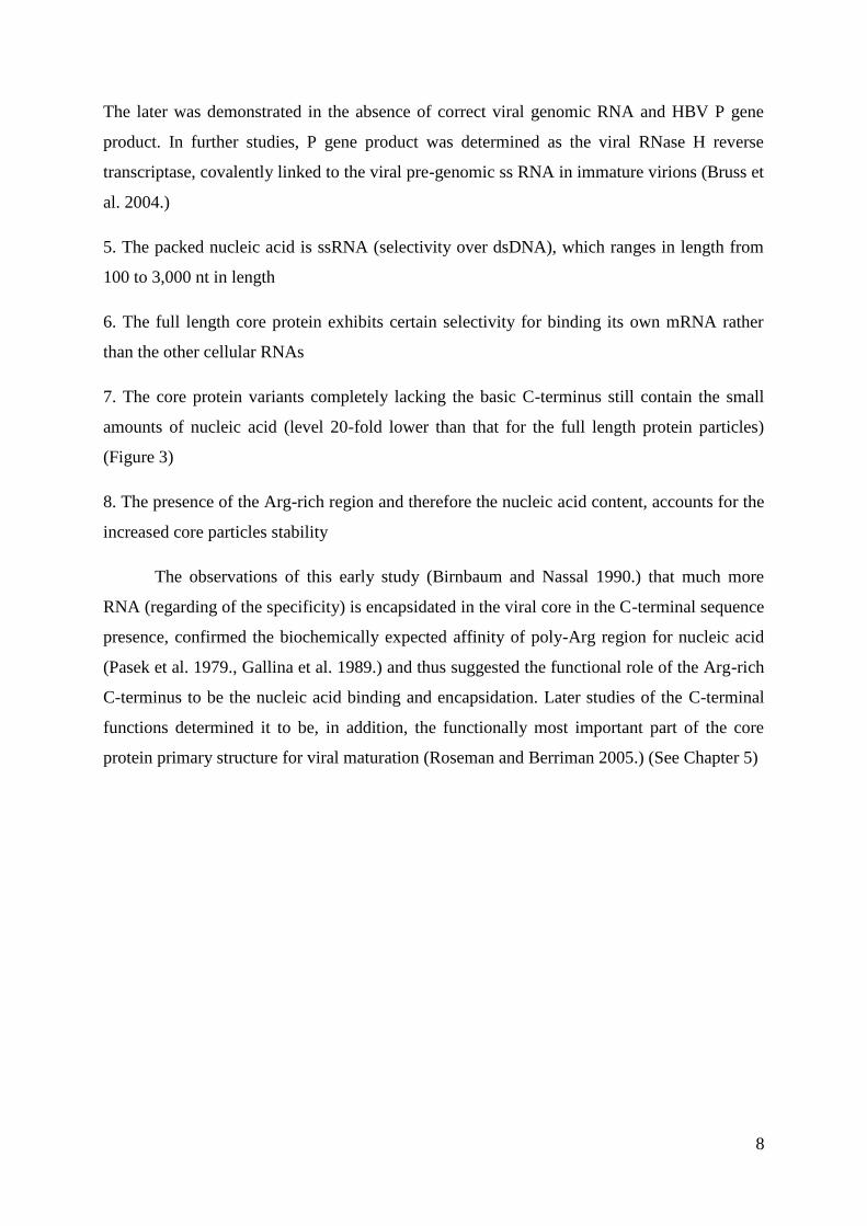
Figure 1: The amino acid sequence of the CW variant of HBV capsid, truncated at amino
acid 149 (indicated by Δ). The 94 full-length human HBV capsid sequences in the SWISS-
PROT and TrEMBL databases were aligned using CLUSTALW. Fully conserved residues
(identical in all 94 sequences) are marked (*). (completely taken from Wynee et al. 1999)
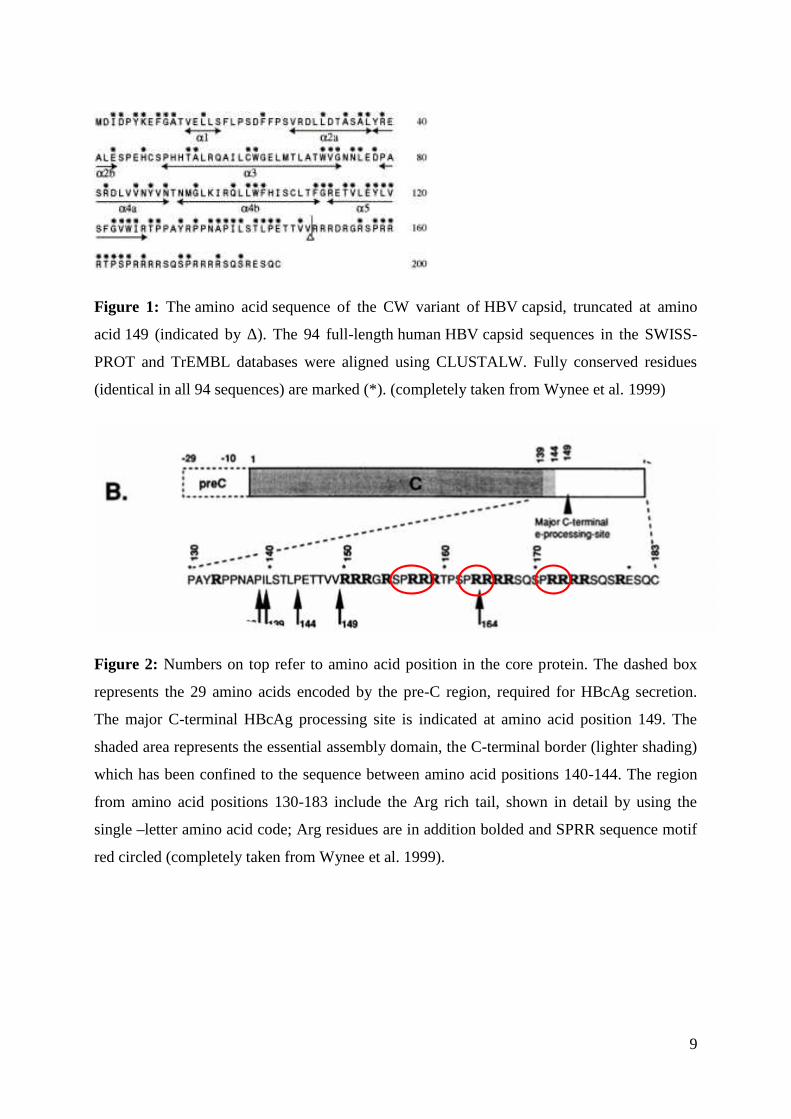
Figure 2: Numbers on top refer to amino acid position in the core protein. The dashed box
represents the 29 amino acids encoded by the pre-C region, required for HBcAg secretion.
The major C-terminal HBcAg processing site is indicated at amino acid position 149. The
shaded area represents the essential assembly domain, the C-terminal border (lighter shading)
which has been confined to the sequence between amino acid positions 140-144. The region
from amino acid positions 130-183 include the Arg rich tail, shown in detail by using the
single –letter amino acid code; Arg residues are in addition bolded and SPRR sequence motif
red circled (completely taken from Wynee et al. 1999).
10
3. The HBV core protein monomer fold
The second chapter introduced the basic primary structure amino-acid sequence of the
HBV core protein (Figure 1), defined the N-terminal and C-terminal regions and drove a bit
more attention to some experimental observations explaining the function of the C-terminal
region (2.1). However, as already introduced there the intra-cytoplasmical HBV core
assembly capability completely resides in the first 140-144 amino acids of the N-terminal
sequence (Birnbaum and Nassal 1990.). This fact implies that all the structural requirements
for the correct monomer fold, as well as the productive intersubunit contacts, are contained in
this N-terminal sequence. The C-terminal region, although as already stressed, extremely
important for interactions with, and encapsidation of, the pre-genome/RT complex (Bruss et
al. 2004), does not seem to play any essential role in the HBV core protein monomer fold.
The first hypothesis on the HBV core protein fold was introduced after its primary
structure and the minimum length of the protein required for the proper core assembly were
determined (Birnbaum and Nassal, 1990). The fact that the length of minimum 140 N-
terminal amino acids provided proper fold of the viral core (thus also the proper fold of all its
constituting monomers) it was suggested that the protein folds into the eight stranded β-barrel
structure. The basis for the conclusion was that the β barrel folding structure of the monomer
core proteins with similar length was already determined for some RNA viruses with
icosahedral core. The minimal length requirement for correct folding in such structure is
estimated to be 150 amino acids (Rosemann and Johnson, 1989).
However, the model of the HBV core protein fold, set after the first early cryo-electron
microscopy studies of the core particles, introduced completely unexpected structural
features. The early cryo-electron microscopy experiment was performed on the series of the
empty HBV core particles constituted from the C-terminally truncated core protein (140
amino acids in length) heterologously expressed in E. coli (Böttcher et al. 1997). The results
obtained described the unexpected largely helical fold of the monomer core protein which
formed T=4 icosahedral HBV core particles. At the time the structure of the HIV-1 core
protein (Momany et al. 1996, Gamble et al. 1997) was also determined to be largely helical,
the HBV and HIV-1 core proteins were among the first proteins of icosahedral viral core
described to have the dominant helical fold instead of mostly observed β-barrel structure. The
conserved amino acid residues in monomer in primary amino acid non-continuing chain
distribution give rise to the conserved fold motif called hydrophobic core (Figure 6). The
11
hydrophobic interactions which mediate the formation of such hydrophobic core constitute the
major driving force in the monomer HBV core protein folding.
3.1. The structure of the HBV core protein (HBc) fold determined by the protein X-ray
protein crystallography
Even more detailed structural information came with the atomic level X-ray microscopy
structural analyses performed after the HBV empty core particles had been successfully
crystallized (Wynee et al. 1999). It not only confirmed the excellent agreement with cryo-
electron microscopic-suggested helical monomer fold, but in addition proved assignment of
the core protein amino acid residues as remarkably accurate (matching the X-ray atomic level
structure by 97%) (Wynee et al. 1999). The initial cryo-EM map model of the helical
monomer fold was used to generate the initial resolution phases to 8 Å and then extended to
better X-ray resolution of 3.3 Å. The X-ray determined 3.3 Å resolution provides remarkable
atomic-level details of all parts of the HBV core protein secondary and tertiary structure,
except for the C-terminal tail region (amino acids 149-181/3) (as the crystallized particles
were empty core particles of C-terminally truncated core protein). In order to fulfill the
deficiencies in the information on the contribution of the C-terminal tail region to the
monomer fold, the region was observed by the later taken cryo-electron microscopy study of
the mature, partially ds DNA containing, core particles (Roseman and Berriman 2005).
The N-terminal region (1-12 amino acid residues) adopts an irregular structure of
extended amino acid chain, which is followed by the short α1 helix (residues 13-17). The next
is 10 amino acid long extended chain after which the longer α2 helix follows (residues 27-43).
The α2 helix is kinked after the residue 37 - the conserved leucine (L) residue (Figure 1). The
chain of amino acids 50-73 constitutes the helix α3, which is joined to helix α4 (residues 79-
110) by a very short amino acid chain (residues 73-79). The α4 helix is also kinked between
the residues 90-92 (the kink divides it to α4 a and α4 b part), the feature which is very
significant in functional aspect, and will therefore be later described (see Chapters 4 and 5).
The C-terminal region follows immediately after α4 helix and folds into the helix α5 (residues
112-127). The helices α4 and α5 are thus divided solely by one amino acid residue, the
conserved glycine residue (G-111) which, as well as the kink in α4 helix, has conserved
functional role (see Chapter 5). After helix α5 an irregular loop (residues 128-136) rich in
conserved proline (P) amino acids follows. The protein sequence finishes with an extended
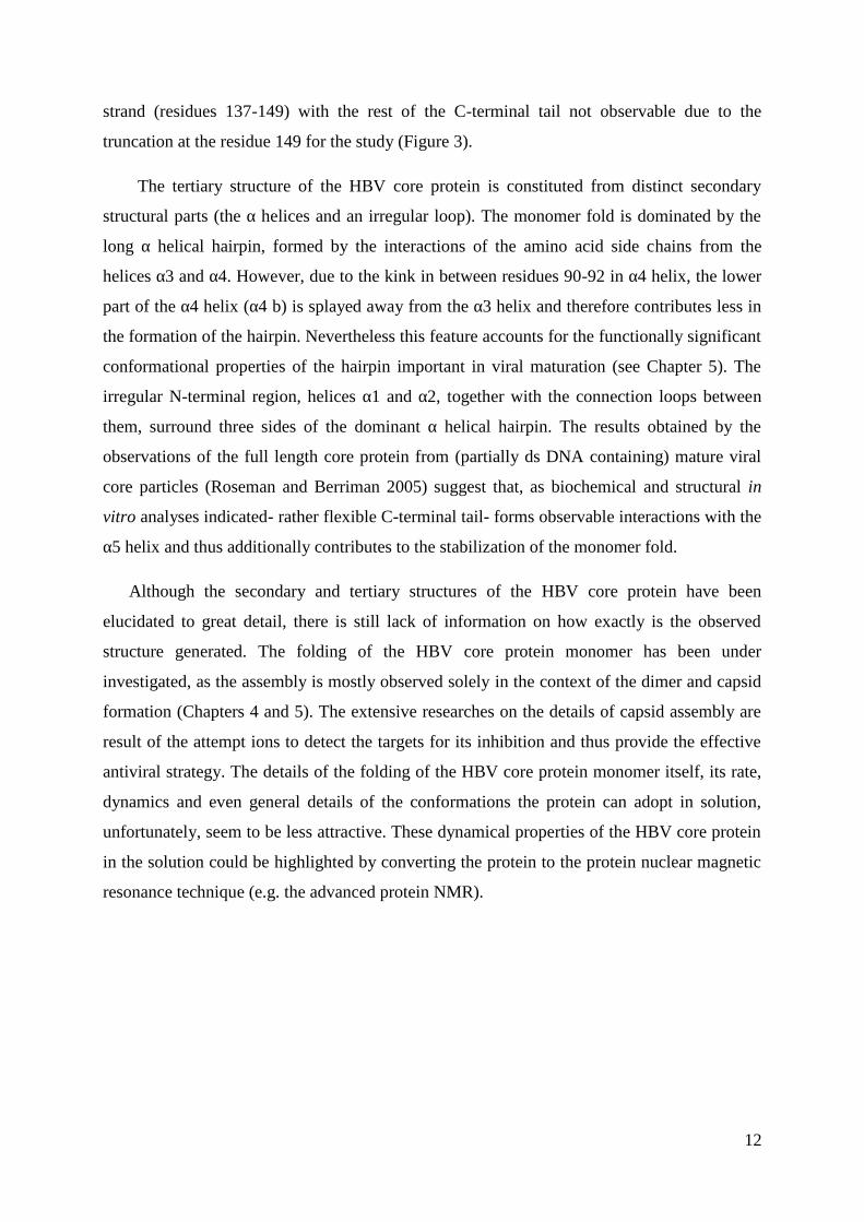
12
strand (residues 137-149) with the rest of the C-terminal tail not observable due to the
truncation at the residue 149 for the study (Figure 3).
The tertiary structure of the HBV core protein is constituted from distinct secondary
structural parts (the α helices and an irregular loop). The monomer fold is dominated by the
long α helical hairpin, formed by the interactions of the amino acid side chains from the
helices α3 and α4. However, due to the kink in between residues 90-92 in α4 helix, the lower
part of the α4 helix (α4 b) is splayed away from the α3 helix and therefore contributes less in
the formation of the hairpin. Nevertheless this feature accounts for the functionally significant
conformational properties of the hairpin important in viral maturation (see Chapter 5). The
irregular N-terminal region, helices α1 and α2, together with the connection loops between
them, surround three sides of the dominant α helical hairpin. The results obtained by the
observations of the full length core protein from (partially ds DNA containing) mature viral
core particles (Roseman and Berriman 2005) suggest that, as biochemical and structural in
vitro analyses indicated- rather flexible C-terminal tail- forms observable interactions with the
α5 helix and thus additionally contributes to the stabilization of the monomer fold.
Although the secondary and tertiary structures of the HBV core protein have been
elucidated to great detail, there is still lack of information on how exactly is the observed
structure generated. The folding of the HBV core protein monomer has been under
investigated, as the assembly is mostly observed solely in the context of the dimer and capsid
formation (Chapters 4 and 5). The extensive researches on the details of capsid assembly are
result of the attempt ions to detect the targets for its inhibition and thus provide the effective
antiviral strategy. The details of the folding of the HBV core protein monomer itself, its rate,
dynamics and even general details of the conformations the protein can adopt in solution,
unfortunately, seem to be less attractive. These dynamical properties of the HBV core protein
in the solution could be highlighted by converting the protein to the protein nuclear magnetic
resonance technique (e.g. the advanced protein NMR).
13
Figure 3: The fold of the HBV core protein monomer determined by the combination of
computed maps constructed upon the cryo-EM and X-ray crystallography studies (completely
taken from Wynee et al. 1999)
14
4. The dimer of the two HBV core protein monomers-the main core structural unit
The early biochemical in vitro studies of the HBV core monomer protein, defined the dimer
association, acquired by the cytoplasmic homodimerization of the two core monomers, as the
only stable intermediate in the HBV core assembly and thus its fundamental structural unit
(Zheng et al. 1992, Zhou and Standring 1992). The fold of the HBV core protein, described in
the last chapter, has introduced some structural characteristics, which are here going to be
further explained as these are very important for the proteins functional role. The chapter 3
has given certain details of the HBV core protein monomer fold, defining α helical hairpin as
its major tertiary structural feature. Formed by the interactions of the helices α3 and α4, the
hairpin presents the central protein fold, around which the rest of the protein’s secondary
structural components (N-terminal region, helices α1 and α2, and the connection loops
between them) are folded.
Early cryo-electron microscopy observations (Böttcher et al., 1997) of the HBV core
particles detected the structure of the compact dimer as the quaternary protein structure
formed by the adjacent core protein monomers. This dimer association accounted for the
observed characteristic spikes decorating the HBV core surface (Figure 4) (Ceres et al, 2004).
The observed spikes were defined as the four-helix bundle, the association of the two α helical
hairpins, one from each of the two associated monomers. Each monomer contributed the
observed structure with its two helices (α3 and α4) (Böttcher et al. 1997). The later taken X-
ray crystallography diffraction analyses (Wynee et al.1999) have confirmed the existence of
the four-helix bundle structure and in addition provided more detailed insight into its
structural characteristics. Nevertheless, the four helix bundle presents only the protruding part
(the spike) of the dimer formed (Figure 4). The whole dimer resembles the “pickaxe”, four
helix bundle corresponding the shaft and the basal region corresponding the head (Wynee et
al.1999) (Figure 5).
4.1. The HBV core protein monomer structural features responsible for the compact dimer
formation
As already introduced in the Chapter 2, the structural characteristics of the monomer fold
and its amino acid side chain distribution is responsible for monomer’s interactions which
give rise to compact, stable dimeric quaternary structure. The major structural feature of the
monomer fold, the helical hairpin is the first to be considered in this aspect. The amino acid
content of the helices α3 and α4, (Figure 1) is diverse. There is an approximately equal
15
amount of the charged and hydrophilic vs. hydrophobic amino acids, which accounts for the
largely amphipatic helical hairpin formed upon their folding into the α3 and α4 helices. The
hydrophobic faces of the two monomer hairpins associate on dimer formation, resulting in the
63% hydrophobic surface (Wynee et al. 1999). Thus formed largely hydrophobic dimer
interface corresponds to the 2000 surface area. These hydrophobic interactions between
the two monomers present the major driving force for the dimer formation (Ceres and
Zlotnick, 2002) (see the subchapter 4.2). Although referred to as the four-helix bundle, an
important feature must be taken into account. The four helices do not contribute equally to the
contacts which give rise to dimer stability. Only two of the four helices, the α3 helices of each
monomer form the extensive interactions, while the α4 helices demonstrate almost no contact
interactions except on the tip of the bundle (Wynee et al. 1999).
Furthermore, the interactions between the two core protein monomers are not constricted
only to the interface between the two amphipatic helical hairpins. The rest of the monomer
fold also accounts for the extensive monomer-monomer interactions, which give rise to the
head of the observed dimer “pickaxe”. The rest of the monomer fold, surrounding the
dominant helical hairpin, constitutes the other, for dimer formation important, structural
element, defined as the hydrophobic core. The core includes the following residues: Tyr-6,
Phe-9, Leu-15, Leu-16, Phe-18, Leu-19, Phe-23, Phe-24, Trp-102, Phe-103, Phe-110, Val-
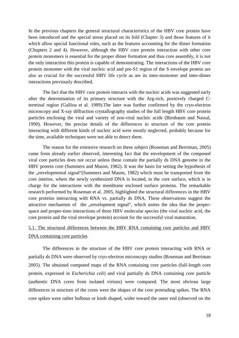
115, Tyr-118, Leu-119, Phe-122, Trp-125 and Leu-140 (Wynee et al.1999) (Figure 6).
The other important monomer residues responsible for forming the interactions in the dimer
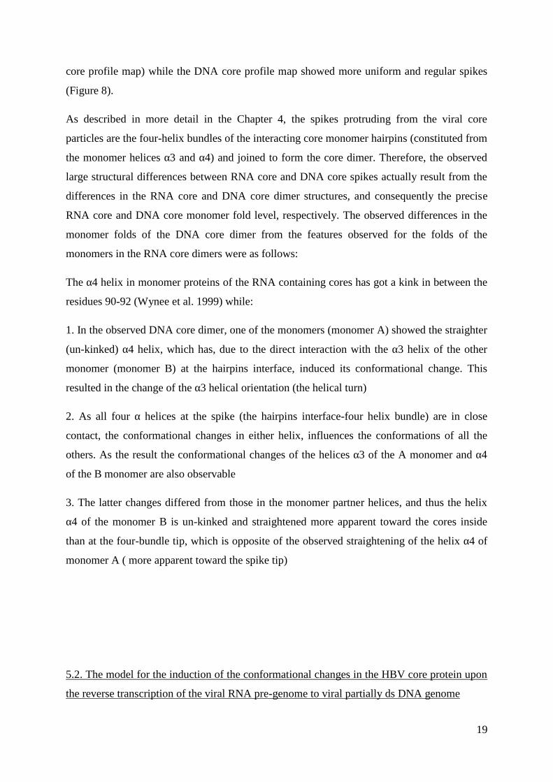
interface are the conserved cysteine residues (Cys-61) in the α3 helices of the two monomers
folds (Figure 7). Although between these two residues there is always observable disulfide
bond at the orthogonal view of the dimer interface (Figure 7), it has been proved that this
interaction does not constitute an essential component of the dimer assembly. Mutations on
this residue, leading to its change to either serine (Ser) or alanine (Ala), did not influence the
stabile dimer and capsid assembly (Nassal et al., 1992; Zheng et al. 1992; Zlotnick et al.
1996). This fact confirms the hypothesis of the dominance that hydrophobic interactions have
in driving the dimer assembly. Although several in vitro studies of the core assembly from
dimers of purified, E.coli expressed, HBV core proteins were taken, the details of the
oligomerization core assembly processes are, unfortunately, still largely unclear(Patient et al.
2009).
16
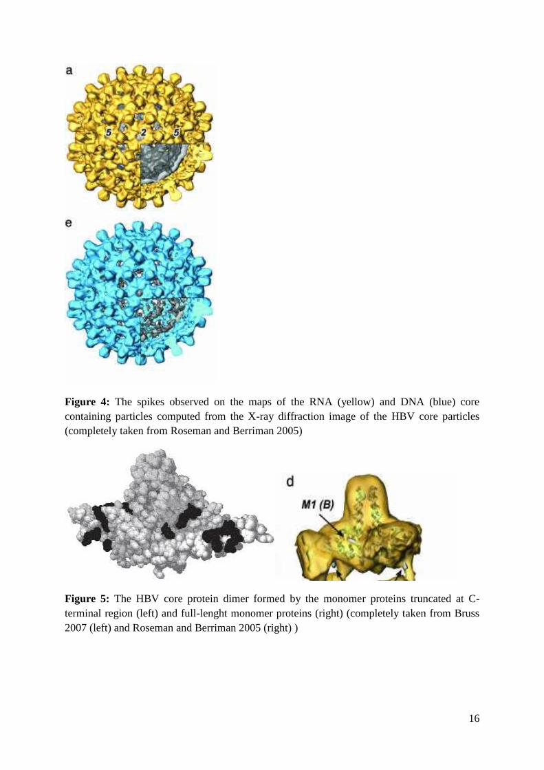
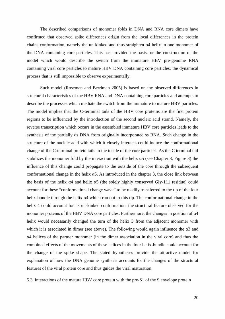
Figure 4: The spikes observed on the maps of the RNA (yellow) and DNA (blue) corecontaining particles computed from the X-ray diffraction image of the HBV core particles(completely taken from Roseman and Berriman 2005)
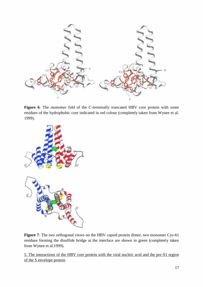
Figure 5: The HBV core protein dimer formed by the monomer proteins truncated at C-terminal region (left) and full-lenght monomer proteins (right) (completely taken from Bruss2007 (left) and Roseman and Berriman 2005 (right) )
17
Figure 6: The monomer fold of the C-terminally truncated HBV core protein with someresidues of the hydrophobic core indicated in red colour (completely taken from Wynee et al.1999).
Figure 7: The two orthogonal views on the HBV capsid protein dimer, two monomer Cys-61residues forming the disulfide bridge at the interface are shown in green (completely takenfrom Wynee et al.1999).
5. The interactions of the HBV core protein with the viral nucleic acid and the pre-S1 regionof the S envelope protein
18
In the previous chapters the general structural characteristics of the HBV core protein havebeen introduced and the special stress placed on its fold (Chapter 3) and those features of itwhich allow special functional roles, such as the features accounting for the dimer formation(Chapters 2 and 4). However, although the HBV core protein interaction with other coreprotein monomers is essential for the proper dimer formation and thus core assembly, it is notthe only interaction this protein is capable of demonstrating. The interactions of the HBV coreprotein monomer with the viral nucleic acid and pre-S1 region of the S envelope protein arealso as crucial for the successful HBV life cycle as are its inter-monomer and inter-dimerinteractions previously described.
The fact that the HBV core protein interacts with the nucleic acids was suggested earlyafter the determination of its primary structure with the Arg-rich, positively charged C-terminal region (Gallina et al. 1989).The later was further confirmed by the cryo-electronmicroscopy and X-ray diffraction crystallography studies of the full length HBV core proteinparticles enclosing the viral and variety of non-viral nucleic acids (Birnbaum and Nassal,1990). However, the precise details of the differences in structure of the core proteininteracting with different kinds of nucleic acid were mostly neglected, probably because forthe time, available techniques were not able to detect them.
The reason for the extensive research on these subject (Roseman and Berriman, 2005)came from already earlier observed, interesting fact that the envelopment of the composedviral core particles does not occur unless these contain the partially ds DNA genome in theHBV protein core (Summers and Mason, 1982). It was the basis for setting the hypothesis ofthe „envelopmental signal“(Summers and Mason, 1982) which must be transported from thecore interior, where the newly synthesized DNA is located, to the core surface, which is incharge for the interactions with the membrane enclosed surface proteins. The remarkableresearch performed by Roseman et al. 2005, highlighted the structural differences in the HBVcore proteins interacting with RNA vs. partially ds DNA. These observations suggest theattractive mechanism of the „envelopment signal“, which unites the idea that the proper-space and proper-time interactions of three HBV molecular species (the viral nucleic acid, thecore protein and the viral envelope protein) account for the successful viral maturation.
5.1. The structural differences between the HBV RNA containing core particles and HBV
DNA containing core particles
The differences in the structure of the HBV core protein interacting with RNA or
partially ds DNA were observed by cryo-electron microscopy studies (Roseman and Berriman
2005). The obtained computed maps of the RNA containing core particles (full-length core
protein, expressed in Escherichia coli) and viral partially ds DNA containing core particle
(authentic DNA cores from isolated virions) were compared. The most obvious large
differences in structure of the cores were the shapes of the core protruding spikes. The RNA
core spikes were rather bulbous or knob shaped, wider toward the outer end (observed on the
19
core profile map) while the DNA core profile map showed more uniform and regular spikes
(Figure 8).
As described in more detail in the Chapter 4, the spikes protruding from the viral core
particles are the four-helix bundles of the interacting core monomer hairpins (constituted from
the monomer helices α3 and α4) and joined to form the core dimer. Therefore, the observed
large structural differences between RNA core and DNA core spikes actually result from the
differences in the RNA core and DNA core dimer structures, and consequently the precise
RNA core and DNA core monomer fold level, respectively. The observed differences in the
monomer folds of the DNA core dimer from the features observed for the folds of the
monomers in the RNA core dimers were as follows:
The α4 helix in monomer proteins of the RNA containing cores has got a kink in between the
residues 90-92 (Wynee et al. 1999) while:
1. In the observed DNA core dimer, one of the monomers (monomer A) showed the straighter
(un-kinked) α4 helix, which has, due to the direct interaction with the α3 helix of the other
monomer (monomer B) at the hairpins interface, induced its conformational change. This
resulted in the change of the α3 helical orientation (the helical turn)
2. As all four α helices at the spike (the hairpins interface-four helix bundle) are in close
contact, the conformational changes in either helix, influences the conformations of all the
others. As the result the conformational changes of the helices α3 of the A monomer and α4
of the B monomer are also observable
3. The latter changes differed from those in the monomer partner helices, and thus the helix
α4 of the monomer B is un-kinked and straightened more apparent toward the cores inside
than at the four-bundle tip, which is opposite of the observed straightening of the helix α4 of
monomer A ( more apparent toward the spike tip)
5.2. The model for the induction of the conformational changes in the HBV core protein upon
the reverse transcription of the viral RNA pre-genome to viral partially ds DNA genome
20
The described comparisons of monomer folds in DNA and RNA core dimers have
confirmed that observed spike differences origin from the local differences in the protein
chains conformation, namely the un-kinked and thus straighten α4 helix in one monomer of
the DNA containing core particles. This has provided the basis for the construction of the
model which would describe the switch from the immature HBV pre-genome RNA
containing viral core particles to mature HBV DNA containing core particles, the dynamical
process that is still impossible to observe experimentally.
Such model (Roseman and Berriman 2005) is based on the observed differences in
structural characteristics of the HBV RNA and DNA containing core particles and attempts to
describe the processes which mediate the switch from the immature to mature HBV particles.
The model implies that the C-terminal tails of the HBV core proteins are the first protein
regions to be influenced by the introduction of the second nucleic acid strand. Namely, the
reverse transcription which occurs in the assembled immature HBV core particles leads to the
synthesis of the partially ds DNA from originally incorporated ss RNA. Such change in the
structure of the nucleic acid with which it closely interacts could induce the conformational
change of the C-terminal protein tails in the inside of the core particles. As the C terminal tail
stabilizes the monomer fold by the interaction with the helix α5 (see Chapter 3, Figure 3) the
influence of this change could propagate to the outside of the core through the subsequent
conformational change in the helix α5. As introduced in the chapter 3, the close link between
the basis of the helix α4 and helix α5 (the solely highly conserved Gly-111 residue) could
account for these “conformational change wave” to be readily transferred to the tip of the four
helix-bundle through the helix α4 which run out to this tip. The conformational change in the
helix 4 could account for its un-kinked conformation, the structural feature observed for the
monomer proteins of the HBV DNA core particles. Furthermore, the changes in position of α4
helix would necessarily changed the turn of the helix 3 from the adjacent monomer with
which it is associated in dimer (see above). The following would again influence the α3 and
α4 helices of the partner monomer (in the dimer association in the viral core) and thus the
combined effects of the movements of these helices in the four helix-bundle could account for
the change of the spike shape. The stated hypotheses provide the attractive model for
explanation of how the DNA genome synthesis accounts for the changes of the structural
features of the viral protein core and thus guides the viral maturation.
5.3. Interactions of the mature HBV core protein with the pre-S1 of the S envelope protein
21
However, one important fact has not yet been explained. How are all these
conformational changes in the HBV core particle, connected to the final stage of virus
maturation, namely its envelopment and budding?
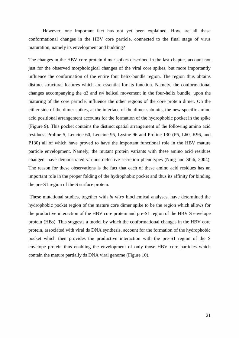
The changes in the HBV core protein dimer spikes described in the last chapter, account not
just for the observed morphological changes of the viral core spikes, but more importantly
influence the conformation of the entire four helix-bundle region. The region thus obtains
distinct structural features which are essential for its function. Namely, the conformational
changes accompanying the α3 and α4 helical movement in the four-helix bundle, upon the
maturing of the core particle, influence the other regions of the core protein dimer. On the
either side of the dimer spikes, at the interface of the dimer subunits, the new specific amino
acid positional arrangement accounts for the formation of the hydrophobic pocket in the spike
(Figure 9). This pocket contains the distinct spatial arrangement of the following amino acid
residues: Proline-5, Leucine-60, Leucine-95, Lysine-96 and Proline-130 (P5, L60, K96, and
P130) all of which have proved to have the important functional role in the HBV mature
particle envelopment. Namely, the mutant protein variants with these amino acid residues
changed, have demonstrated various defective secretion phenotypes (Ning and Shih, 2004).
The reason for these observations is the fact that each of these amino acid residues has an
important role in the proper folding of the hydrophobic pocket and thus its affinity for binding
the pre-S1 region of the S surface protein.
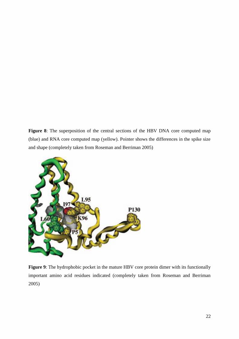
These mutational studies, together with in vitro biochemical analyses, have determined the
hydrophobic pocket region of the mature core dimer spike to be the region which allows for
the productive interaction of the HBV core protein and pre-S1 region of the HBV S envelope
protein (HBs). This suggests a model by which the conformational changes in the HBV core
protein, associated with viral ds DNA synthesis, account for the formation of the hydrophobic
pocket which then provides the productive interaction with the pre-S1 region of the S
envelope protein thus enabling the envelopment of only those HBV core particles which
contain the mature partially ds DNA viral genome (Figure 10).
22
Figure 8: The superposition of the central sections of the HBV DNA core computed map
(blue) and RNA core computed map (yellow). Pointer shows the differences in the spike size
and shape (completely taken from Roseman and Berriman 2005)
Figure 9: The hydrophobic pocket in the mature HBV core protein dimer with its functionally
important amino acid residues indicated (completely taken from Roseman and Berriman
2005)
23
Figure 10: The model of the mature HBV core particle envelopment (completely taken fromBruss 2007)
6. Conclusion:
24
The structure of the HBV core, and thus the core monomer protein fold, described in this
seminar was obtained by the cryo-EM and protein X-ray crystallography studies on purified,
C-terminally truncated protein variants heterologously expressed in E.coli. It is therefore
questionable to which extent obtained structural information are comparable to the structure
of the HBV core protein in immature and mature core particles assembled in vivo in infected
hepatocyte. However, the extensive research done by Roseman and Berriman 2005, compared
the structures of the several HBV core variants; the bacterial RNA containing (E.coli
expressed and assembled HBV core protein), the viral RNA containing and the viral mature
(partially ds DNA containing) HBV core particles. The structural comparison of the general
core morphology, core dimer and monomer folds, concluded that there are no detectable
differences between the two RNA containing core types. The later implies that the monomer
protein fold is largely determined by the in primary structure contained information and thus
the possible influences of the cellular factors in HBV core monomer folding could be to some
extent neglected. The precise information on the dynamics of the core monomer folding, the
subsequent dimer and core assembly are still largely unknown, as the features have been
under investigated.
The differences between the viral RNA and mature DNA containing particles showed
to be the differences in the conformation of the dimer core subunits with the stressed
difference in the hydrophobic pocket accessibility. The later structural information was used
for the construction of the model for the core protein interactions with pre-S1 region of the S
envelope protein and consequent HBV maturation. However, the information obtained must
be considered with caution for several reasons. First of all, the viral RNA containing core
particles, although hosting the viral encoded RNA, did not contain the exact viral pgRNA/RT
complex found in the immature core particles assembled in infected hepatocyte. Therefore, it
is questionable to which extent the core, dimer and monomer structural features of such
particles are relevant for the comparison with the mature viral isolated cores. The lack of the
ways to experimentally track the dynamical structural changes in HBV core dimers in the
exact moment when the process of reverse transcription and consequent pgRNA-> partially ds
DNA switch occurs, unfortunately, leaves only the possibility to observe the fragments of
structural information (with questionable relevance) to deduce on these events.
7. References:
25
Birnbaum, F., Nassal, M. (1990) Hepatitis B virus nucleocapsid assembly; primary structure
requirements in the core protein. J Virol; 64: 3319-3330
Beck, J., Nassal, M. (2007) Hepatitis B virus replication. World J Gastroenterol; 13: 48-64
Böttcher, B., Wynee, S.A., Crowther, R.A. (1997) Determination of the fold of the core
protein of hepatitis B virus by electron cryomicroscopy. Nature; 386: 88-91
Bruss, V. (2007) Hepatitis B virus morphogenesis. World J Gastroenterol; 13(1): 65-73
Ceres, P., Zlotnick, A. (2002) Weak protein-protein interactions are sufficient to drive
assembly of hepatitis B virus capsids. Biochemistry; 41: 11525-11531
Chain, B.M., Myers, R. (2005) Variability and conservation in hepatitis B virus core protein.
BMC Microbiol; 5:33
Conway, J.F., Cheng, N., Zlotnick, A., Wingfield, P.T., Stahl, S.J., Steven, A.C. (1997)
Visualization of a 4-helix bundle in the hepatitis B virus capsid by cryo-electron microscopy.
Nature; 386: 91-94
Crowther, R.A., Kiselev, N.A., Böttcher, B., Berriman, J.A., Borisova, G.P., Ose V.,
Pumpens, P. (1994) Three-dimensional structure of hepatitis B virus core particles determined
by electron cryomicroscopy. Cell; 77: 943-950
Dane, DS., Cameron, CH., Briggs M. (1970) Virus like particles in serum of patients with
Australia-antigen-associated hepatitis Lancet; 1: 695-698
Gallina, A., Bonelli, F., Zentilin, L., Rindi, G., Muttini, M., Milanesi, G. (1989) A
recombinant hepatitis B core antigen polypeptide with the protamine-like domain deleted self-
assembles into capsid particles but fails to bind nucleic acids. J Virol; 63: 4645-4652
Ganem, D., Varmus, H.E. (1987) The molecular biology of hepatitis B viruses. Annu Rev
Biochem; 56:651-693
König, S., Beterams, G., Nassal, M. (1998) Mapping of homogous interaction sites in the
hepatitis B virus core protein. J Virol; 72: 4997-5005
Newman, M., Suk, F.M., Cajimat, M., Chua, P.K., Shih, C. (2003) Stability and morphology
comparison of self-assembled virus like particles from wild-type and mutant human hepatitis
B virus capsid proteins. J Virol; 77: 12950-12960
26
Pasek, M., Goto T., Gilbert, W., Zink, B., Schaller, H., MacKay, P., Lead-better, G., Murray,
K. (1979) Hepatitis B virus genes and their expression in Escherichia coli. Nature; 282: 575-
579
Patient, R., Hourioux, C., Roingeard, P. (2009) Morphogenesis of hepatitis B virus and its
subviral envelope particles. Cell Microbiol; 11(11): 1561-1570
Perz, JF., Armstrong, GL., Farrington, LA., Hutin, YJ., Bell, BP. (2006) The contribution of
hepatitis B and hepatitis C virus infections to cirrhosis and primary liver cancer worldwide. J
Hepatol; 45: 529-538
Roseman, A.M., Berriman, J.A., Wynee, S.A., Butler, P.J.G., Crowther, R.A.(2005) Structural
model for maturation of the hepatitis B virus core. PNAS; 102: 15821-15826
Wingfield, P.T., Stahl, S.J., Williams, R.W., Steven, A.C.(1995) Hepatitis core antigen
produced in Escherichia coli: subunit composition, conformational analysis, and in vitro
capsid assembly. Biochemistry; 34: 4919-4932
Wynee, S.A., Crowther, R.A., Leslie, A.G. (1999) The crystal structure of the human hepatitis
B virus capsid. Mol Cell; 3:771-780
Zheng, J., Schödel, F., Peterson, D.L. (1992) The structure of hepadnaviral core antigens. J
Biol Chem 267: 9422-9429
Zlotnick, A., Cheng, N., Stahl, S.J., Conway, J.F., Steven, A.C., Wingfield, P.T. (1997)
Localization of the C terminus of the assembly domain of hepatitis B virus capsid
protein:implications for morphogenesis and organization of encapsidated RNA. Proc Natl
Acad Sci USA; 94: 9556-9561
8. Summary:
27
The hepatitis B virus (HBV) is small, enveloped virus, member of the Hepadna viral family
with unusual, partially double-stranded DNA genome. The predominant viral core form in
vivo is 34 nm particle with T=4 icosahedral symmetry, constructed from 240 copies of the
HBV core protein (HBc). The HBV core protein is 22 kD, 183-185 amino acids long protein
with primary structure composed of two structurally and functionally distinct regions. The N-
terminal region is 149-151 amino acids long assembly domain, capable of directing the
correct fold of the core monomer. The 34 residues long C-terminal domain, completely
dispensable for core assembly, is extremely rich in conserved arginine (R) residues and takes
part in the viral pre-genome/RT binding and encapsidation. The cryo-electron microscopy and
X-ray crystallography studies on the HBV core particles revealed an unexpectedly large
helical protein structure, with the long α helical hairpin dominating the entire monomer fold.
The amphipatic helical hairpins of two core monomers interact forming the compact dimer,
whose interface covers the large hydrophobic surfaces of the individual monomers. The
hydrophobic interactions thus constitute the major driving force for the dimer assembly and
account for the assembled core stability. The four helix bundle of the dimer is a distinct core
structural feature, with important functional role. This is well observed upon the maturation of
the immature pre-genomic RNA/RT complex-containing HBV core particle. The unusual
reverse transcription of the pre-genomic RNA generates the partially ds DNA of mature
virion, the event which induces the sequence of the conformational changes in the dimer core
protein with which the nucleic acid interacts. The changes are best observable on the four-
helix bundle dimer structure and insure the generating of the dimer hydrophobic pocket on
core surface which subsequently interacts with the pre-S1 region of the HBV S envelope
protein. This mechanism ensures that only the mature viral core particles (with encapsidated
dsDNA) are capable of the successful envelopment and budding at an intracellular hepatocyte
membrane.
9. Sažetak:
28
Virus hepatitisa B (HBV) je član Hepadna virusne obitelje s ovojnicom i neobičnim,djelomično dvostrukim DNA genomom. Prevladavajući oblik virusne srži, in vivo, je sferičnaproteinska čestica, 34 nm u promjeru, T = 4 ikosaedarne simetrije, građena od 240 monomeraproteina srži HBV(HBc). Protein srži ima molekulsku težinu 22 kD te 183-185 duge sekvenceaminokiselina koja se sastoji od dvije strukturno i funkcionalno različite regije. N-terminalnuregiju, nazvanu i domena slaganja, čine prvih 149-151 aminokiselina. Ova je regija posvesamostalna u usmjeravanju funkcionalnog smatanje monomera proteina srži i posljedičnosklapanje same srži. 34 aminokiseline duga C-terminalna domena, posve je neophodna zasmatanje proteina i sklapanje srži te budući da je izuzetno bogata konzerviranim argininskim(R) amino kiselinama ima ulogu u slaganju kompleksa pre-genomske RNA/reverznatranskriptaze u sklapajuću proteinsku srž. Krio-elektronsko mikroskopske analize i proteinskaX-ray kristalografija na srži HBV ukazala su na neočekivanu dominaciju α zavojnica utercijarnoj strukturi monomera, s dugom α-helikalnom ukosnicom dviju zavojnica (α3 i α4)kao njezinom osnovom. Amfipatske α-helikalne ukosnice dvaju monomera, interakcijomtvore kompaktan dimer, čije sučelje pokriva veliku površinu hidrofobnih bočnih lanacaindividualnih monomera. Hidrofobne interakcije, predstavljaju glavnu pokretaču snagu zaspontano sklapanje dimera te ujedno čine osnovu stabilnosti čitave srži. Dvije α-helikalneukosnice okupljaju u dimeru četiri α uzvojnice, koje tako čine četvero-helikalni snop, osobitustrukturu srži s važnom funkcionalnom ulogom. Uloga strukture četvero-helikalnog snopanajbolje se očituje u sazrijevanju nezrelih čestica srži HBV, koje sadrže kompleks pre-genomske RNA/RT. Neobičajenom reverznom transkripcijom pre-genomske RNA generira sedjelomično dvolančana DNA, što je okidač za slijed konformacijskih promjena u dimerimasrži. Promjene se najbolje očituju na strukturi četvero-helikalnog snopa dimera gdjeosiguravaju izbočivanje hidrofobnog džepa sučelja dimera. Hidrofobni džep tako biva izloženna površini sada zrele srži te osigurava njezinu interakciju s pre-S1 regijom S proteina HBVovojnice. Ovaj mehanizam osigurava da se samo zrelim česticama srži (s uklopljenomdjelomičnom dvolančanom DNA) HBV-a omogućuje uspješna interakcija s proteinimaovojnice, što rezultira virusnim omatanjem i pupanjam s unutarstanične membrane hepatocita.