Embed Size (px)
Citation preview
Title Lenticulo-Striate Arteries in Hypertensive IntracerebralHemorrhage as Demonstrated by Angiography
Author(s) KANAYA, HARUYUKI; ONO, SETSUO; HORIE,YUTAKA; OANA, KATSUMARO
Citation 日本外科宝函 (1969), 38(1): 3-11
Issue Date 1969-01-01
URL http://hdl.handle.net/2433/207536
Right
Type Departmental Bulletin Paper
Textversion publisher
Kyoto University
Arch. Jap. Chir. 38(1), 3~11, Jan. JCJl)CJ
原著
Lenticulo・StriateArteries in Hypertensive Intracerebral
Hemorrhage as Demonstrated by Angiography
by
HARUYUKI KANAYA, SETSUO ONO, YUTAKA HORIE
and KATSUMARO 0ANA
Department of Neurosurgery, Iwate Medical University, School of Medicine
Received for Publication Nけv.12, J'”が
Among the arteries perfusing the internal and external capasules as well as the basal
ganglia, two perforaturs branching from the anterior cerebral artery and middle cerebral
artery are distinguished , the former being called the recurrent artery of Heubner or A.
striatica rostralis, the latter the lenticulostriate artery, strio-thalamic artery or A. striatica
caudalis. No uniform terminology is presently available.
The lenticulo・striate artery has frequently an abnormal course in association with
lesions in the deep part of the brain, so that the cerebral angiographic diagnosis becomes
important. Only the description by HEUBNER4>, WESTBERG8>, ANDERSEN°2>, and other is
available. Since the report of Charcot and BOUCHARD (1868)” stating that this artery was
invariable found ruptured miliary aneurysma on its smaller arterial branches, particularly the
lenticulostriate arteries in hypertensive and arteriosclerotic apopletic brains, the artery
causing apoplexy has drawn widespread attention (A. apoplectica). In actual autopsy,
the site of hemorrhage was frequently found in the perfusing area of this artery.
Although the importance of this artery in diagnosis is recognized, its detailed angiographic
studies were not reported.
Since 1956 weりainhave continued studies on the surgical therapy of severe hyper-
tensive intracerebral hemorrhage. The rate of salvage is 72% in the capsular type accord-
ing to my classification and much lower in other types. A high correlation was found
between posthemorrhagic vital sign and neurologic signs and the location and volume of
the hematoma. It is therefore very important to know the location and volume of the
hematoma before choosing surgical intervention.
The present communication deals with the diagnositic significance of cerebral angio-
graphy of the lenticulo-striate artery in cases of hypertensive intracerebral hemorrhage with
hematoma of known location and volume, confirmed by autopsy and operative findings,
the pontine and cere恥liarhemorrhage is out of consideration.
4 日・外・ '.t'. 第38巻第1号(昭和44年1月)
MATERIALS AND METHODS
Sixty-six cases of hypertensive intracerebral hemorrhage were selected. The location
of the hematoma was classified into 3 following types from the standpoint of surgical
management. The回 psulartype which was named for hematoma in the internal capsule
and its lateral side comprising internal and external capsule, caudate nucleus, putamen and
globus pallidus was seen in 41 cases. The thalamic type, hematoma in the thalamus and
hypothalamus was seen in 1 case. The capsulothalamic type in which the hematoma
extended to the surface of the thalamus and hypothalamus was seen in 25 cases. As
controls, '19αses of arteri osclerosis according to the WHO classification and 100 normal
young subjects without intracranial lesions and arteriosclerosis were selected.
Carefid angiography was carried, using about 10 cc of 60% urografin. Angiography
W出回rriedout in hypertensive intracerebral hemorrhage one day after stroke in 45
%,三 days after stroke in 18 %, 3 days
after stroke in 13 %, and beyond this stage
in 24%.
For the estimation of filling frequency,
clear visualization of the lenticulo・striateartery
with diagnostic usefulness was called “fαii
fill印g'', visualization of the main artery
without diagnostic use on account of the
unclear course of the vessel was called “poor filling”, and failure of visualization of the
main cerebral artery was called non-filling.
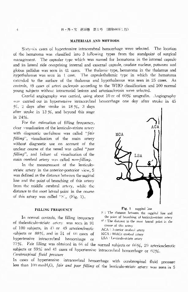
In the measurement of the lenticulo-
striate artery in the anterior-posterior view, S
was defined as the distance between the sagittal
line and the point of branching of this artery
from the middle cerebral ar!t'ry, while the
distance to the most lateral point in the course
of this artery was called uメ・,, (Fig. 1).
/、iACA
FILLING FREQUENCY Fig, 1. saggital line 計: Thedistance betw田nthe s"ggital line and
In normal controls, the filling frequency 、athe悶 ntof branching of lenticulo-striate artery
of thelenticulo-striate arterv was seen in 91 ゾ :Thedistance to the most lateral point in the 世 ofthiお arten
of 100 subjects, in 4:1 or 49 arteriosclerotic ACA: Anterior ~;;;ふl a町 V
subjects or 88%, and in 51 of l)(i cases of MCA: Middle cereb叫 artery.
hypertensive intracerebral hemorrhage or LSA Lenticul口・str以eartary
77%. Fair filling was obtained in制 ofthe normal subjects or州%, 29 arteriosclerotic
subj町 tsor 59% and 41四 sesof hypertensive 凶 racerebralhemorrh~g~ or山%.Cerebrospinal fluid pressure
In cases of hypertensive intracerebral hemorrhage with cerebrospinal fluid pressure
less than 1 r)り mmH20,fair a仰dpoor filling of the lenticulo・striateartery was seen in 5
LENTICULO・STRIATEARTERIES IN HYPERTENSIVE HEMORRHAGE 5
of 6四 S白 or83%, while the corresponding figures were 14 of 18αses or 78% in those
with a pressure of 101 200 mmH20, and 10 of 12αS白 or 83 % in those with a
pressure of 201-300 mmH20, and 13 of 21団 ses or 62 % in those with a pr白 sure
more than 301 mmH20, suggesting a marked decrease more than 301 mmH20.
Hematomαlocatio仰
The frequency of fair and poor filling in the capsulothalamic type was 16 of 25回 ses
or 64%, while the corresponding figure was 35 of 40田 sesor 88 % in the capsular type,
and 1 of 1 case of the thalamic type. The frequency of fair filling alone was seen in
13 cases or 52% of the the capsulothalamic type, 26αses or 65 % of the capsular type,
sugg白 ting a higher frequency of visualization in the回 psulartype than in the capsul-
othalamic type.
Hematoma volume
The fair filling was obtained in 32 of 44 cases or 73% in cas白 witha hematoma
volume of less than 100 cc while a similar finding was obtained in 7 of 19 cases or
37% when the hematoma was more than 101 cc, suggesting a decrease in the rate of
fair filling as the volume of hematoma increased.
Secondary ventricular he1何orrhαge
In hypertensive intracerebral hemorrhage accompanied by intraventricular hemorrhage,
fairαud poor filling was obtained in 39 of 50 cases or 78%, while the corresponding
figure in cases without intraventricular hemorrhage was 11 of 16 cases or 69%. Fair
fillig was seen in 8 cases or 50% of cases without intraventricular hemorrhage, suggest-
ing a higher frequency of visualization inαS白 withintraventricular hemorrhage.
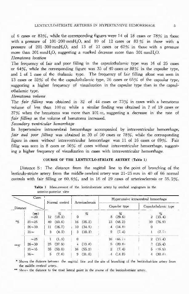
COURSE OF THE LENTICULO-STRIATE ARTERY (Table 1)
Distance S : The distance from the sagittal line to the point of branching of the
lenticulo-striate arteηfrom the middle cerebral artery was 21-25 mm in 40 of 66 normal
controls with fair filling or 60. 6%, and in 16 of 29 cases of arteriosclerosis or 55. 2%.
Table 1 Measurement of the lenticulostriate artery by cerebral angiogram in the
antenor-postenor view
Cふ示! 「 | I I Hypertensive intracerebral hemorrhage
Normal control I Arteriosclerosis I 一一÷一一一一一一一一一一D附悶 ~I [ I Ca戸ularty戸 j Capsu alar ty戸
(mm) I % I % ・ % . % ~20 I 12 (18. 2) I o . s (29. 6) 2 (15. 4)
*S I 2日 5 I 40 (60. 6) I 16 (55. 2) I 13 (48. 2) 10 (76. 9)
! 26~却 I11 (16. 7) i 10 (34. 5) ! 4 (14. 8 I 0
31~ I 3 (4.5) i 3 c10.3) ; 2 c1.4) i c1.11
|~25 i 1 (1. 5) i 0 16 I 66. 7 I i 2 (15. 4)
件目 I 2焔~30 I 25 (37. 9) • (13. s) 5 (20. s 1 2 (15. 4) )[… I 35 (53. 0) : 16 (55. 2) 2 (7. 4) i 5 (川
36~ I 5 c1. 6) I 9 (31. o) 1 4 c14. s) 1 4 (3o. s 1
ホ Showsth唱 distancebetween the sagittal line and the site of branching of the lenticulostriate artery from
the middle cerebral artery.
** Sho"' the distance to the most lateral伊 intin the course of the lenticulostriate arten.
6 日・外・宝第38巻第1号(昭和44年 1月)
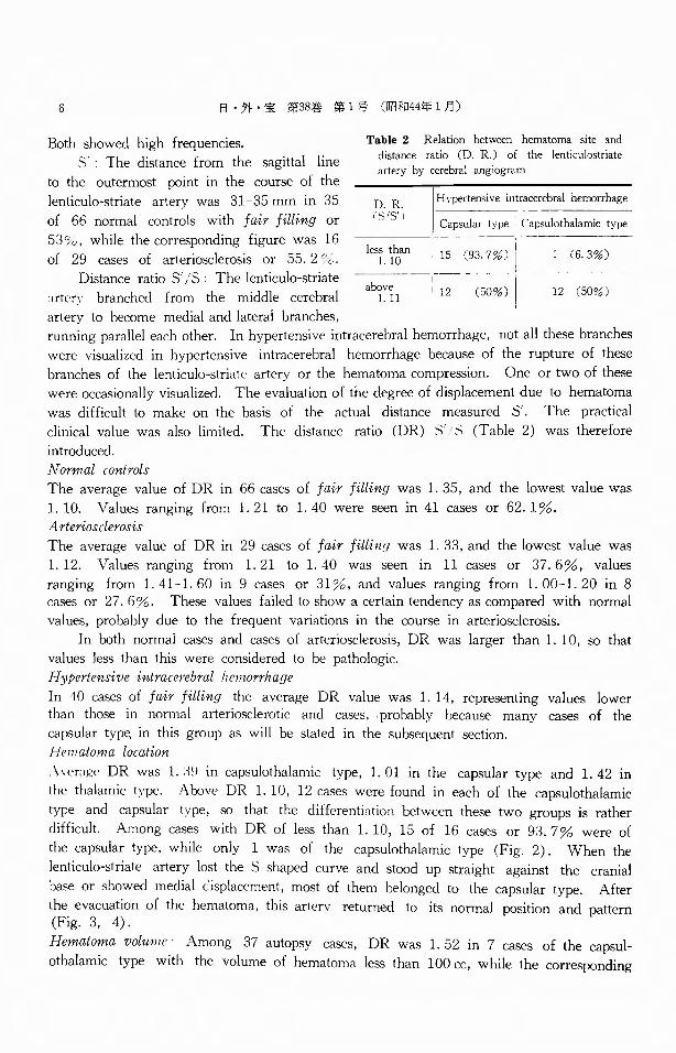
Both showed high frequencies.
S' : The distance from the sagittal line
to the outermost point in the course of the
lenticulo・striate artery was 31 35 mm in 35
of 66 normal controls with fair filling or
53%, while the corresponding figure was 16
of 29 cases of arteriosclerosis or 55. 2 %.
Distance ratio S' /S ・ The lenticulo・striate
;irtery branched from the middle cerebral
artery to become medial and lateral branches,
Table 2 Relation betw田nhematoma site and distance ratio (D. R〕ofthe lenticulostriate artery by cerebral angiogram
D.R. (S1S’}
日、pertensiveintracerebral hemorrhage
Capsular type Capsulothalamic type
le~ . it;Jn 15 (竺ゴ 1 (6. 3%)
a~~~l : 12 σ 12 (50%)
running parallel each other. In hypertensive intracerebral hemorrhage, not all these branches
were visualized in hァpertensive intracerebral hemorrhage because of the rupture of these
branches of the lenticulo-striate artery or the hematoma compression. One or two of these
were occasionally visualized. The evaluation of the degree of displacement due to hematoma
was difficult to make on the basis of the actual distance measured S'. The practical
clinical value was also limited. The distance ratio (DR) S' 1S (Table 2) was therefore
introduced.
Normal controls
The average value of DR in 66伺 sesof fair filling was 1. 35, and the lowest value was
1. 10. Values ranging from 1. 21 to 1. 40 were seen in 41αses or 62. 1%.
Arteriosclerosis
The average value of DR in 29 cases of fair filling was 1. 33, and the lowest value was
1. 12. Values ranging from 1. 21 to 1. 40 was seen in 11回 ses or 37. 6%, values
ranging from 1. 41-1. 60 in 9 cases or 31%, and values ranging from 1. 00-1. 20 in 8
cases or 27. 6%. These values failed to show a certain tendency as compared with normal
values, probably due to the frequent variations in the course in arteriosclerosis.
In both normal cases and田 sesof arteriosclerosis, DR was larger than 1. 10, so that
values less than this were considered to be p<1thologic.
Hypertensive intracerebral lieniorrhage
In 40 cases of fair filling the average DR value was 1. 14, representing values lower
than those in normal arteriosclerotic and cases, . probably because many cases of the
capsular typl; in this group as will be stated in the subsequent section.
Henzatoma locat骨on
入Vぞれl貯 DR was 1. :1~ in capsulothalamic type, 1. 01 in the capsular type and 1. 42 in
the thalamic t:-・pe. Above DR 1. 10, 12 cases were found in each of the capsulothalamic
type and capsular type, so that the differentiation between these two groups is rather
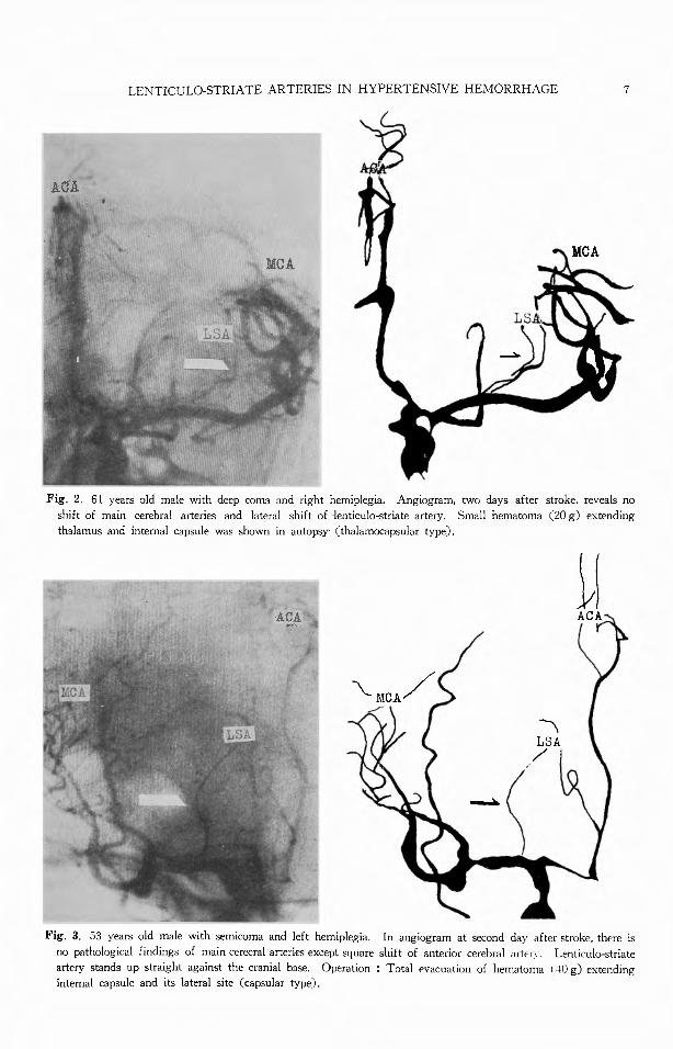
difficult. Among cases with DR of less than 1. 10, 15 of 16 cases or 93. 7% were of
the capsular type, while only 1 was of the capsulothalamic type (Fig. 2). When the
lenticulo・striateartery lost the S shaped curve and stood up straight against the cranial
base or showed medial displacement, most of them belonged to the capsular type. After
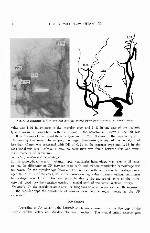
the evacuation of the hematoma, this artery returned to its normal position and pattern (Fig. 3, 4).
Hematoma volume . Among 37 autopsy cases, DR was 1. 52 in 7 cases of the capsuト
othalamic type with the volume of hematoma less than 100 cc, while the corresponding
LENTICULO・STRIATEARTERIES IN HYPERTENSIVE HEMORRHAGE 7
:2 ACA
Fig. 2. 61 years old male with deep coma and right hemiplegia. Angiogram, two days after stroke, reveals no
shift of main cerebral arteries and lateral shift of、lenticulo-striateartery. Small hematoma (20 g〕extending
thalamus and internal capsule was shown in autopsy. (thalamocapsular type).
,
一〈
~\
LSA /
Fig. 3. 53 years old male with semicoma and left hemiplegia. In angiogram at second day after stroke, there is
no pathological findings of main cerecral arteries except square shift of anterior cerebral <lftE'n・. Lenticul仁時triate
artery stands up straight against the cranial base. Operation : Total evacuation of hematoma I 40 g) extending
internal capsule and its lateral site (capsular type).
8 II・外・宝第38巻第 1号(昭和44年 1月)
Fi耳 4. In angiogram at fifty days after operation, lenticulo-striate arter¥' returns t" its normal pattern.
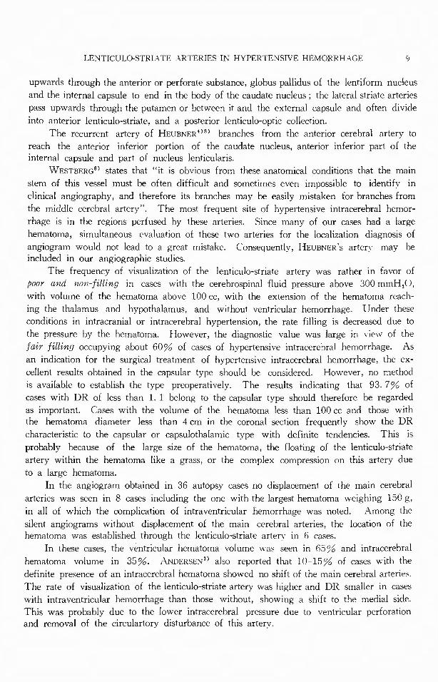
value was 1. 01 inヅlcases of the capsular type, and 1. 42 in one case of the thalamic
type, showing a correlation with the volume of the hematoma. Above 101 cc DR was
1. 20 in 6 cases of the capsulothalamic type and 1. 07 in 3 cases of the capsular type.
Diameter of hematoniα:In autopsy, the largest transverse diameter of the hematoma of
less than 40 mm was associated with DR of 0. 71 in the capsular type and 1. 73 in the
capsulothalamic type. Above 41 mm, no correlation was found between this and trans-
刊 rsぞ diameterof hematoma.
ヌC((l11c/αrヲ ve竹tγicularhemorrhage
In the capsulothalamic and thalamic types, ventricular hemorrhage was seen in all cases,
so that the difference in DR between cases with and without ventricular hemorrhage was
unknown. In the capsular type, however, DR in回 seswith ventricular hemorrhage aver-
aged 0. 97 in 17 of 2.:J cases, while the corresponding value in cases without ventricular
hemorrhage was 1. 12. This was probably due to the rupture of some of the intra-
cerebral blood into the ventricle causing a medial shift of the lenticulo-striate artery.
Prognosis: In the capsulothalamic type, the prognosis became poorer as the DR increased.
In the capsular type the disturbance of consciousness became more intense as the DR
decreased.
DISCUSSION
According to ANDERSEN1>, the lenticulo-striate artery arises from the first part of the
middle cerebral artery and divides into two branches. The medial striate arteries pass
LENTICULO-STRIATE ARTERIES IN HYPERTENSIVE HEMORRH主GE 9
upwards through the anterior or perforate substance, globus pallidus of the lentiform nucleus
and the internal capsule to end in the body of the caudate nucleus ; the lateral striate arteries
pass upwards through the putamen or between it and the external capsule and often divide
into anterior lenticulo・striate,and a posterior lenticulo・opticcollection.
The recurrent artery of HEUBNER4>8> branches from the anterior cerebral artery to
reach the anterior inferior portion of the caudate nucleus, anterior inferior part of the
internal capsule and part of nucleus lenticularis.
川TESTBERG8)states that“it is obvious from these anatomical conditions that the main
stem of this vessel must be often difficult and sometimes even impossible to identify in
clinical angiography, and therefore its branches may be easily mistaken for branches from
the middle cerebral artery”. The most frequent site of hypertensive intracerebral hemor-
rhage is in the regions perfused by these arteries. Since many of our cases had a large
hematoma, simultaneous evaluation of these two arteries for the localization diagnosis of
angiogram would not lead to a great mistake. Consequently, HEUBNER’s arterv may be included in our angiographic studies.
The frequency of visualization of the lenticulo-striate artery was rather in favor of
poor and non-filling in cases with the cerebrospinal fluid pressure above 300 mmH2C l,
with volume of the hematoma above 100 cc, with the extension of the hematoma reach-
ing the thalamus and hypothalamus, and without ventricular hemorrhage. Under these
conditions in intracranial or intracerebral hypertension, the rate filling is decreased due to
the pressure by the hematoma. However, the diagnostic value was large in view of the
fair filling occupying about 60% of cases of hypertensive intracerebral hemorrhage. As
an indication for the surgical treatment of hypertensive intracerebral hemorrhage, the ex-
cellent results obtained in the capsular type should be considered. However, no method
is available to establish the type preoperatively. The results indicating that 93. 7% of
cases with DR of less than 1. 1 belong to the capsular type should therefore be regarded
as important. Cases with the volume of the hematoma less than 100 cc and those with
the hematoma diameter less than 4 cm in the coronal section frequently show the DR
characteristic to the capsular or capsulothalamic type with definite tendencies. This is
probably because of the large size of the hematoma, the floating of tb.e lenticulo-striate
artery within the hematoma like a grass, or the complex compression on this artery due
to a large hematoma.
In the angiogram obtained in 36 autopsy cases no displacement of the main cerebral
arteries was seen in 8 cases including the one with the largest hematoma weighing 150 g,
in all of which the complication of intraventricular hemorrhage was noted. Among the
silent angiograms without displacement of the main cerebral arteries, the location of the
hematoma was established through the lenticulo-striate artery in 6 cases.
In these cases, the ventricular hematoma volume was seen in 65% and intracerebral
hematoma volume in 35%. ANDERSEN2> also reported that 10 15% of cases with the
definite presence of an intracerebral hematoma showed no shift of the main cerebral arterie~.
The rate of visualization of the lenticulo・striateartery was higher and DR smaller in cases
with intraventricular hemorrhage than those without, showing a shift to the medial side.
This was probably due to the lower intracerebral pressure due to ventricular perforation and removal of the circulartory disturbance of this artery.
10 日・外・宝第38巻 第1号(昭和44年1月〕
SUMMARY
The location of the lenticulo・striateartery in angiography was studied in 49 cases of
acute hypertensive intracerebral hemorrhage. In the antero-posterior view of the angio-
gram in hypertensive intracerbral hemorrhage, DR showed a marked correlation with the
location and volume of hematoma and the presence of intraventricular hemorrhage. When
DR was smaller than 1. 10, the capsular type occupied the major part, giving a diagnostic
significance.
REFERENCE
1) Andersen, P. E. : The lenticulo-striate arteries and their diagnostic value. A preliminary report. Acta
radio!., 50 : 84-91, 1958.
2) Andersen, P. E. : Angiographic localization of small intracerebral hematomas. Acta radial.. Diagnosis, N.
s .. 1 : 173-181, 1963.
3) Charcot, J. M. and Bouchard, B. C.: Cited by Murphy, J.P.: Cerebral vascular disease. Year Book Fub .. Chicago, 1954.
4) Heubner, A. : Zur Topographie der Ernahrungsgebiete der einzelnen Hirnarterien. Zbl. med.羽Tiss.,52 :
817, 1872.
5) Kanaya, H., Ono, S. and Horie, Y. : Diagnostic value of Ienticulo-striate arteries in hypertensive intra-cerebral hemorrhage. J. Jap. Coll. Angiol., 8 : 68, 1967.
6) Kanaya, H., Ohsawa, K., Ishikawa, Y., Onodera, E., Ono, S., Sano, Y.. Oana, K., Saiki, I. and Horie, Y. : Surgical indication of hypertensive intracerebral hemorrhage. Iji-shinpo, 2260 : 12-26, 1967.
7) Misuno, T., Kanaya, H., Shirakata, S., Ohsawa, K. and Ishikawa, Y. : Surgical treatment of hypertensive
intracerebral hemorrhage. J. Nuerosurg., 24 : 70-76, 1966.
8)羽Testberg,G. : The recurrent atrtery of Heubner and the arteries of the central ganglia. Acta rediol., Dia-gnosis, N. S. 1 : 949 95』, 1963.
LENTICULO・STRIATEARTERIES IN HYPERTENSIVE HEMORRHA心E 11
和文抄録
高血圧性脳出血に於ける線状体動脈群の
脳血管所見について
岩手医科大学金谷外科
金谷春之・小野勢津男・堀江 寛・小穴勝!戸;
脳血管写上, 高血圧性脳出血の血腫局在診断のた
め,線状{本動脈の所見について,正常例,脳動脈硬化
症例を対照として検討した.線状体動脈の距離比(頭
蓋正中線より本動脈分岐部までの距離分の本動脈最大
偏位部までの距離)は血腫の局在, E,拡がりと密接
な関係があった.特に距離比が 1.1以下,即ち本動脈
特有の S-shapedcurveが失われ,直立するか,内側
偏{立を示す場合,その大部分が血騒が視床,視床下部
より外側にあるものであり,手術適応上の意味は大き
~ '•