Embed Size (px)
Citation preview

Endodontic Surgery
Surgical methods of preserving the
teeth
Apicoectomy (root-end resection)
Retrograde root canal filling
Transdental fixation
Tooth replantation
Tooth transplantation
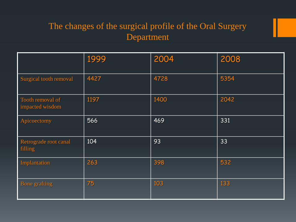
The changes of the surgical profile of the Oral Surgery
Department
1999 2004 2008
Surgical tooth removal 4427 4728 5354
Tooth removal of
impacted wisdom
1197 1400 2042
Apicoectomy 566 469 331
Retrograde root canal
filling
104 93 33
Implantation 263 398 532
Bone grafting 75 103 133
The history of the use of microscope in surgery
In 1953 Carl-Zeiss company made the first binocular operating microscope
In 1981 Apotheker and Jakob planned and started to sell the first operating
microscope for dentistry with the name Dentiscope
In March 1993 the first symposium of endodontic surgery was organized
at the dentistry department of Pennsylvanian University
Operating microscope The benefits of usage of operating microscope in
endodontics:
• Accurate localization of the root apex • Smaller „bone window” necessary during the
operation • The resection angle is smaller, than 10º • Ability to inspect, prepare and seal the isthmus area • More accurate preparation • Precise retrograde root filling
•
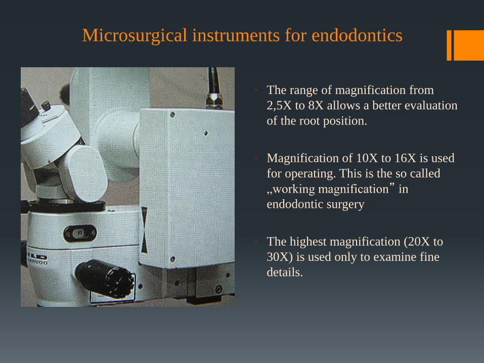
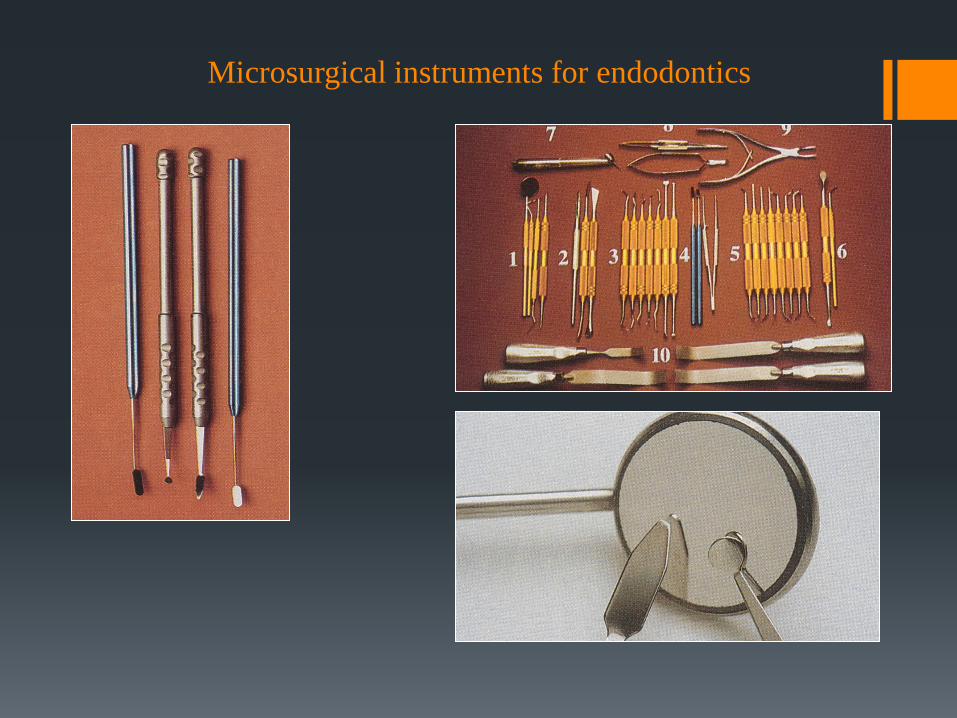
Microsurgical instruments for endodontics
The range of magnification from
2,5X to 8X allows a better evaluation
of the root position.
Magnification of 10X to 16X is used
for operating. This is the so called
„working magnification” in
endodontic surgery
The highest magnification (20X to
30X) is used only to examine fine
details.
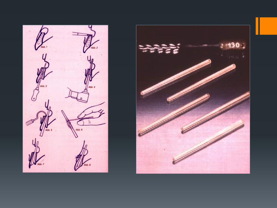
Microsurgical instruments for endodontics
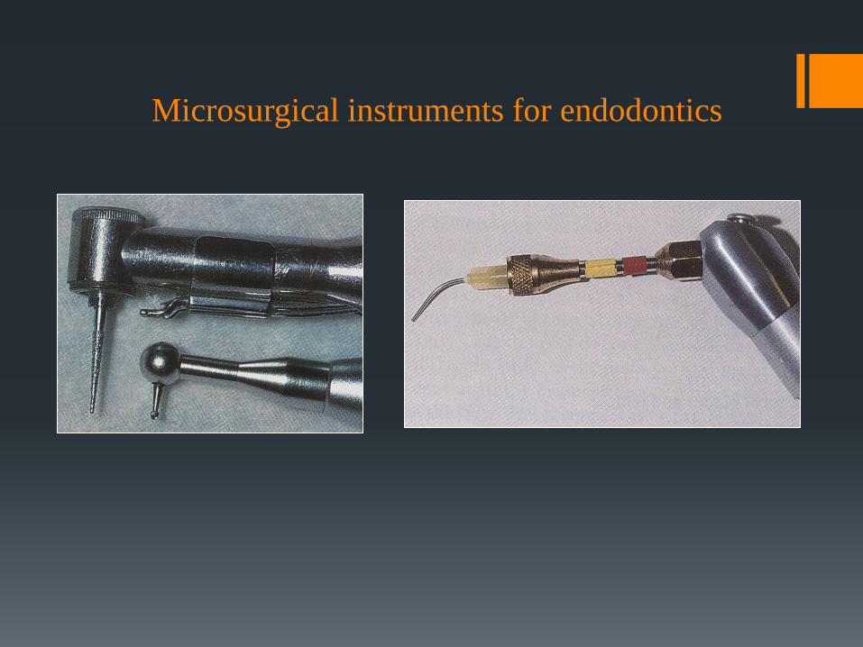
Microsurgical instruments for endodontics
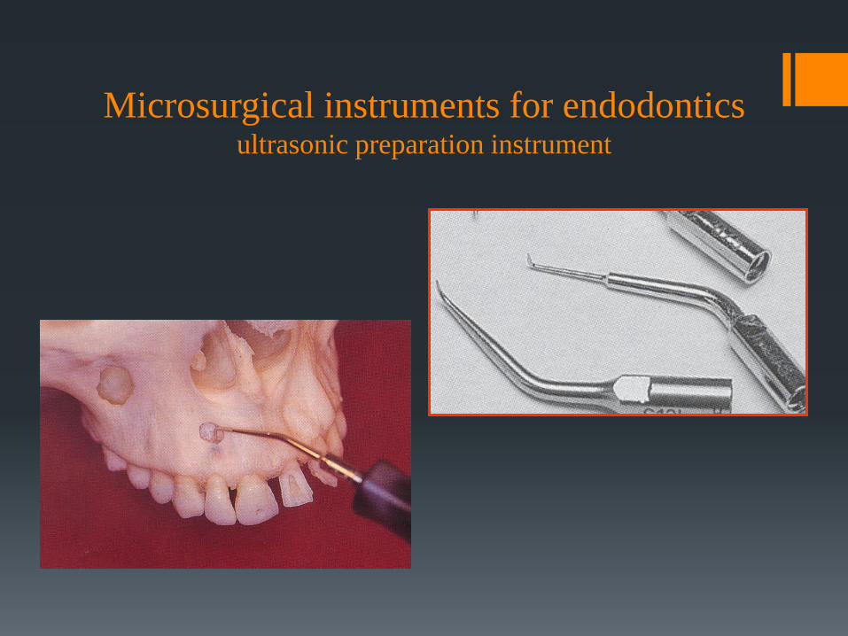
Microsurgical instruments for endodontics ultrasonic preparation instrument
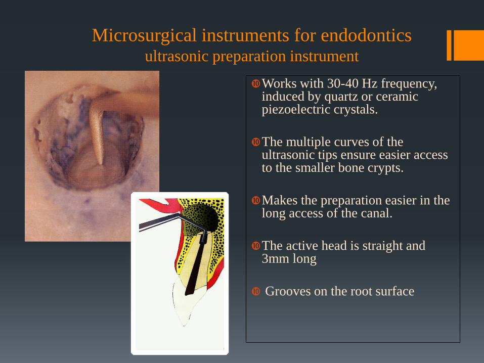
Microsurgical instruments for endodontics ultrasonic preparation instrument
Works with 30-40 Hz frequency, induced by quartz or ceramic piezoelectric crystals.
The multiple curves of the ultrasonic tips ensure easier access to the smaller bone crypts.
Makes the preparation easier in the long access of the canal.
The active head is straight and 3mm long
Grooves on the root surface
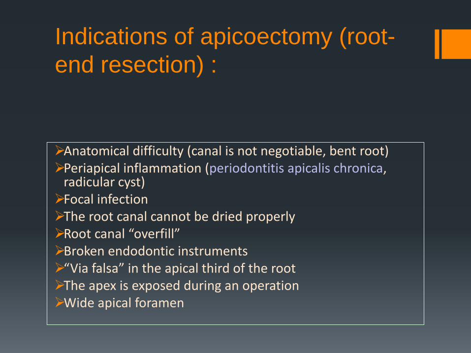
Indications of apicoectomy (root-
end resection) :
Anatomical difficulty (canal is not negotiable, bent root) Periapical inflammation (periodontitis apicalis chronica,
radicular cyst) Focal infection The root canal cannot be dried properly Root canal “overfill” Broken endodontic instruments “Via falsa” in the apical third of the root The apex is exposed during an operation Wide apical foramen
Contraindications of apicoectomy:
Acute purulent inflammation
General surgical contraindications
Teeth with a weakened periodontium
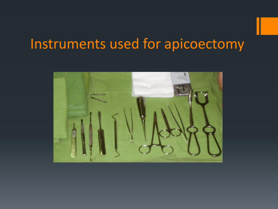
Instruments used for apicoectomy
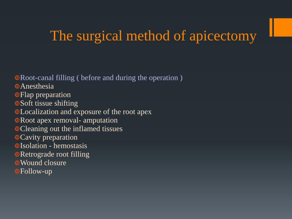
The surgical method of apicectomy
Root-canal filling ( before and during the operation ) Anesthesia Flap preparation Soft tissue shifting Localization and exposure of the root apex Root apex removal- amputation Cleaning out the inflamed tissues Cavity preparation Isolation - hemostasis Retrograde root filling Wound closure Follow-up
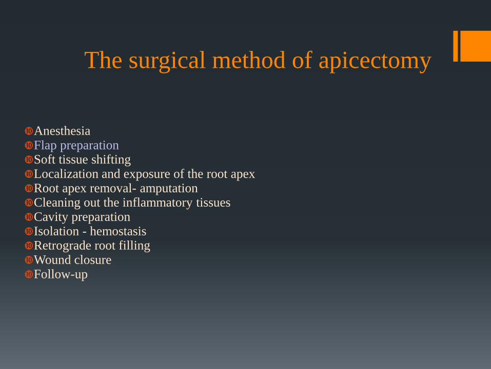
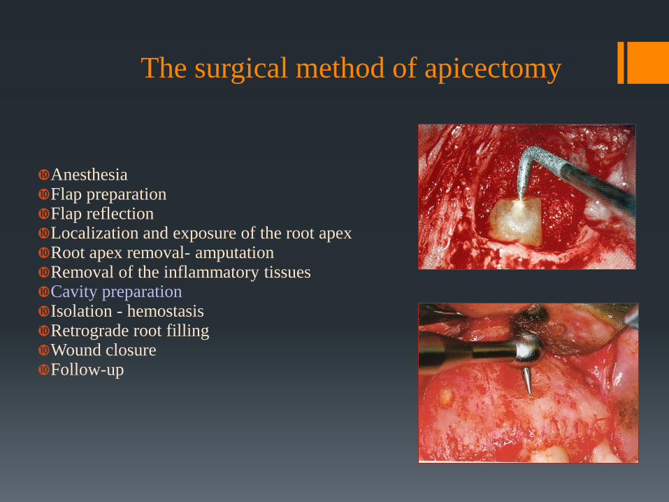
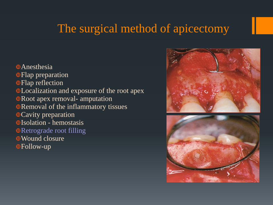
The surgical method of apicoectomy
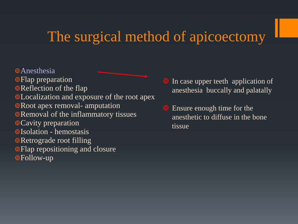
Anesthesia Flap preparation Reflection of the flap Localization and exposure of the root apex Root apex removal- amputation Removal of the inflammatory tissues Cavity preparation Isolation - hemostasis Retrograde root filling Flap repositioning and closure Follow-up
In case upper teeth application of
anesthesia buccally and palatally
Ensure enough time for the
anesthetic to diffuse in the bone
tissue
The surgical method of apicectomy
Anesthesia Flap preparation Soft tissue shifting Localization and exposure of the root apex Root apex removal- amputation Cleaning out the inflammatory tissues Cavity preparation Isolation - hemostasis Retrograde root filling Wound closure Follow-up
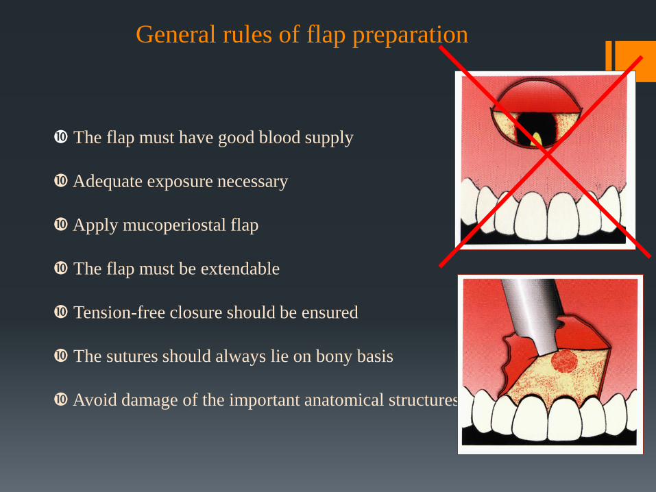
The flap must have good blood supply
Adequate exposure necessary
Apply mucoperiostal flap
The flap must be extendable
Tension-free closure should be ensured
The sutures should always lie on bony basis
Avoid damage of the important anatomical structures
General rules of flap preparation
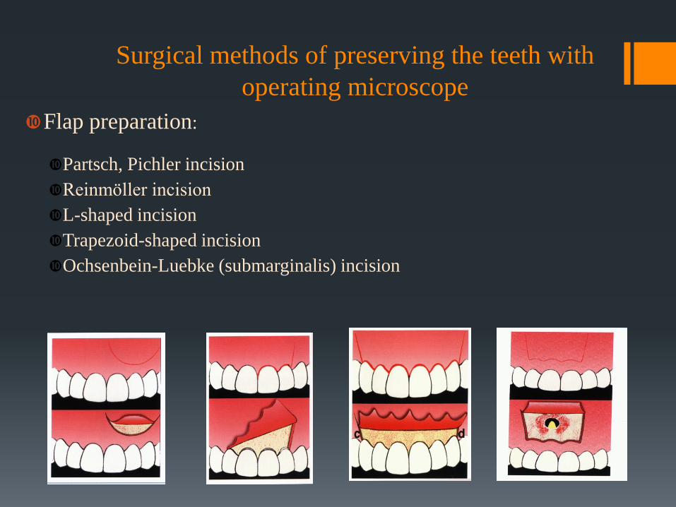
Surgical methods of preserving the teeth with
operating microscope
Flap preparation:
Partsch, Pichler incision
Reinmöller incision
L-shaped incision
Trapezoid-shaped incision
Ochsenbein-Luebke (submarginalis) incision
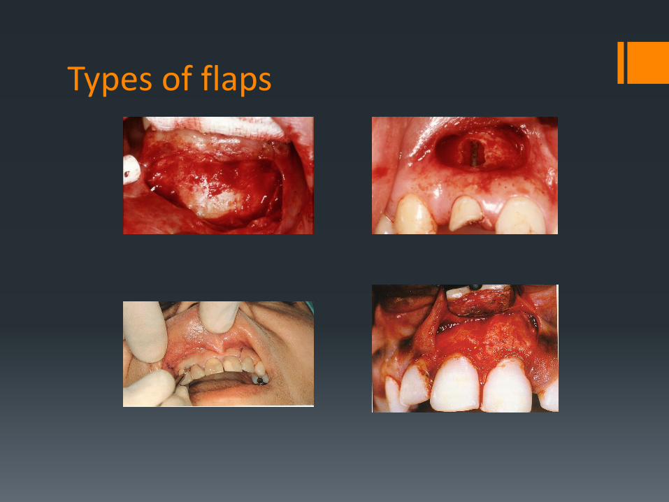
Types of flaps
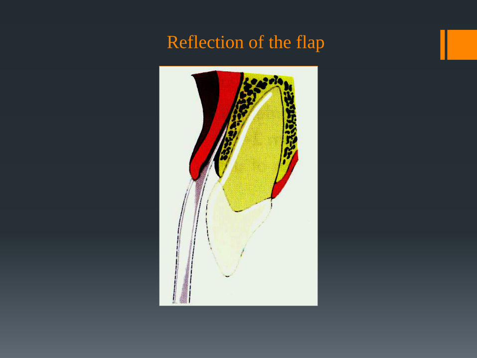
Reflection of the flap

The surgical method of apicectomy
Anesthesia Flap preparation Soft tissue shifting Localization and exposure of the root apex Root apex removal- amputation Cleaning out the inflammatory tissues Cavity preparation Isolation - hemostasis Retrograde root filling Wound closure Follow-up
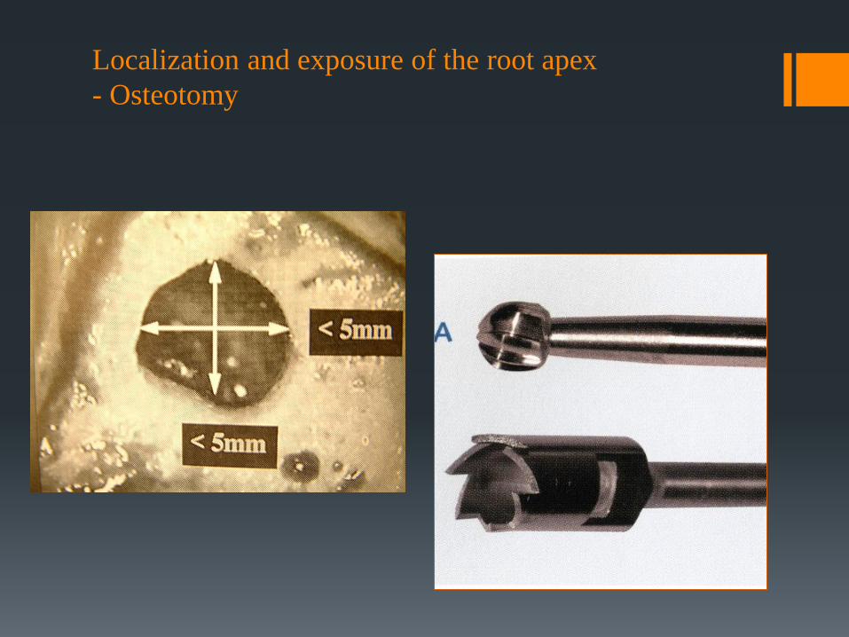
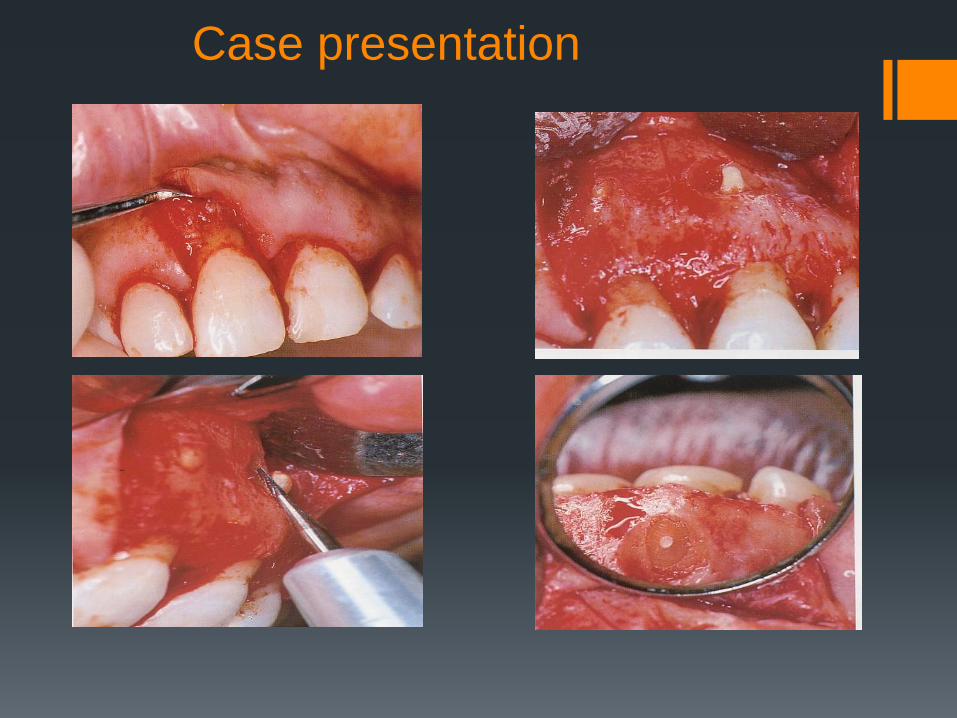
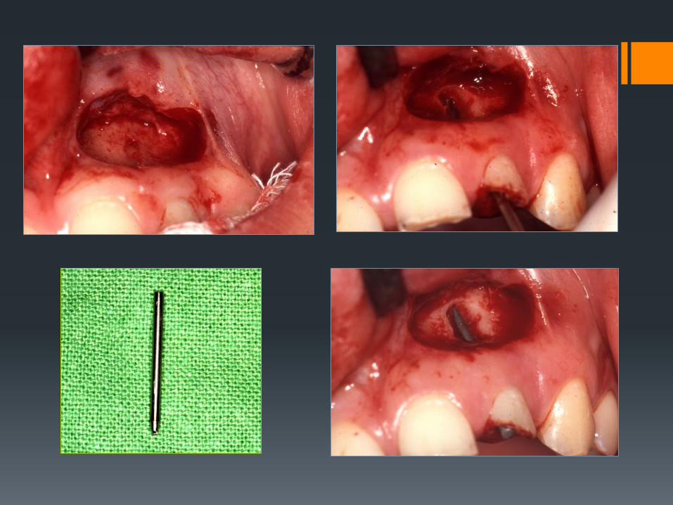
Localization and exposure of the root apex
- Osteotomy
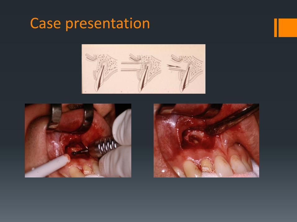
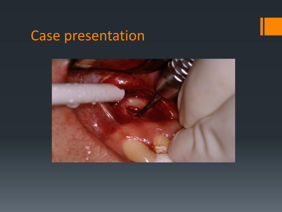
Case presentation

The surgical method of apicectomy
Anesthesia Flap preparation Soft tissue shifting Localization and exposure of the root apex Root apex removal- amputation Cleaning out the inflammatory tissues Cavity preparation Isolation - hemostasis Retrograde root filling Wound closure Follow-up
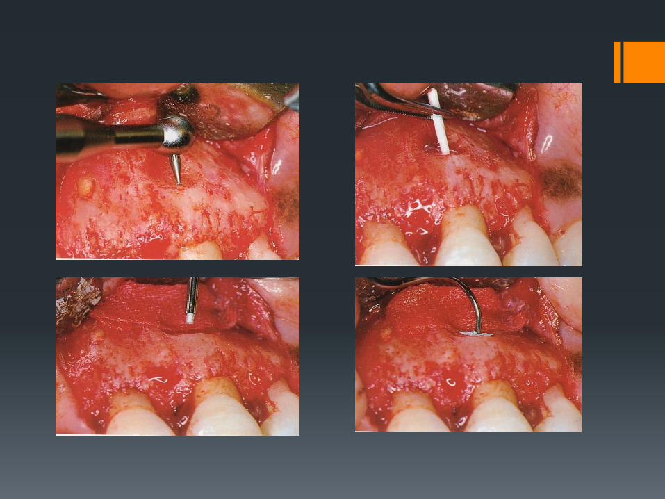
Case presentation
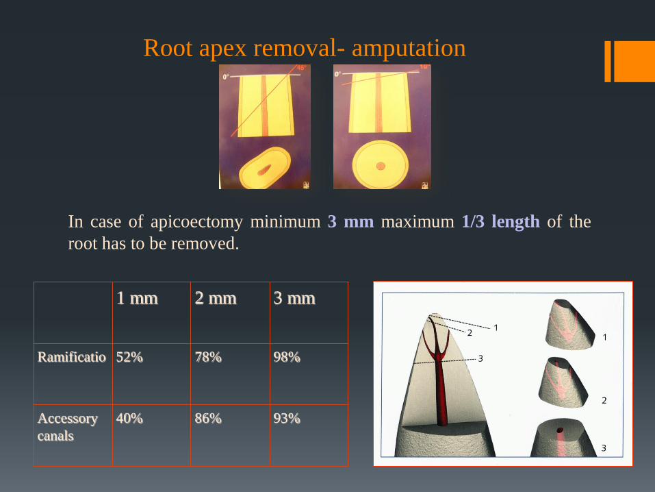
Root apex removal- amputation
1 mm 2 mm 3 mm
Ramificatio 52% 78% 98%
Accessory
canals
40% 86% 93%
In case of apicoectomy minimum 3 mm maximum 1/3 length of the
root has to be removed.
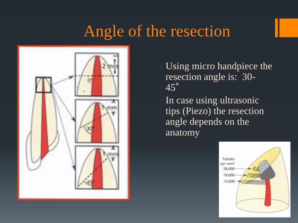
Angle of the resection
Using micro handpiece the resection angle is: 30-45°
In case using ultrasonic tips (Piezo) the resection angle depends on the anatomy
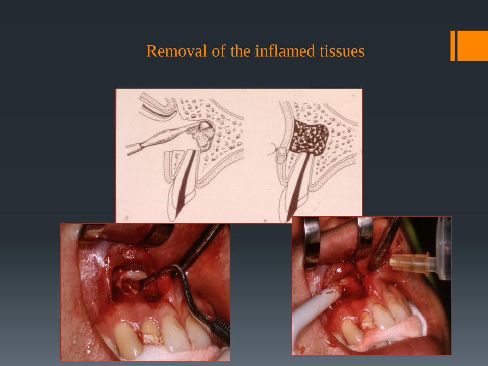
Removal of the inflamed tissues
The surgical method of apicectomy
Anesthesia Flap preparation Flap reflection Localization and exposure of the root apex Root apex removal- amputation Removal of the inflammatory tissues Cavity preparation Isolation - hemostasis Retrograde root filling Wound closure Follow-up
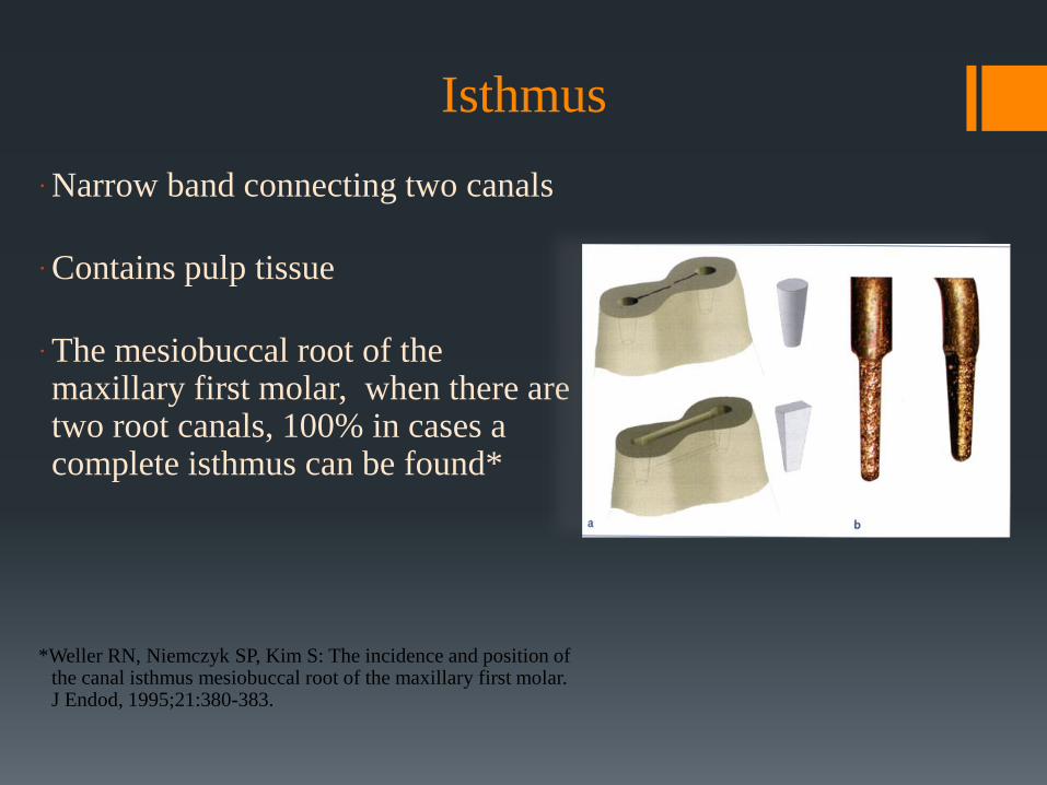
Isthmus
Narrow band connecting two canals
Contains pulp tissue
The mesiobuccal root of the maxillary first molar, when there are two root canals, 100% in cases a complete isthmus can be found*
*Weller RN, Niemczyk SP, Kim S: The incidence and position of
the canal isthmus mesiobuccal root of the maxillary first molar. J Endod, 1995;21:380-383.
RETROGRADE CAVITY PREPARATION
Preparation of cavity of appropriate size.
The walls are parallel with the long axis of the root
The cavity must be in the central position compared to
the cross-section of the root
Proper cavity depth for the retrograde filling
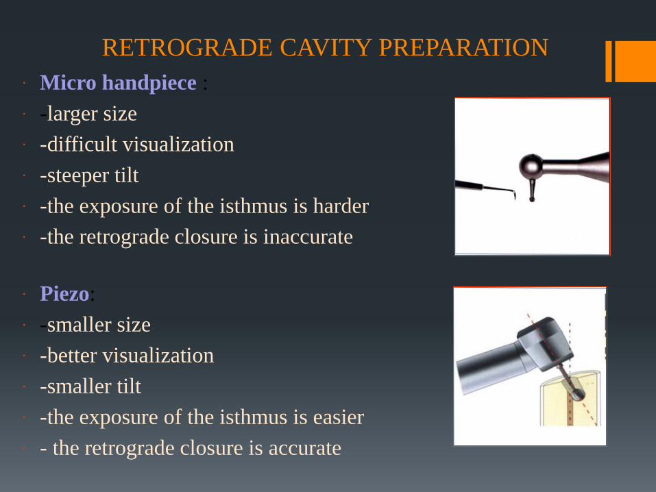
RETROGRADE CAVITY PREPARATION
Micro handpiece :
-larger size
-difficult visualization
-steeper tilt
-the exposure of the isthmus is harder
-the retrograde closure is inaccurate
Piezo:
-smaller size
-better visualization
-smaller tilt
-the exposure of the isthmus is easier
- the retrograde closure is accurate
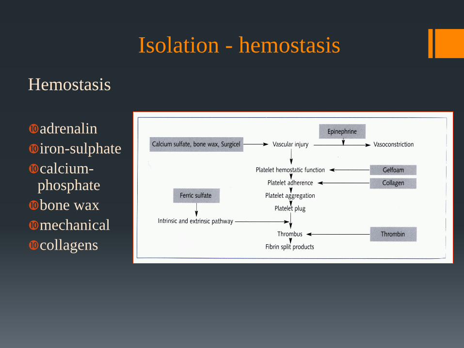
Isolation - hemostasis
Hemostasis
adrenalin
iron-sulphate
calcium-phosphate
bone wax
mechanical
collagens
The surgical method of apicectomy
Anesthesia Flap preparation Flap reflection Localization and exposure of the root apex Root apex removal- amputation Removal of the inflammatory tissues Cavity preparation Isolation - hemostasis Retrograde root filling Wound closure Follow-up
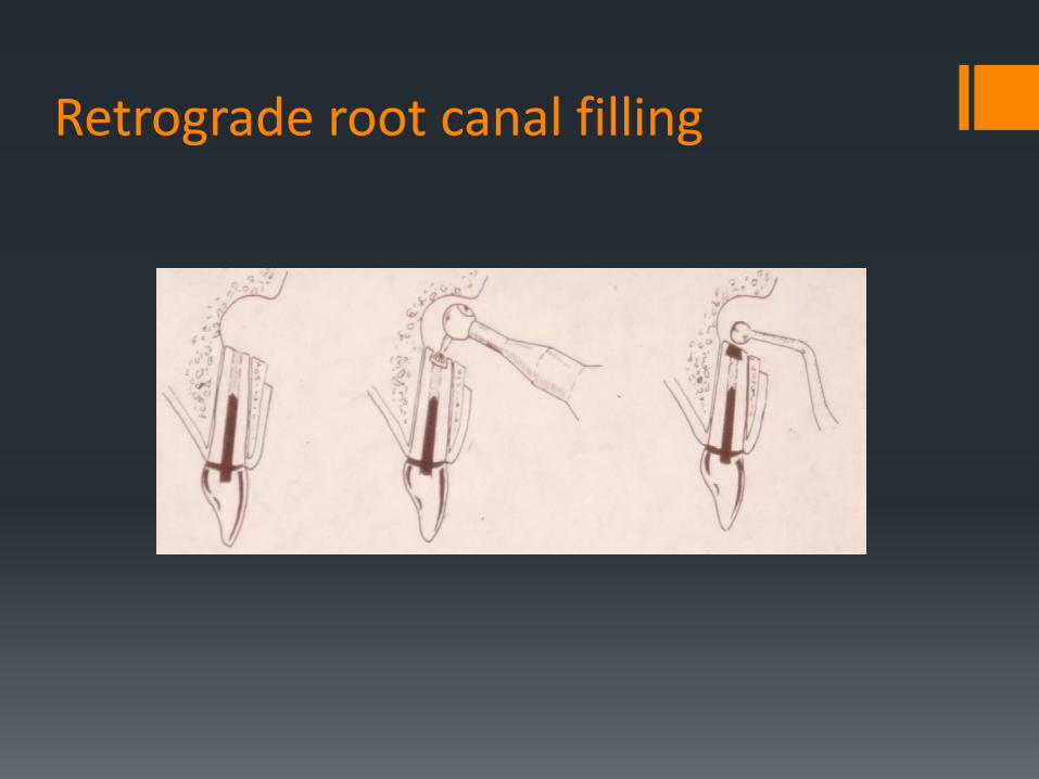
Retrograde root canal filling
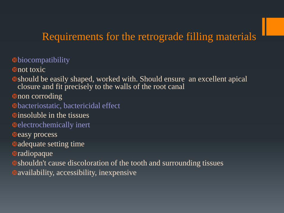
Requirements for the retrograde filling materials
biocompatibility
not toxic
should be easily shaped, worked with. Should ensure an excellent apical closure and fit precisely to the walls of the root canal
non corroding
bacteriostatic, bactericidal effect
insoluble in the tissues
electrochemically inert
easy process
adequate setting time
radiopaque
shouldn't cause discoloration of the tooth and surrounding tissues
availability, accessibility, inexpensive
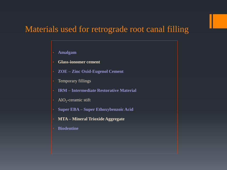
Materials used for retrograde root canal filling
• Amalgam
• Glass-ionomer cement
• ZOE – Zinc Oxid-Eugenol Cement
• Temporary fillings
• IRM – Intermediate Restorative Material
• AlO2-ceramic stift
• Super EBA – Super Ethoxybenzoic Acid
• MTA – Mineral Trioxide Aggregate
• Biodentine
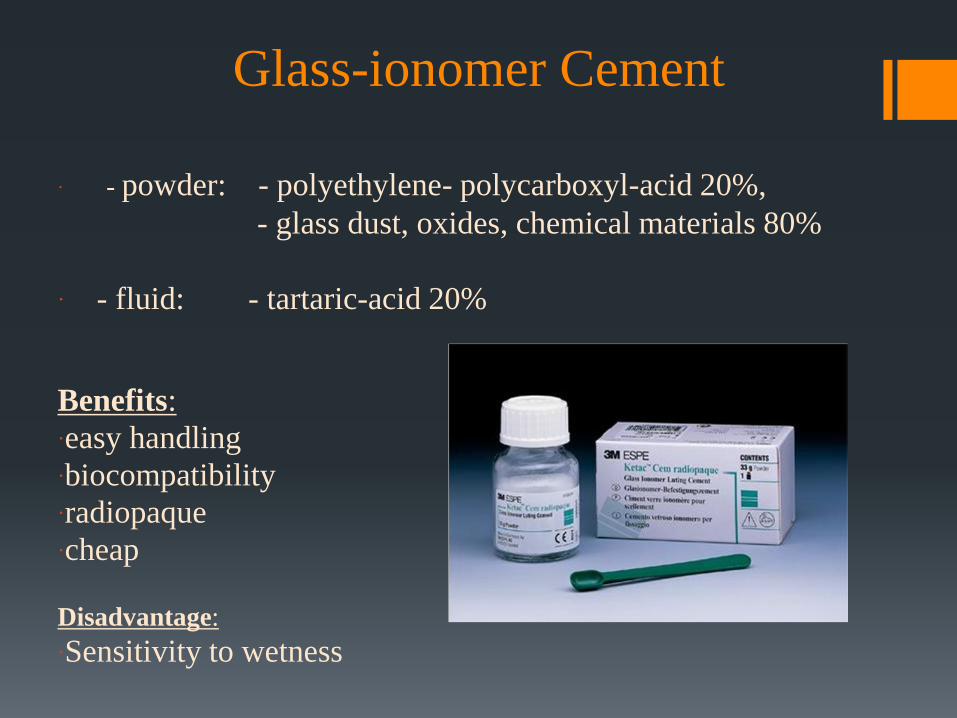
Glass-ionomer Cement
- powder: - polyethylene- polycarboxyl-acid 20%,
- glass dust, oxides, chemical materials 80%
- fluid: - tartaric-acid 20%
Benefits:
easy handling
biocompatibility
radiopaque
cheap
Disadvantage:
Sensitivity to wetness
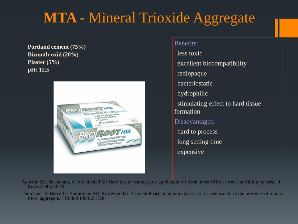
MTA - Mineral Trioxide Aggregate
Portland cement (75%)
Bizmuth-oxid (20%)
Plaster (5%)
pH: 12,5
Apaydin ES, Shabahang S, Torabinejad M: Hard tissue healing after application of fresh or set MTA as root-end filling material. J Endod 2004;30:21.
Thomson TS, Berry JE, Somerman MJ, Kirkwood KL: Cementoblasts maintain expression of osteocalcin in the presence of mineral trioxi aggregate. J Endod 1999;25:728.
Benefits:
less toxic
excellent biocompatibility
radiopaque
bacteriostatic
hydrophilic
stimulating effect to hard tissue
formation
Disadvantages:
hard to process
long setting time
expensive

Biodentin
Tricalcium-silicate powder
Calcium-chloride
Biocompatibility
Radiopaque
Physical properties similar to
dentin
Other applications:
-pulp capping,
-restore root perforations
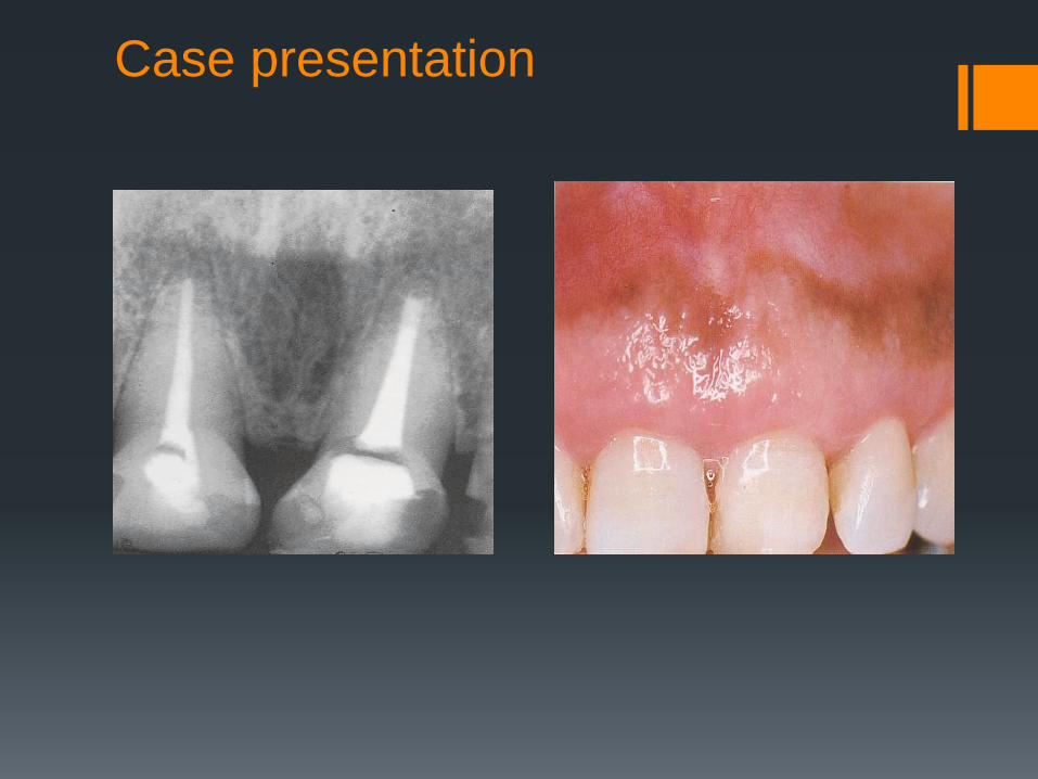
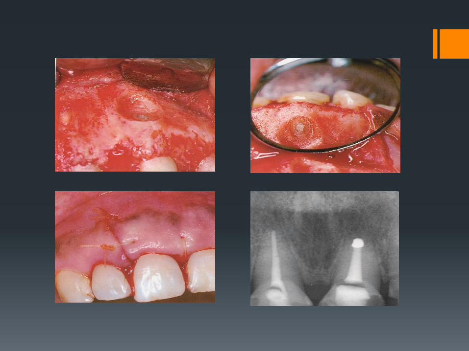
Case presentation
Case presentation
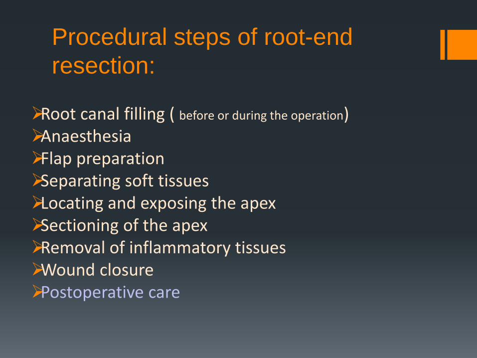
Procedural steps of root-end
resection:
Root canal filling ( before or during the operation) Anaesthesia Flap preparation Separating soft tissues Locating and exposing the apex Sectioning of the apex Removal of inflammatory tissues Wound closure Postoperative care
Postoperative care
Cooling
Proper oral hygiene (Corsodyl, camomilla)
Removal of sutures on the 7.-8. day
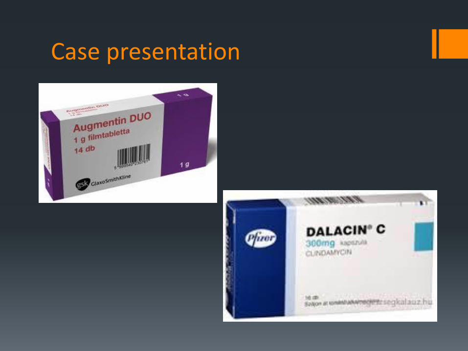
Antibiotics, pain killers if necessary
Controll periapical X-ray ( immediately after operation, 6, 12 month later )
Case presentation
Postoperative complications:
Pain
Swelling
Inflammation
Bleeding
Intraoral haematome
Soft tissue injury
Foreign body in the operated area
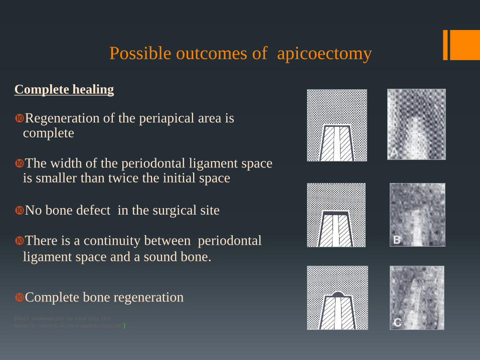
Possible outcomes of apicoectomy
Complete healing
Regeneration of the periapical area is
complete
The width of the periodontal ligament space is smaller than twice the initial space
No bone defect in the surgical site
There is a continuity between periodontal
ligament space and a sound bone.
Complete bone regeneration (Rud,J., Andreasen,J.O.: Int.J.Oral Surg, 1972
Molven O., Halsen A.:Int.J.Oral Maxillofac.Surg.1987)
Possible outcomes of apicoectomy
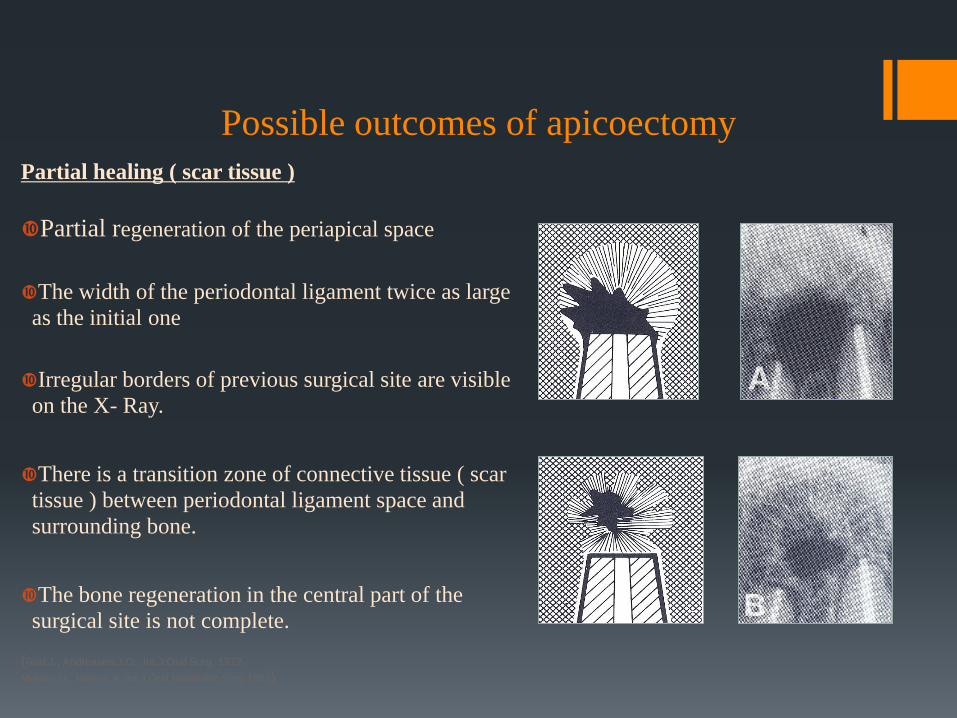
Partial healing ( scar tissue )
Partial regeneration of the periapical space
The width of the periodontal ligament twice as large
as the initial one
Irregular borders of previous surgical site are visible
on the X- Ray.
There is a transition zone of connective tissue ( scar
tissue ) between periodontal ligament space and
surrounding bone.
The bone regeneration in the central part of the
surgical site is not complete. (Rud,J., Andreasen,J.O.: Int.J.Oral Surg, 1972
Molven O., Halsen A.:Int.J.Oral Maxillofac.Surg.1987)

Possible outcomes of apicoectomy
Uncertain healing
Partial regeneration of the periapical space.
The width of the periodontal ligament space
is twice the size of the initial one.
The surgical site gives round shape shadow
on an X-Ray.
The cone shape bone defect around resected
root is visible.
The bone regeneration in the center of the
surgical site is absent
(Rud,J., Andreasen,J.O.: Int.J.Oral Surg, 1972
Molven O., Halsen A.:Int.J.Oral Maxillofac.Surg.1987)
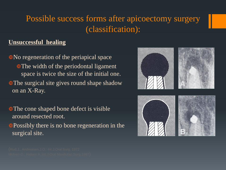
Possible success forms after apicoectomy surgery
(classification):
Unsuccessful healing
No regeneration of the periapical space
The width of the periodontal ligament
space is twice the size of the initial one.
The surgical site gives round shape shadow
on an X-Ray.
The cone shaped bone defect is visible
around resected root.
Possibly there is no bone regeneration in the
surgical site.
(Rud,J., Andreasen,J.O.: Int.J.Oral Surg, 1972
Molven O., Halsen A.:Int.J.Oral Maxillofac.Surg.1987)
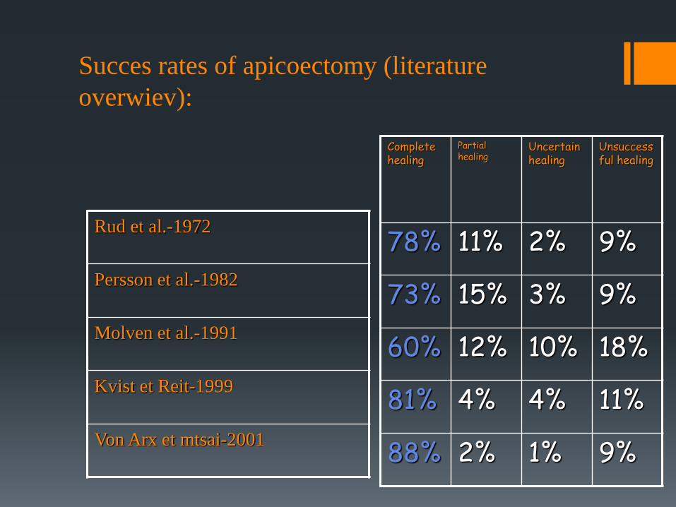
Rud et al.-1972
Persson et al.-1982
Molven et al.-1991
Kvist et Reit-1999
Von Arx et mtsai-2001
Complete healing
Partial healing
Uncertain healing
Unsuccessful healing
78% 11% 2% 9%
73% 15% 3% 9%
60% 12% 10% 18%
81% 4% 4% 11%
88% 2% 1% 9%
Succes rates of apicoectomy (literature
overwiev):
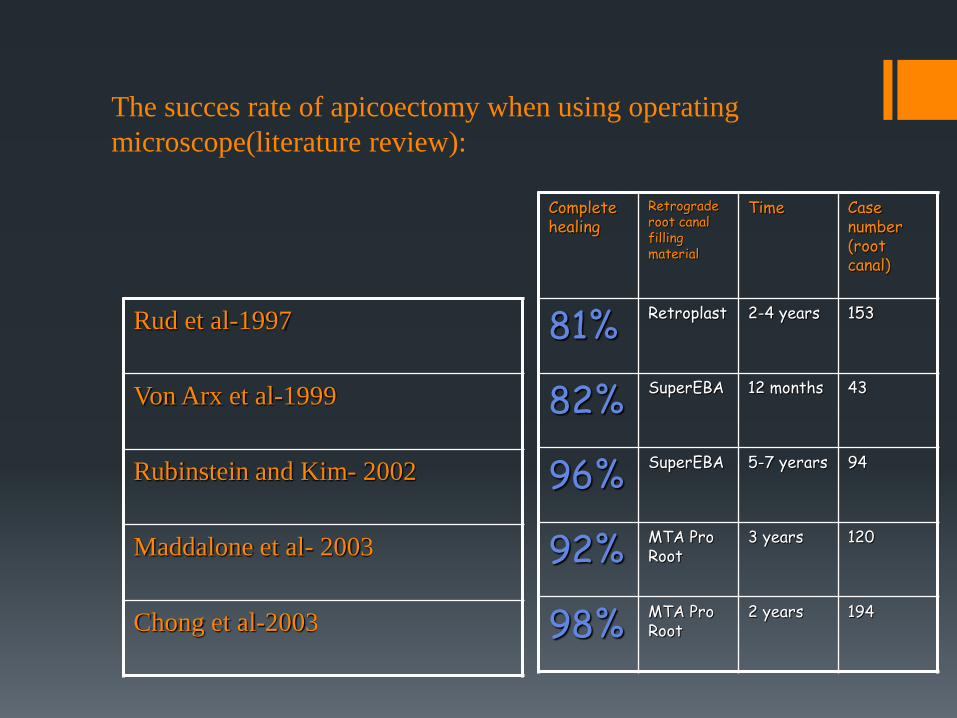
Rud et al-1997
Von Arx et al-1999
Rubinstein and Kim- 2002
Maddalone et al- 2003
Chong et al-2003
Complete healing
Retrograde root canal filling material
Time Case number (root canal)
81% Retroplast 2-4 years 153
82% SuperEBA 12 months 43
96% SuperEBA 5-7 yerars 94
92% MTA Pro Root
3 years 120
98% MTA Pro Root
2 years 194
The succes rate of apicoectomy when using operating
microscope(literature review):
Bibliography

Surgical Methods of the
Conservative Treatment of Teeth
Apicoectomy (root-end resection)
Retrograde root canal filling
Transdental fixation
Tooth replantation
Tooth transplantation
Surgical Methods of the
Conservative Treatment of Teeth
Apicoectomy (root-end resection)
Retrograde root canal filling
Transdental fixation
Tooth replantation
Tooth transplantation
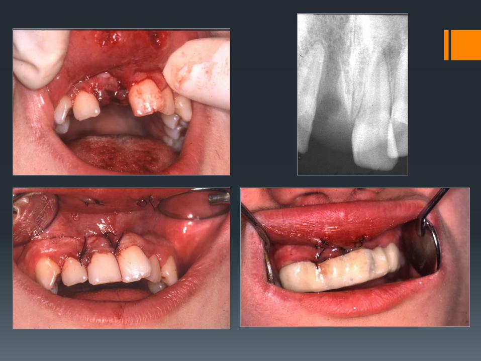
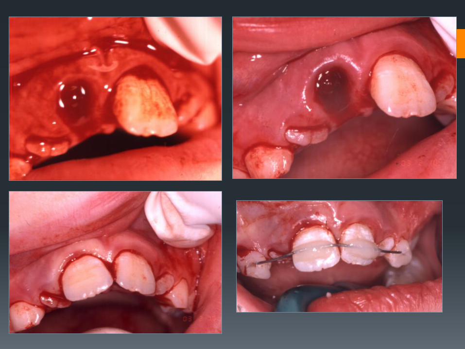
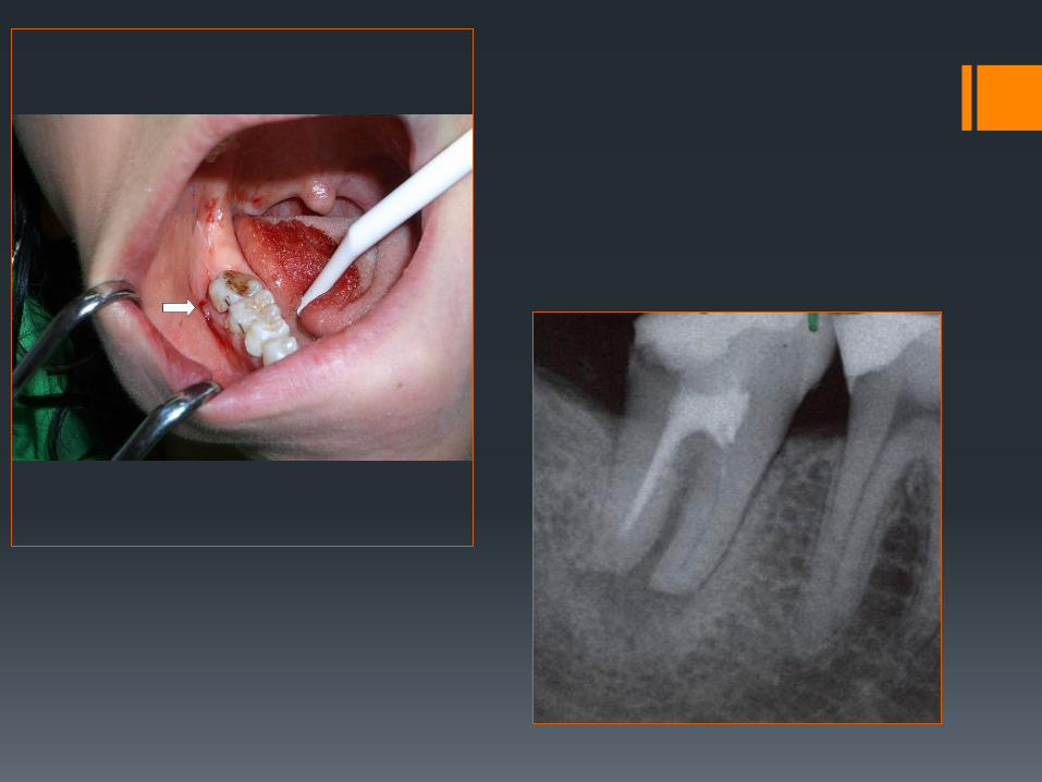
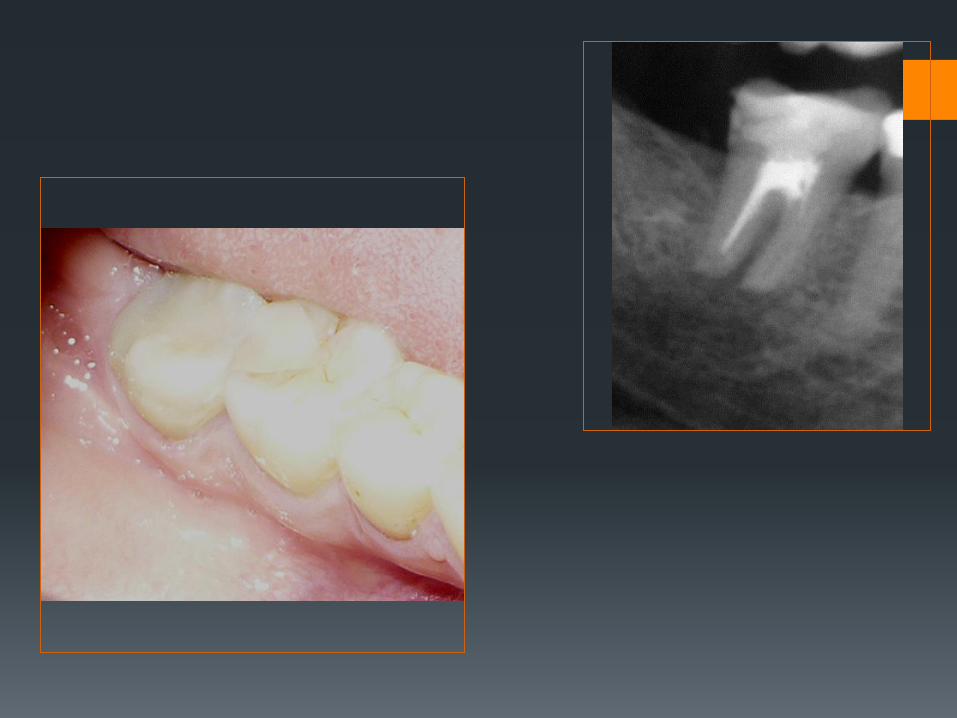
Tooth replantation
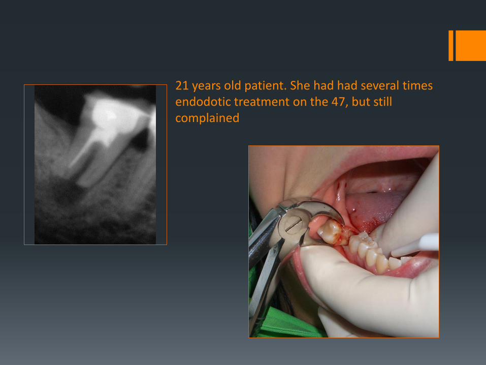
21 years old patient. She had had several times endodotic treatment on the 47, but still complained
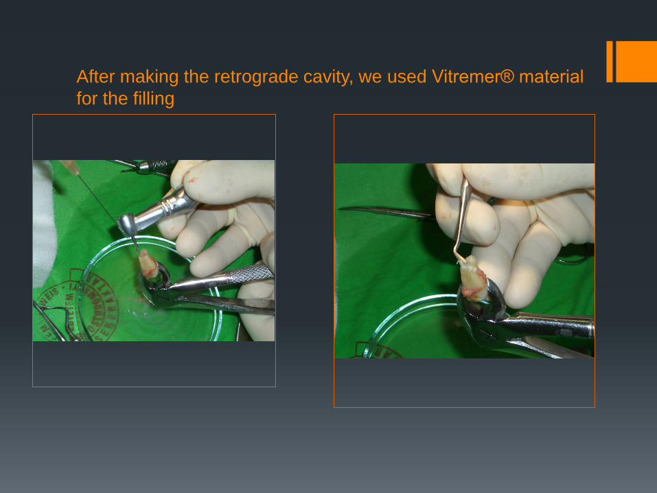
After making the retrograde cavity, we used Vitremer® material
for the filling

Surgical Methods of the
Conservative Treatment of Teeth
Apicoectomy (root-end resection)
Retrograde root canal filling
Transdental fixation
Tooth replantation
Tooth transplantation
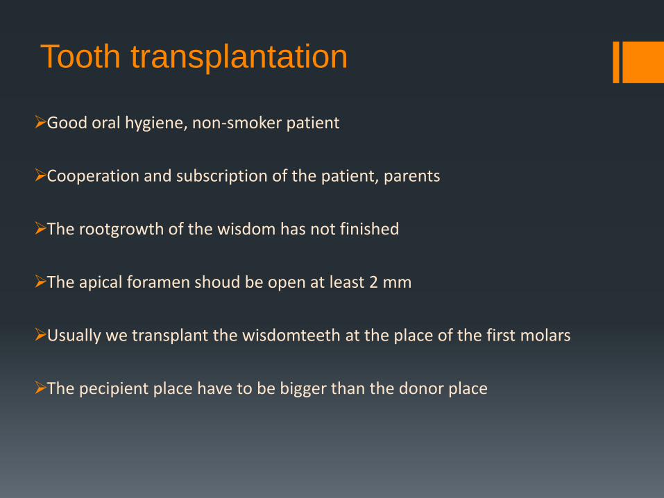
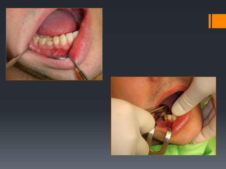
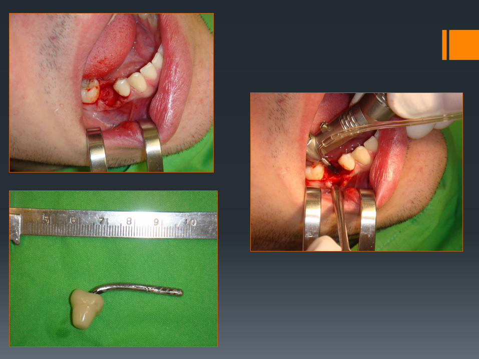
Tooth transplantation Good oral hygiene, non-smoker patient
Cooperation and subscription of the patient, parents
The rootgrowth of the wisdom has not finished
The apical foramen shoud be open at least 2 mm
Usually we transplant the wisdomteeth at the place of the first molars
The pecipient place have to be bigger than the donor place
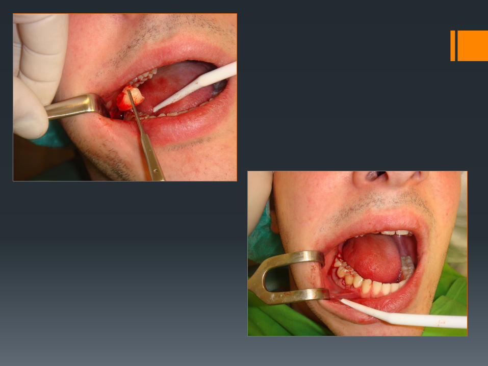
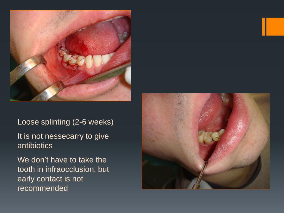
Loose splinting (2-6 weeks)
It is not nessecarry to give
antibiotics
We don’t have to take the
tooth in infraocclusion, but
early contact is not
recommended

Thank You for your attention