Embed Size (px)
Citation preview
1
PHYSIOLOGY OF THE LIVER
BY Abdelaziz Hussein
Assistant Lecturer of Physiology

2
Gross Anatomy
q The Liver is the largest gland in the body ( weighing about 1.5 Kg in adults; representing 2% of the TBW)
q It is essential to life. q It is situated in the right upper quadrant of the abdomen.
q It is is covered by Glisson's capsule, a visceral continuation of the peritoneum.

3
Hepatic lobes q The two major lobes, right and left, and 2 accessory lobes, quadrate and caudate q The right lobe is six times larger than left lobe

4
Hepatic lobes (cont.)

5
MICROSCOPIC STRUCTURE
Functionally the liver consists of 3 systems;
q Liver Cell (Hepatocyte) Systems → arranged in hexagonal and pentagonal units called hepatic lobules.
q Biliary System. q Blood Circulatory System.

6
Hepatic Lobule
q The hepatic lobule is the structural unit of the liver which is hexagonal or pentagonal in shape (1 st described by Malpighian 1666).
q Each lobule consists of radiating columns( 2 or more rows of cells) of hepatocytes around a central vein and surrounded by 4 to 6 portal tracts (bile duct, branches of the hepatic artery and portal vein, along with nerves and lymphatics).
q The human liver contains about 50000100000 lobules.
7
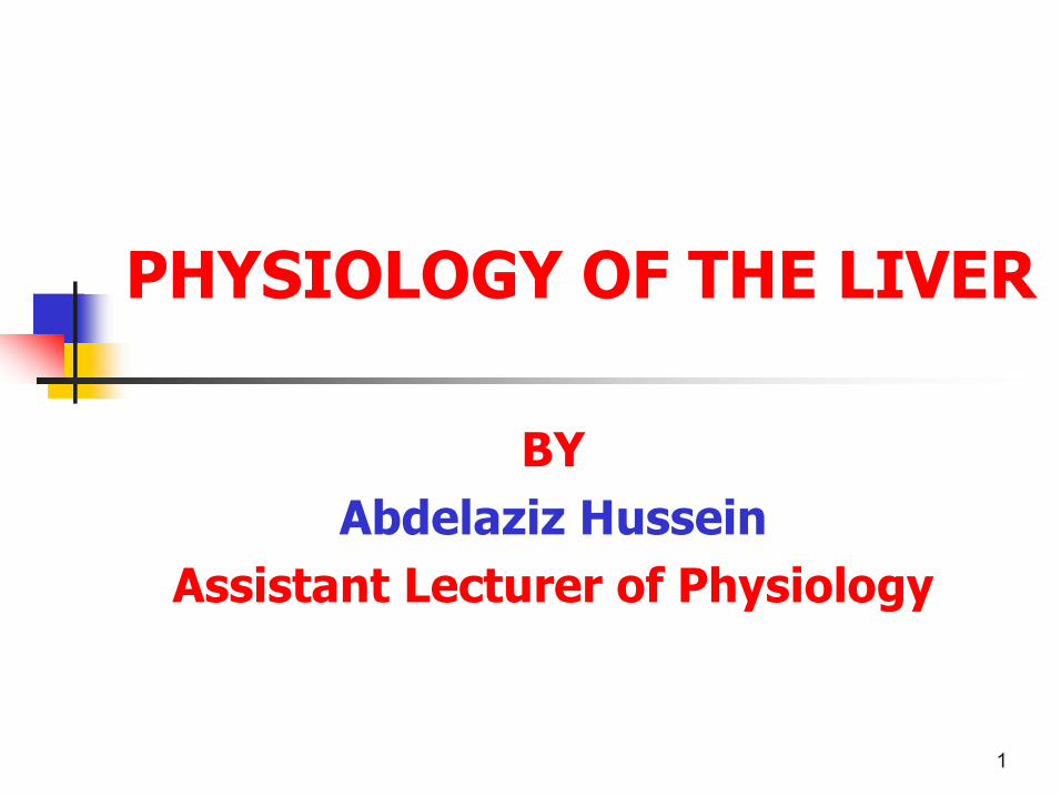
Liver acinus
q The acinus is a diamond – shaped mass of liver parenchyma from 2 adjacent hepatic lobules.
q It is subdivided into 3 zones; v Zone 1 cells form the most active core of the acinus and are the last to die and the first to regenerate.
v Zone 3 cells are the most prone to toxic, viral, or anoxic injury.
8
Ultrastructure of Hepatocytes n Hepatocytes represent 94 % of liver parenchyma n Hepatocytes are covered by specific membranes which have 3 surfaces :
1) Sinusoidal (70 % of surface area) for exchange of material between the Disse space and intracellular compartment (endo and exocytosis).
2) Canalicular membrane (15 %) for exchange with the biliary canaliculi or hemicanals.
3) Lateral membrane (15 %) separated from neighboring hepatocytes by tight junctions and involved in intercellular transport between hepatocytes.
9
Ultrastructure of Hepatocytes
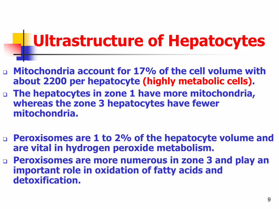
q Mitochondria account for 17% of the cell volume with about 2200 per hepatocyte (highly metabolic cells).
q The hepatocytes in zone 1 have more mitochondria, whereas the zone 3 hepatocytes have fewer mitochondria.
q Peroxisomes are 1 to 2% of the hepatocyte volume and are vital in hydrogen peroxide metabolism.
q Peroxisomes are more numerous in zone 3 and play an important role in oxidation of fatty acids and detoxification.

10
Ultrastructure of Hepatocytes
Also hepatocytes contains; n Lysosomes are electrondense cytoplasmic organelles responsible for degrading biological material using acid hydrolases.
n The endoplasmic reticulum constitutes 19% of the cell volume and is the site of protein synthesis.
n The Golgi apparatus is responsible for processing of macromolecules.

11
Biliary System n Bile secreted through the canalicular membrane of the hepatocyte collects in biliary canaliculi.
n These small biliary canaliculi form channels continuous with the short duct of Hering that join the cholangioles at the limiting plate of the portal areas.
n These cholangioles then merge into larger bile ducts

12
Hepatic vascular system n The liver receives about 1.5 L blood / minute (about 25 % of the COP) from 2 sources;
n The portal vein which is formed by the confluence of the superior mesenteric vein and the splenic veins → 75% of HBF
n The hepatic artery arises from the coeliac trunk → 25% of HBF
n These vessels pour their blood into the sinusoids which drain into central veins and these coalesce forming hepatic veins which drain into the inferior vena cava.

13
Blood Sinusoids n Sinusoids are specialized capillaries without a basement membrane and lined with endothelial lining cells through which proteins of low molecular weight may percolate into the space of Disse.
n The sinusoidal endothelial cells lack a basement membrane and are perforated by abundant small fenestrae (average diameter 100 nm) in clusters called sieve plates.

14
Hepatic sinusoid lining cells
q There are 4 types of hepatic sinusoid lining cells
q They make up 6 % of all liver parenchyma
q They include endothelial cells, Kupffer cells, hepatic stellate cells (Ito cells, fatstoring cells), and pit cells (intrahepatic lymphocytes)

15
Kupffer cells q These cells represent part of the mononuclear phagocyte system and are adherent to the sinusoidal surface of endothelial lining cells, predominantly in a periportal distribution.
q 2 % of the total liver parenchyma cells. q Their main function is to phagocytose a range of particulate material including cellular debris, senescent red blood cells, parasites, bacteria, endotoxin, and tumour cells. Phagocytosis is via a range of mechanisms including coated pits, macropinocytotic vesicles, and phagosomes aided by opsonization of particles by fibronectin or opsonin.
q Also they secrete 10 % of erythropoietin hormone.

16
Hepatic stellate cells n Stellate cells (Ito cells, fatstoring cells) have a similar morphology to fibroblasts with the addition of fat droplets, and are located within the Disse space.
n Stellate cells contain most of the body's stores of vitamin A.
n These cells are central to the process of hepatic fibrogenesis, responding to mediators released by parenchymal and Kupffer cells, causing transformation into myofibroblasts.
n Activation of stellate cells is also an important mechanism for control of sinusoidal perfusion, through cytoskeletal actin within branching cellular processes beneath the endothelium.

17
Pit cells
n Pit cells are large granular lymphocytes which have natural killer cell properties with spontaneous activity against tumour cells in the absence of prior activation.
n They may also play a role in hepatic regeneration

18
Lymphatics q The liver has a high blood flow and a highly permeable microcirculation. The consequent production of interstitial fluid, intrahepatic lymph, is formed in the perisinusoidal space of Disse between the hepatocytes and sinusoidal lining endothelium.
q Lymphatic vessels drain via the portal tracts, closely applied to the hepatic arterial branches, to the hilum and thence to the thoracic duct.
q some interstitial fluid drains through Glisson's capsule into the peritoneum.
q The lymph flow rate in mammalian liver is approximately 0.5 ml/kg of liver perminute making up 25 to 50 per cent of thoracic duct lymph flow.
q

19
Hepatic blood flow Hepatic blood flow is about 1500 ml blood / minute v It increases after feeding and with expiration. v It decreases with standing, inspiration, and sleep.
Regulation of HBF: A) Autoregulation: v The portal venous system is passive, without pressure dependent autoregulation, and the major physiological factors controlling flow are those modulating supply to the intestines and spleen
v Vascular autoregulation of hepatic arterial blood flow mediated by adenosine is present, but may not be of great physiological importance.

20
Hepatic blood flow A) Autoregulation: v Changes in hepatic oxygen consumption do not seem to
control hepatic blood flow. v There is an important reciprocity between portal venous and
hepatic arterial flow with a reduction in portal venous input being associated with significant compensatory decrease in hepatic arterial resistance and rise in arterial flow.
v The mechanism for this relationship is unproven but may be due to adenosinemediated arterial vasodilatation.
B) Nervous regulation : v Sympathetic nerve stimulation may reduce hepatic blood
volume by up to 50 per cent.

21
Sinusoidal perfusion n Blood pressure in sinusoids ranges from 4.8 to 1.7 mmHg,
with flows of 270 to 410 ml/s. n The unidirectional sinusoidal flow can be controlled for by
either passive (haemodynamic) or active mechanisms;
Passive control mechanisms include: (i) the arterial input pressure and flow at the level of the
arteriosinous twig at the origin of the sinusoid; and (ii) changes in right atrial pressure, central venous pressure, and
hepatic venous pressure that are transmitted to the sinusoid from the centrilobular veins.

22
Sinusoidal perfusion Active control mechanisms include: (i) the presence of ‘functional’ sphincters at the inlet and outlet of the sinusoid due to indentations by the cell bodies of sinusoidal lining cells, which under different physiological stimuli may change dimension and alter sinusoidal perfusion.
(ii) plugging by leucocytes, which are less compressible than erythrocytes and may under physiological stimuli adhere to endothelial lining cells.
(iii) activation of Kupffer cells within sinusoids and release of other vasoactive mediators including nitric oxide, cytokines, and prostanoids.
(iv) transformation of hepatic stellate cells into activated contractile myofibroblasts that constrict the sinusoidal lumen.

23
Overview of Liver Functions

24
1) Metabolic Functions:
q Hepatic metabolic processes have a central role in protein, carbohydrate, and lipid metabolism and fuel economy, orchestrating a diverse interplay between central splanchnic and peripheral organs.
q Interruption to these processes results in the major metabolic consequences of acute and chronic liver disease.

25
1) Metabolic Functions (Cont.):
A) Carbohydrate metabolism n The liver has a central role in maintaining blood glucose within a narrow margin→ Glucostat.
n During fasting, hepatic glucose release is contributed to by both glycogenolysis (by glucagon and catecholamines) and gluconeogenesis (by glucocorticoids) from lactate, pyruvate, glycerol, and the glucogenic amino acids alanine and glutamine.
n After meals the excess blood glucose is converted into glycogen by insulin.

26
1) Metabolic Functions (Cont.): B) Protein metabolism The liver manufactures and exports; 1) Most of plasma proteins except gamma globulins
(approximately 15 50 gm/day)→ if ½ plasma proteins lost it can be replaced within 12 weeks.
2) Enzymes e.g. transaminases and alkaline phosphatase. 3) With the exception of factor VIII, the blood clotting factors
are made exclusively in hepatocytes. Biosynthesis of factors II, VII, IX, and X depends on vitamin K 4) A variety of carrier proteins e.g.
transcortin,transferrin,cruloplasmin,haptoglobin and haemopexin.
27
1) Metabolic Functions (Cont.):
C) Amino acid and ammonia metabolism: q The liver is the most important organ in controlling the plasma concentration of amino acids.
q During prolonged starvation, hepatic proteolysis stimulated by glucagon increases splanchnic export of amino acids, whereas during the postprandial absorptive state, amino acid uptake is significantly increased.
q Formation of essential amino acids by transamination. q Conversion of amino acids to CHO or fats by deamination. q The liver has a critical role in clearing portal venous ammonia generated within the gut lumen, by both formation of carbamoyl phosphate and entry into the urea cycle in periportal hepatocytes, and glutamine synthetasedriven glutamine synthesis in perivenous hepatocytes.

28
1) Metabolic Functions (Cont.):
D) Lipid metabolism
i) Oxidation of fatty acids to supply energy.
ii) Synthesis of cholesterol , lipoproteins and phospholipids.
iii) Lipogenesis→ synthesis of fats from carbohydrates and proteins.
29
1) Metabolic Functions (Cont.):
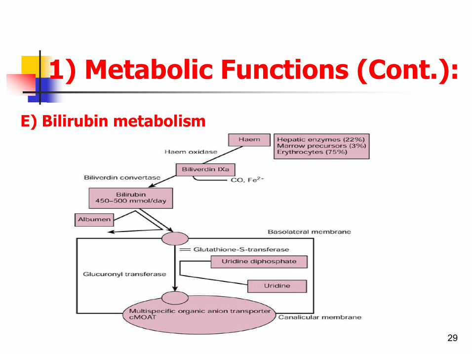
E) Bilirubin metabolism
30
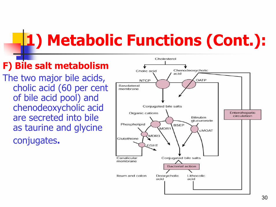
1) Metabolic Functions (Cont.): F) Bile salt metabolism The two major bile acids, cholic acid (60 per cent of bile acid pool) and chenodeoxycholic acid are secreted into bile as taurine and glycine conjugates.

31
2)Detoxication Functions: a) Metabolism of many drugs: Hepatic drug metabolism, or biotransformation, is divided into two broad
aspects: activation (phase I) and detoxification (phase II). q The hemoprotein cytochromes of the P450 system are associated with
most phase I reactions q Phase II detoxifying reactions are performed by different enzymes
including glutathione Stransferases, glucuronosyl transferases, epoxide hydrolase, sulfotransferases, and Nacetyltransferases. These catalyze reactions to complete the transformation of hydrophobic compounds to hydrophilic ones that can be excreted into the urine or bile.
b) Metabolism of alcohol by oxidation to acetaldehydes and acetic acid.
c) Metabolism of Hormones: Liver inactivates many hormones e.g. insulin,cortisol
,aldosterone,testosterone,estrogens and thyroid hormones.

32
3) Storage Functions: a) Vitamins: q B12 for 13 years q A for 10 months q D for 34 months b) Iron as ferritin→ blood iron buffer function. c) Glycogen (about 100gm) and fats. d) Blood→ blood reservoir function q Blood is retained in liver sinusoids when the hepatic vein
pressure increased. q 4 mmHg rise in hepatic vein pressure cause 200 ml blood to
be stored in the liver. q Blood is returned to circulation again when hepatic vein
pressure drop to normal again.
33
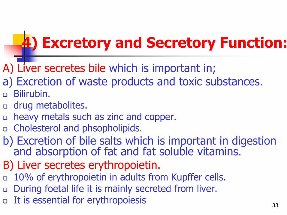
4) Excretory and Secretory Function:
A) Liver secretes bile which is important in; a) Excretion of waste products and toxic substances. q Bilirubin. q drug metabolites. q heavy metals such as zinc and copper. q Cholesterol and phsopholipids. b) Excretion of bile salts which is important in digestion and absorption of fat and fat soluble vitamins.
B) Liver secretes erythropoietin. q 10% of erythropoietin in adults from Kupffer cells. q During foetal life it is mainly secreted from liver. q It is essential for erythropoiesis

34
5) Blood Filtration Functions:
q The Von Kupffer cells ( hepatic macrophages) phagocytose and digest 99% of the bacteria that enter the portal blood from intestine.
q Also they remove foreign and unrequired substances e.g. small blood clots and Hb released from disintegrated RBCs

35
EVALUATION OF LIVER FUNCTION “liver function tests”
Several biochemical tests are useful in the evaluation and management of patients with hepatic dysfunction.
These tests can be used to; (1) detect the presence of liver disease, (2) distinguish among different types of liver disorders,
(3) gauge the extent of known liver damage. (4) follow the response to treatment.

36
A) Tests based on detoxication and Excretory Functions:
1) Serum Bilirubin: The normal total serum bilirubin concentration is <17 µmol/L (1 mg/dL).
Up to 30%, or 5.1 µmol/L (0.3 mg/dL), of the total is direct reacting (or conjugated) bilirubin.
q An isolated elevation of unconjugated bilirubin is seen primarily in hemolytic disorders and in a number of genetic conditions such as CriglerNajjar and Gilbert's syndromes
q In contrast, conjugated hyperbilirubinemia almost always implies liver or biliary tract disease.
37
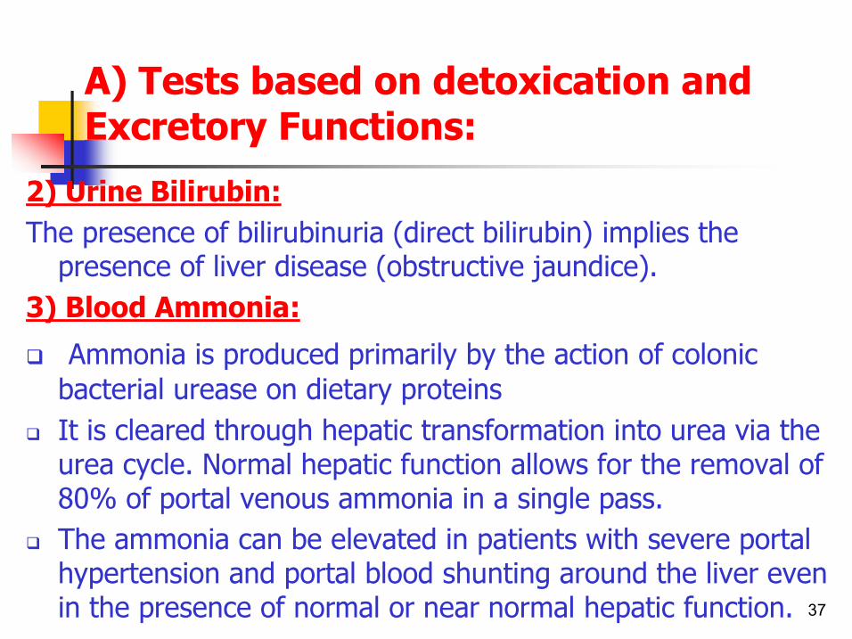
A) Tests based on detoxication and Excretory Functions:
2) Urine Bilirubin: The presence of bilirubinuria (direct bilirubin) implies the presence of liver disease (obstructive jaundice).
3) Blood Ammonia:
q Ammonia is produced primarily by the action of colonic bacterial urease on dietary proteins
q It is cleared through hepatic transformation into urea via the urea cycle. Normal hepatic function allows for the removal of 80% of portal venous ammonia in a single pass.
q The ammonia can be elevated in patients with severe portal hypertension and portal blood shunting around the liver even in the presence of normal or near normal hepatic function.

38
A) Tests based on detoxication and Excretory Functions:
4) Serum Enzymes: The liver contains thousands of enzymes, some of which are also
present in the serum in very low concentrations. Serum enzyme tests can be grouped into 2 categories: (1) enzymes whose elevation in serum reflects damage to
hepatocytes. (2) enzymes whose elevation in serum reflects cholestasis.

39
A) Tests based on detoxication and Excretory Functions:
a) Enzymes that reflect damage to hepatocytes: q The aminotransferases (transaminases) are sensitive indicators of liver cell injury and are most helpful in recognizing acute hepatocellular diseases such as hepatitis.
q They include the aspartate aminotransferase (AST) and the alanine aminotransferase (ALT).

40
A) Tests based on detoxication and Excretory Functions:
b) Enzymes that reflect Cholestasis : q They includes three enzymes; v alkaline phosphatase, v 5′nucleotidase, and v gamma glutamyl transpeptidase (GGT) q Their activities are usually elevated in cholestasis.

41
A) Tests based on detoxication and Excretory Functions:
5) Radionuclide hepatobiliary excretion scans:
q intravenously injection of 99mTcHIDA or its derivatives q It assesses the liver's ability to extract a material from the blood and then excrete it via the biliary tract.
q This test can be used to evaluate the patency of the extrahepatic biliary tree or the functional contraction and excretion capacity of the gallbladder.
q Thus, it is useful in the evaluation of cholestasis, bile duct obstruction, and cholecystitis.
42
B) Tests that measure biosynthetic functions :
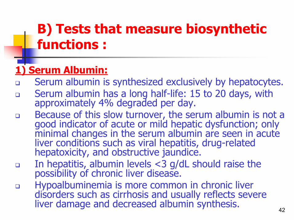
1) Serum Albumin: q Serum albumin is synthesized exclusively by hepatocytes. q Serum albumin has a long halflife: 15 to 20 days, with
approximately 4% degraded per day. q Because of this slow turnover, the serum albumin is not a
good indicator of acute or mild hepatic dysfunction; only minimal changes in the serum albumin are seen in acute liver conditions such as viral hepatitis, drugrelated hepatoxicity, and obstructive jaundice.
q In hepatitis, albumin levels <3 g/dL should raise the possibility of chronic liver disease.
q Hypoalbuminemia is more common in chronic liver disorders such as cirrhosis and usually reflects severe liver damage and decreased albumin synthesis.

43
B) Tests that measure biosynthetic functions :
2) Serum Globulins: q Serum globulins are a group of proteins made up of γ globulins (immunoglobulins) produced by B lymphocytes and alpha and beta globulins produced primarily in hepatocytes.
q Gamma globulins are increased in chronic liver disease, such as chronic hepatitis and cirrhosis.
q In cirrhosis, the increased serum gamma globulin concentration is due to the increased synthesis of antibodies, some of which are directed against intestinal bacteria.
q This occurs because the cirrhotic liver fails to clear bacterial antigens that normally reach the liver through the hepatic circulation.

44
B) Tests that measure biosynthetic functions :
3) Prothrombin time: q It collectively measures clotting factors II, V, VII, and X. q The prothrombin time may be elevated in hepatitis and cirrhosis as well as in disorders that lead to vitamin K deficiency such as obstructive jaundice or fat malabsorption of any kind.

45
C) Radiological investigations:
q Ultrasonography q Computed tomography (CT) q Magnetic resonance imaging (MRI) q Endoscopic retrograde cholangiopancreatography (ERCP)
q Percutaneous transhepatic cholangiography (PTC)

46
Liver Test Patterns in Hepatobiliary Disorders
n
Elevated, often >4 times normal elevation Fractionate, or confirm liver origin with 5′ nucleotidase or gamma glutamyl transpeptidase
Elevated, often >4 times normal elevation
Normal to <3 times normal elevation
Normal to <3 times normal elevation
Normal to <3 times normal elevation
Normal
Alkaline Phosphatase
Normal
Normal, unless chronic
Often decreased
Often decreased
Normal
Normal
Albumin
Normal Normal to slight elevation
Usually normal Infiltrative diseases (tumor, granulomata); partial bile duct obstruction
Normal If prolonged, will correct with parenteral vitamin K
Normal to moderate elevation Rarely >500 IU
Both fractions may be elevated Bilirubinuria
Intra and extra hepatic cholestasis
Often prolonged Fails to correct with parenteral vitamin K
AST:ALT > 2 suggests alcoholic hepatitis or cirrhosis
Both fractions may be elevated Bilirubinuria
Alcoholic hepatitis Cirrhosis
Often prolonged Fails to correct with parenteral vitamin K
Elevated, but usually <300 IU
Both fractions may be elevated Bilirubinuria
Chronic hepatocellular disorders
Usually normal. Elevated, often >500 IU ALT >AST
Both fractions may be elevated
Acute hepatocellular necrosis (viral and drug hepatitis, hepatotoxins, acute heart failure)
Normal Normal Normal to 5 mg/dl)
85% due to indirect fractions/ No bilirubinuria
Hemolysis/ Gilbert's syndrome
Prothrombin Time
Aminotransferase Bilirubin Type of Disorder

47
THANK YOU