Embed Size (px)
Citation preview
Antonino StipoUOC Emodinamica e Cardiologia Interventistica
Ospedale “S. Maria Goretti”-Latina
UPDATE SU SINDROMI CORONARICHE ACUTE
CLASSIFICAZIONE SINDROMI CORONARICHE
ACUTE ACC/AHA/ESC
Aprilia 4-XII-2010 Clinica “Città di Aprilia”
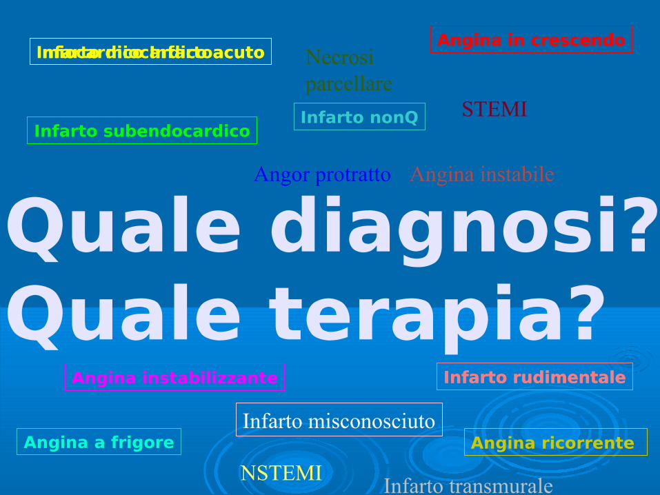
Infarto miocardico acuto
Quale diagnosi?Quale terapia?
miocardico InfartoacutoAngina in crescendo
Infarto nonQ
Infarto rudimentale
Infarto misconosciuto
Angina instabilizzante
Infarto subendocardico
Angina a frigore Angina ricorrente
Infarto rudimentale
Angina in crescendo
STEMI
NSTEMI
Angina instabile
Infarto transmurale
Necrosi parcellare
Angor protratto
Cardiovascular diseases are presently the leading causes of death in industrialized countries and expected to become so in emerging countries by 2020.
Murray CJ, Lopez AD. Alternative projections of mortality and disability by cause 1990–2020: Global Burden of Disease Study. Lancet 1997;349: 1498–1504
Among these, coronary artery disease (CAD)is the most prevalent manifestationand is associated with high mortality and morbidity.
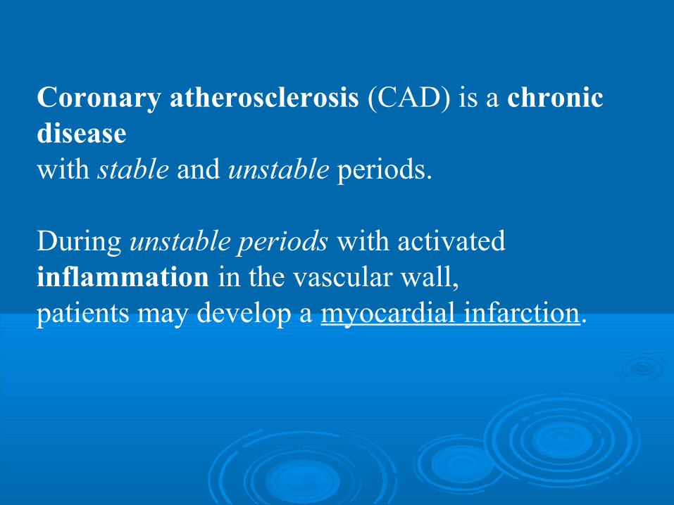
Coronary atherosclerosis (CAD) is a chronic diseasewith stable and unstable periods.
During unstable periods with activated inflammation in the vascular wall, patients may develop a myocardial infarction.
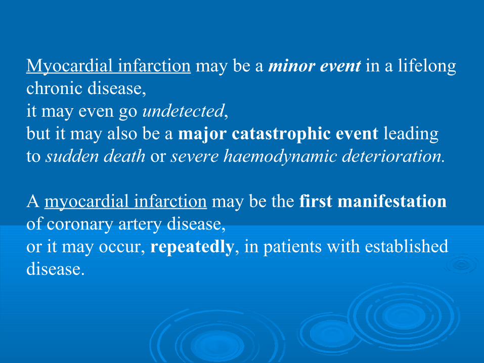
Myocardial infarction may be a minor event in a lifelong chronic disease, it may even go undetected, but it may also be a major catastrophic event leading to sudden death or severe haemodynamic deterioration.
A myocardial infarction may be the first manifestation of coronary artery disease, or it may occur, repeatedly, in patients with established disease.

Coronary Artery Disease (CAD)Coronary Artery Disease (CAD)
Ischaemic Heart Disease (IHD)Ischaemic Heart Disease (IHD)
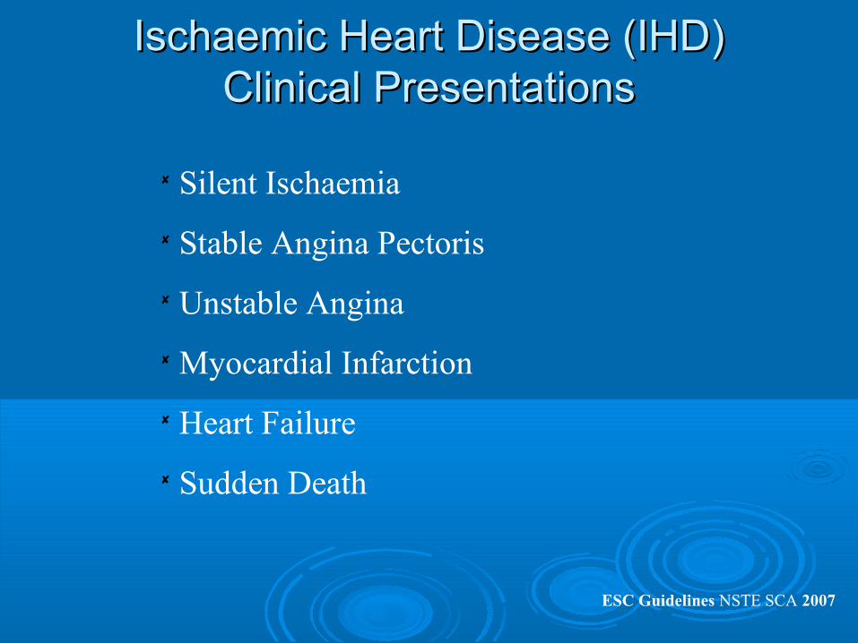
Ischaemic Heart Disease (IHD)Ischaemic Heart Disease (IHD)Clinical PresentationsClinical Presentations
Silent Ischaemia
Stable Angina Pectoris
Unstable Angina
Myocardial Infarction
Heart Failure
Sudden Death
ESC Guidelines NSTE SCA 2007
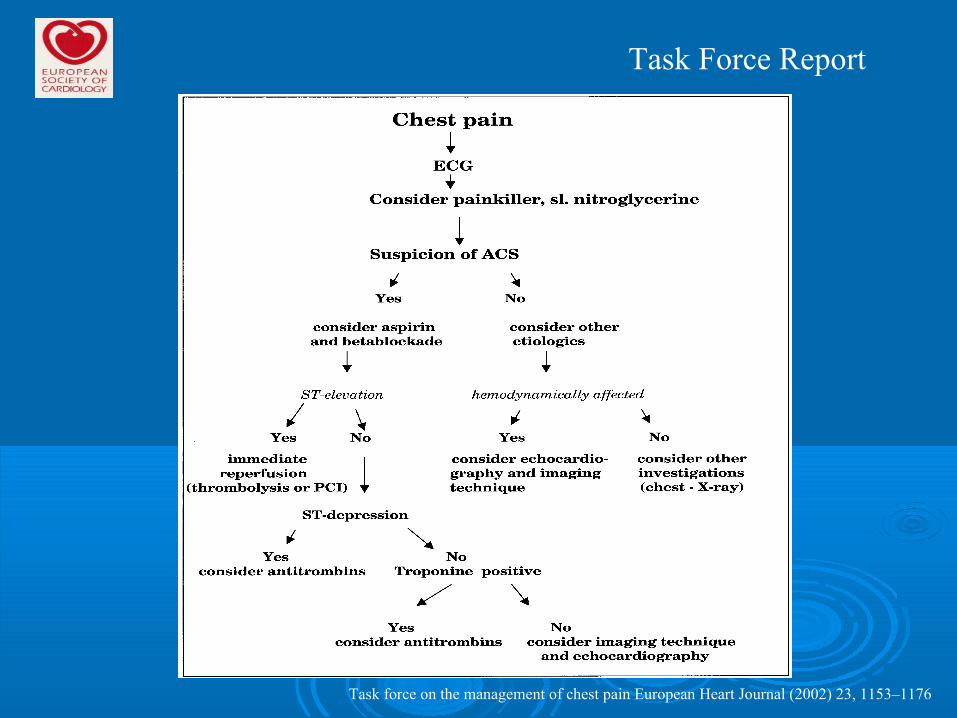
Task force on the management of chest pain European Heart Journal (2002) 23, 1153–1176
Task Force Report
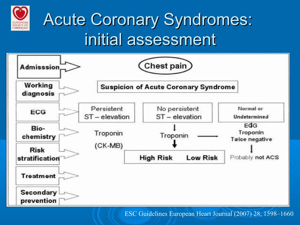
Acute Coronary Syndromes: Acute Coronary Syndromes: initial assessmentinitial assessment
ESC Guidelines European Heart Journal (2007) 28, 1598–1660

Guidelines for the identification of ACS patients by ED registration clerks or Triage Nurses
Registration/Clerical StaffPatients with the following chief complaints require immediate assessment by the triage nurse and should be referred for further evaluation:Chief Complaint• Chest pain, pressure, tightness, or heaviness; pain that radiates to neck, jaw, shoulders, back, or 1 or both arms• Indigestion or “heartburn”; nausea and/or vomiting associated with chest discomfort• Persistent shortness of breath• Weakness, dizziness, lightheadedness, loss of consciousness
Triage NursePatients with the following symptoms and signs require immediate assessment by the triage nurse for the initiationof the ACS protocol:
Chief Complaint• Chest pain or severe epigastric pain, nontraumatic in origin, with components typical of myocardial ischemiaor MI:Central/substernal compression or crushing chest painPressure, tightness, heaviness, cramping, burning, aching sensationUnexplained indigestion, belching, epigastric painRadiating pain in neck, jaw, shoulders, back, or 1 or both arms• Associated dyspnea• Associated nausea and/or vomiting• Associated diaphoresisIf these symptoms are present, obtain stat ECG.Medical HistoryThe triage nurse should take a brief, targeted, initial history with an assessment of current or past history of:• CABG, angioplasty, CAD, angina on effort, or AMI• NTG use to relieve chest discomfort• Risk factors, including smoking, hyperlipidemia, hypertension, diabetes mellitus, family history, and cocaine useThis brief history must not delay entry into the ACS protocol.Special ConsiderationsWomen may present more frequently than men with atypical chest pain and symptoms.Diabetic patients may have atypical presentations due to autonomic dysfunction.Elderly patients may have atypical symptoms such as generalized weakness, stroke, syncope, or a change in mental status.
ACC/AHA Guidelines 2000

Acute Coronary SyndromeAcute Coronary SyndromeAny constellation of clinical signs or symptoms suggestive of AMI or UA. This syndrome includes patients with AMI, STEMI, NSTEMI, enzymediagnosed MI, biomarker-diagnosed MI, late ECG-diagnosed MI, and UA.
This term is useful to generically refer to patients who ultimately prove to have 1 of these diagnoses to describe management alternatives at a time before the diagnosis is ultimately confirmed.
This term is also used prospectively to identify those patients at a time of initial presentation who should be considered for treatment of AMI or UA.
ACC/AHA Guidelines 2000
Acute Coronary SyndromeAcute Coronary Syndrome
Probable acute coronary syndrome is a term that is commonly used, and this represents the primary consideration of patients on initial presentation.
Possible acute coronary syndrome is useful as a secondary diagnosis when an alternate diagnosis seems more likely but an acute ischemic process has not been excluded as a possible cause of the presenting symptoms.
ACC/AHA Guidelines 2000

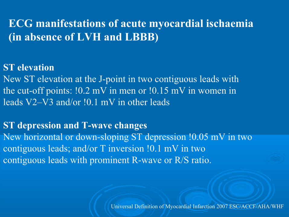
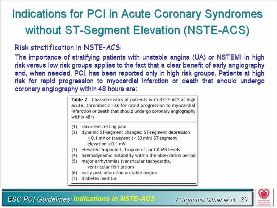
ST elevation New ST elevation at the J-point in two contiguous leads with the cut-off points: !0.2 mV in men or !0.15 mV in women in leads V2–V3 and/or !0.1 mV in other leads ST depression and T-wave changes New horizontal or down-sloping ST depression !0.05 mV in two contiguous leads; and/or T inversion !0.1 mV in two contiguous leads with prominent R-wave or R/S ratio.
ECG manifestations of acute myocardial ischaemia(in absence of LVH and LBBB)
Universal Definition of Myocardial Infarction 2007 ESC/ACCF/AHA/WHF
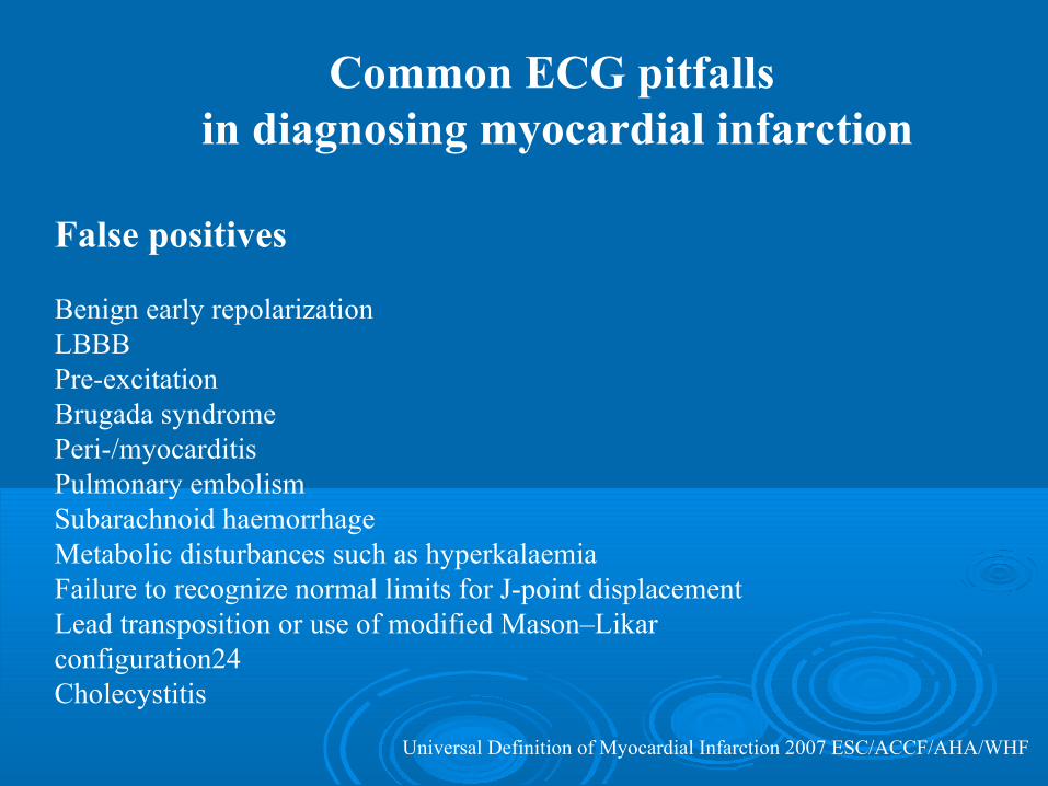
Common ECG pitfalls in diagnosing myocardial infarction
False positives
Benign early repolarizationLBBBPre-excitationBrugada syndromePeri-/myocarditisPulmonary embolismSubarachnoid haemorrhageMetabolic disturbances such as hyperkalaemiaFailure to recognize normal limits for J-point displacementLead transposition or use of modified Mason–Likarconfiguration24Cholecystitis
Universal Definition of Myocardial Infarction 2007 ESC/ACCF/AHA/WHF

Common ECG pitfalls in diagnosing myocardial infarction
False negatives
Prior myocardial infarction with Q-waves and/or persistentST elevationPaced rhythmLBBB
Universal Definition of Myocardial Infarction 2007 ESC/ACCF/AHA/WHF
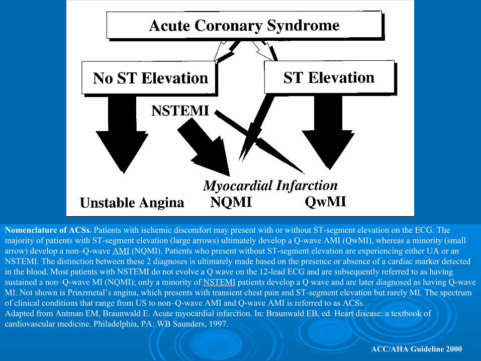
Nomenclature of ACSs. Patients with ischemic discomfort may present with or without ST-segment elevation on the ECG. The majority of patients with ST-segment elevation (large arrows) ultimately develop a Q-wave AMI (QwMI), whereas a minority (small arrow) develop a non–Q-wave AMI (NQMI). Patients who present without ST-segment elevation are experiencing either UA or an NSTEMI. The distinction between these 2 diagnoses is ultimately made based on the presence or absence of a cardiac marker detected in the blood. Most patients with NSTEMI do not evolve a Q wave on the 12-lead ECG and are subsequently referred to as having sustained a non–Q-wave MI (NQMI); only a minority of NSTEMI patients develop a Q wave and are later diagnosed as having Q-wave MI. Not shown is Prinzmetal’s angina, which presents with transient chest pain and ST-segment elevation but rarely MI. The spectrum of clinical conditions that range from US to non–Q-wave AMI and Q-wave AMI is referred to as ACSs. Adapted from Antman EM, Braunwald E. Acute myocardial infarction. In: Braunwald EB, ed. Heart disease: a textbook of cardiovascular medicine. Philadelphia, PA: WB Saunders, 1997.
ACC/AHA Guideline 2000
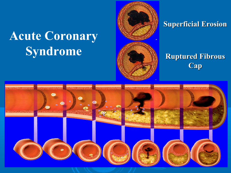
Ruptured Fibrous Ruptured Fibrous CapCap
Superficial ErosionSuperficial Erosion
Acute Coronary Syndrome
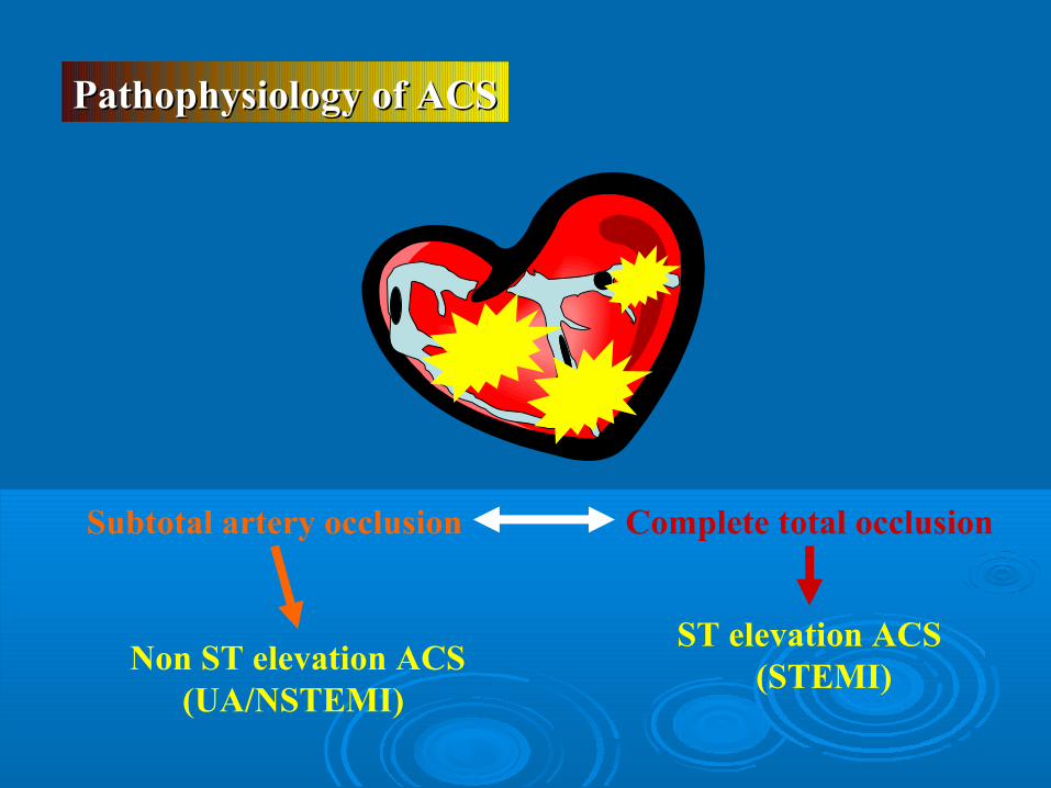
Subtotal artery occlusion Complete total occlusion
Non ST elevation ACS (UA/NSTEMI)
ST elevation ACS (STEMI)
Pathophysiology of ACSPathophysiology of ACS

Patients with typical acute chest pain and persistent (>20 min) ST-segment elevation. This is termed ST-elevation ACS (STE-ACS) and generally reflects an acute total coronary occlusion. Most of these patients will ultimately develop an ST-elevation MI (STEMI). The therapeutic objective is to achieve rapid, complete, and sustained reperfusion by primary angioplasty or fibrinolytic therapy.
ESC Guidelines NSTE-ACS Eur Heart J (2007) 28, 1598–1660
definitions

Patients with acute chest pain but without persistent ST-segment elevation. They have rather persistent or transient ST-segment depression or T-wave inversion, flat T-waves, pseudonormalization of T-waves, or no ECG changes at presentation. The initial strategy in these patients is to alleviate ischaemia and symptoms, to monitor the patient with serial ECG, and to repeat measurements of markers of myocardial necrosis.
ESC Guidelines NSTE-ACS Eur Heart J (2007) 28, 1598–1660
definitions

At presentation, the working diagnosis of non-STE-ACS (NSTE-ACS), based on the measurement of troponins, will be further qualified into non-STelevation MI (NSTEMI) or unstable angina. In a certain number of patients, CAD will subsequently be excluded as the cause of symptoms. The therapeutic management is guided by the final diagnosis.
ESC Guidelines NSTE-ACS Eur Heart J (2007) 28, 1598–1660
definitions
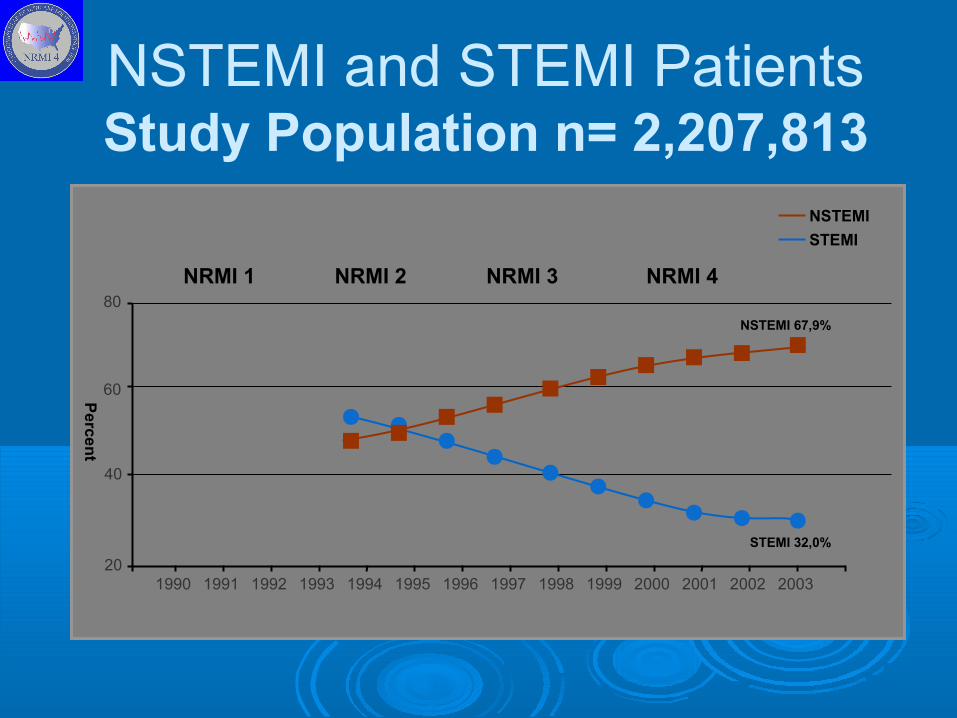
NSTEMI and STEMI PatientsStudy Population n= 2,207,813
20
40
60
80
1990 1991 1992 1993
Pe
rce
nt
NSTEMI
STEMI
NSTEMI 67,9%
NRMI 1
1994 1995 1996 1997 1998 1999 2000 2001 2002 2003
STEMI 32,0%
NRMI 2 NRMI 3 NRMI 4

Hospital mortality is higher in patients with STEMI than among those with NSTE-ACS (7 vs. 5%, respectively), but, at 6 months, the mortality rates are very similar in both conditions (12 vs. 13%, respectively).
NSTEMI and STEMI
Savonitto S et al. Prognostic value of the admission electrocardiogram in acute coronary syndromes. JAMA 1999;281: 707–713.Volmink JA et al , Coronary event and case fatality rates in an English population: results of the Oxford myocardial infarction incidence study. The Oxford Myocardial Infarction Incidence Study Group. Heart 1998;80:40–44.
NSTEMI and STEMI
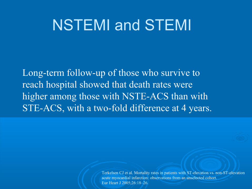
Long-term follow-up of those who survive to reach hospital showed that death rates werehigher among those with NSTE-ACS than with STE-ACS, with a two-fold difference at 4 years.
Terkelsen CJ et al. Mortality rates in patients with ST-elevation vs. non-ST-elevation acute myocardial infarction: observations from an unselected cohort. Eur Heart J 2005;26:18–26.
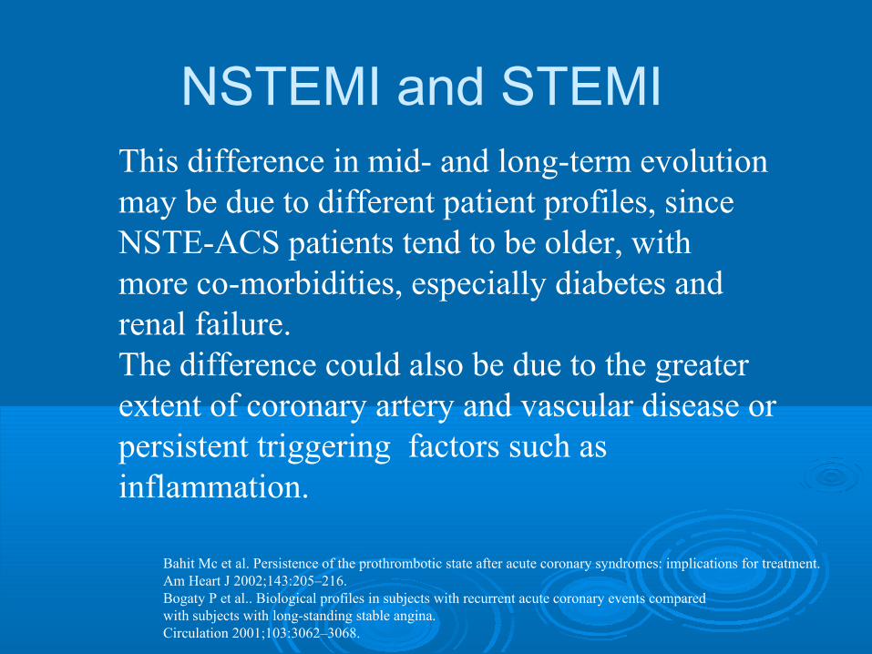
NSTEMI and STEMIThis difference in mid- and long-term evolution may be due to different patient profiles, since NSTE-ACS patients tend to be older, withmore co-morbidities, especially diabetes and renal failure.The difference could also be due to the greater extent of coronary artery and vascular disease or persistent triggering factors such as inflammation.
Bahit Mc et al. Persistence of the prothrombotic state after acute coronary syndromes: implications for treatment. Am Heart J 2002;143:205–216.Bogaty P et al.. Biological profiles in subjects with recurrent acute coronary events comparedwith subjects with long-standing stable angina. Circulation 2001;103:3062–3068.
NSTEMI and STEMI
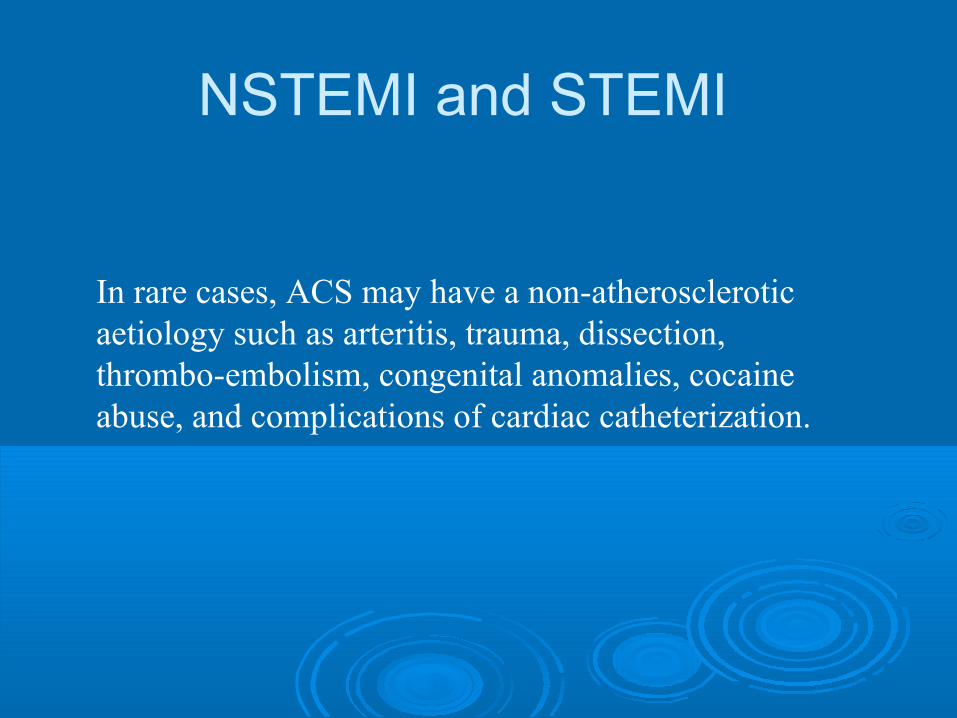
In rare cases, ACS may have a non-atherosclerotic aetiology such as arteritis, trauma, dissection, thrombo-embolism, congenital anomalies, cocaine abuse, and complications of cardiac catheterization.

NSTEMISecondary mechanisms
increase in myocardial oxygen consumption are fever, tachycardia, thyrotoxicosis, a hyperadrenergic state, sudden emotional stress, and increased left ventricular (LV) afterload (hypertension, aortic stenosis)

NSTEMISecondary mechanisms
reduced myocardial oxygen delivery areanaemia, methaemoglobinaemia, and hypoxaemia
NSTEMI
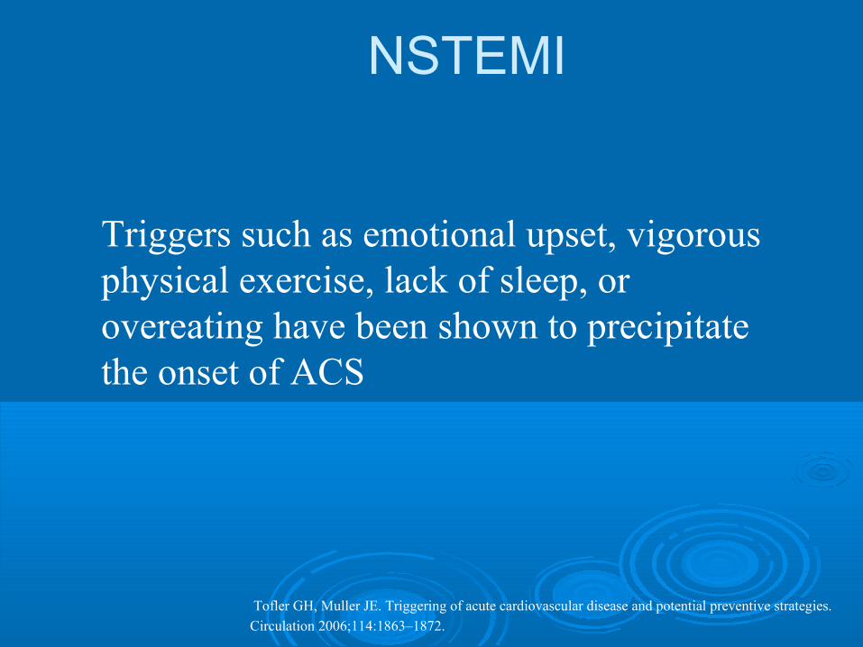
Triggers such as emotional upset, vigorous physical exercise, lack of sleep, or overeating have been shown to precipitate the onset of ACS
Tofler GH, Muller JE. Triggering of acute cardiovascular disease and potential preventive strategies.
Circulation 2006;114:1863–1872.
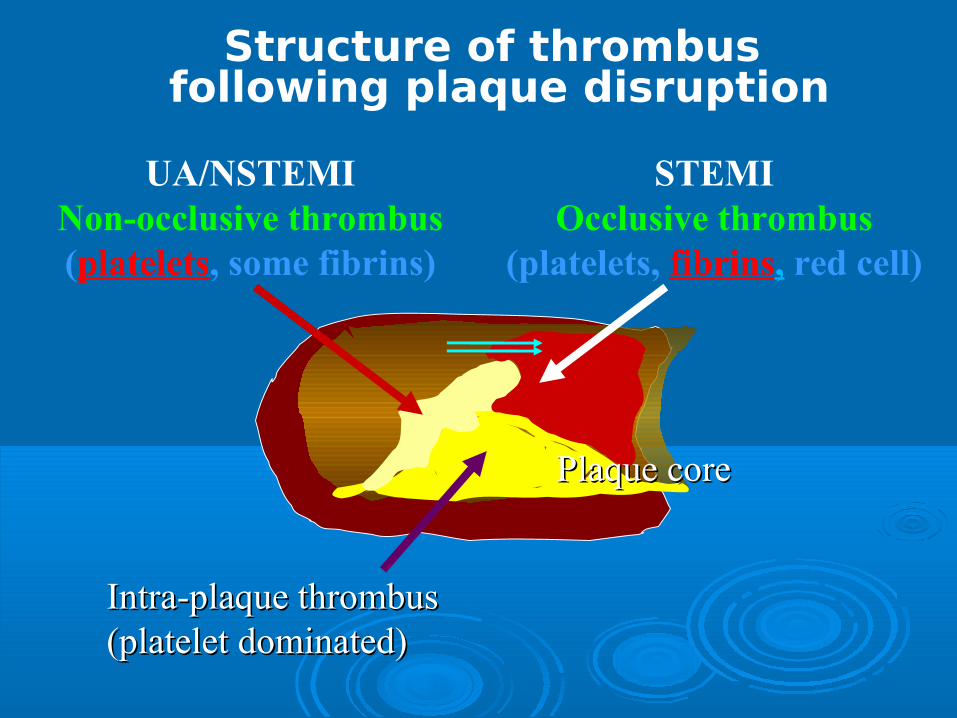
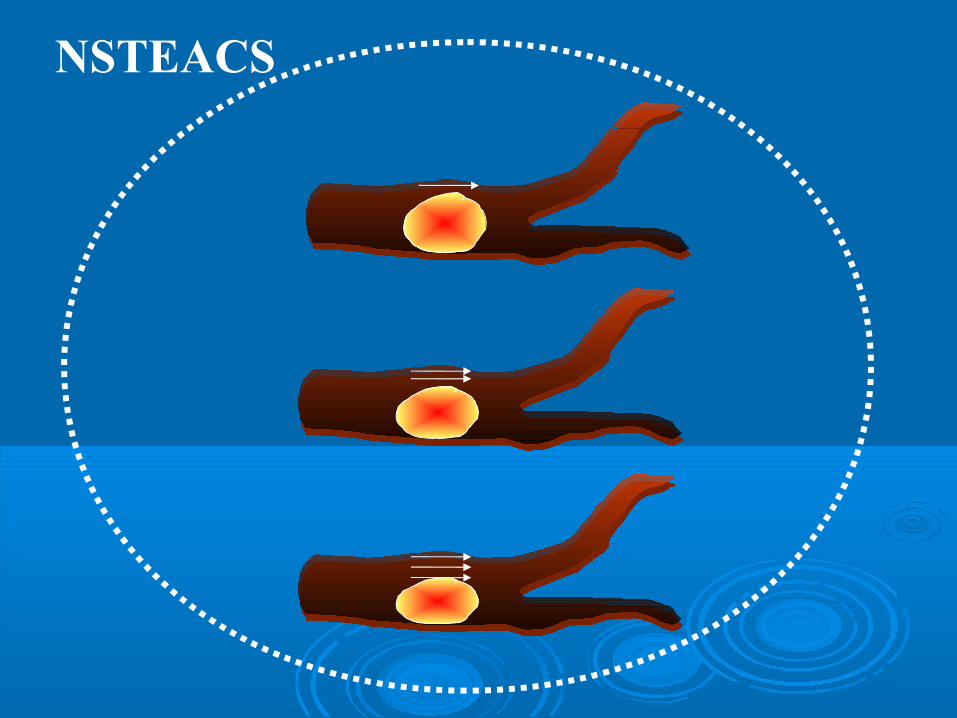
Structure of thrombus following plaque disruption
UA/NSTEMINon-occlusive thrombus(platelets, some fibrins)
STEMIOcclusive thrombus
(platelets, fibrins, red cell)
Intra-plaque thrombusIntra-plaque thrombus(platelet(platelet dominated)dominated)
Plaque corePlaque core

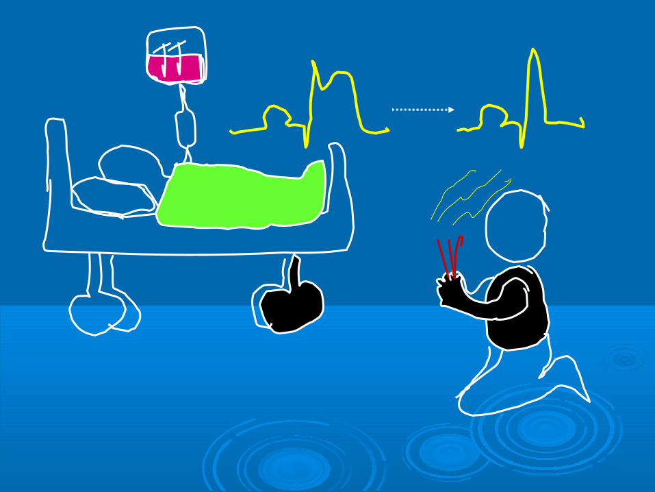
Treatment of the ACS: Perspective on the FutureTreatment of the ACS: Perspective on the Future
STE-ACS
Fibrinolysis vs primary PCI
“Time is muscle”
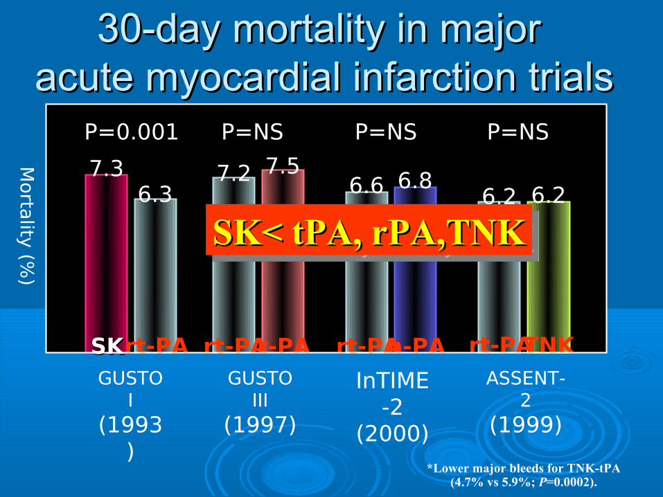
30-day mortality in major 30-day mortality in major acute myocardial infarction trialsacute myocardial infarction trials
Morta
lity (%
)
7.3
SKSK
6.3
rt-PA
P=0.001
GUSTO I
(1993)
7.57.2
rt-PAr-PA
P=NS
GUSTO III
(1997)
6.8
n-PA
6.6
rt-PA
P=NS
InTIME-2
(2000)
6.26.2
rt-PATNK
P=NS
ASSENT-2
(1999)
*Lower major bleeds for TNK-tPA(4.7% vs 5.9%; P=0.0002).
SK< tPA, rPA,TNKSK< tPA, rPA,TNK
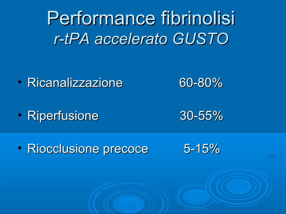
Performance fibrinolisiPerformance fibrinolisi r-tPA accelerato GUSTOr-tPA accelerato GUSTO
• Ricanalizzazione 60-80%Ricanalizzazione 60-80%
• Riperfusione 30-55%Riperfusione 30-55%
• Riocclusione precoce 5-15%Riocclusione precoce 5-15%
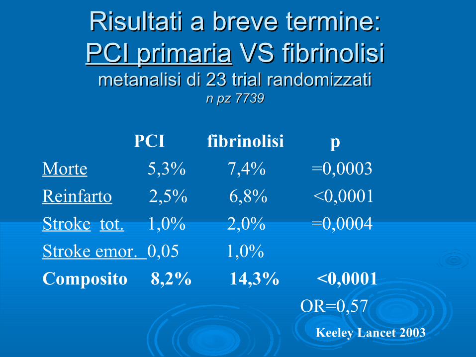
Risultati a breve termine:Risultati a breve termine:PCI primariaPCI primaria VS fibrinolisi VS fibrinolisi
metanalisi di 23 trial randomizzatimetanalisi di 23 trial randomizzatin pz 7739n pz 7739
PCI fibrinolisi p
Morte 5,3% 7,4% =0,0003
Reinfarto 2,5% 6,8% <0,0001
Stroke tot. 1,0% 2,0% =0,0004
Stroke emor. 0,05 1,0%
Composito 8,2% 14,3% <0,0001
OR=0,57 Keeley Lancet 2003
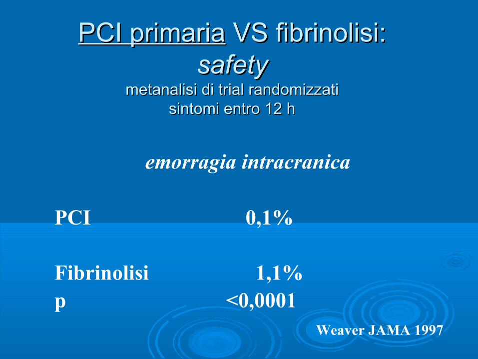
PCI primariaPCI primaria VS fibrinolisi: VS fibrinolisi:safetysafety
metanalisi di trial randomizzatimetanalisi di trial randomizzatisintomi entro 12 hsintomi entro 12 h
emorragia intracranica
PCI 0,1%
Fibrinolisi 1,1% p <0,0001 Weaver JAMA 1997

Primary PCI vs. Thrombolysis: Primary PCI vs. Thrombolysis: Clinical OutcomesClinical Outcomes
Keeley et al. Lancet. 2003.
Frequ
ency, %
5500
4400
3300
2200
1100
00
2255
2200
1155
1100
55
00
Death Death,Excluding
SHOCK Data
Non-fatalMyocardialInfarction
RecurrentIschemia
TotalStroke
Haemorrhagic
Stroke
MajorBleed
Death, Non-fatalReinfarction,
or Stroke
Long-Term Outcomes
Short-Term Outcomes
P = 0.0019P = 0.0053P < 0.0001
P < 0.0001
P < 0.0001
P = 0.0002P = 0.0003P < 0.0001
P < 0.0001
P < 0.0001
P = 0.0032P < 0.0001P = 0.0004
- - -
PCIPCI
Thrombolytic therapyThrombolytic therapy
Death Death,Excluding
SHOCK Data
Non-fatalMyocardialInfarction
RecurrentIschemia
TotalStroke
Haemorrhagic
Stroke
MajorBleed
Death, Non-fatalReinfarction,
or Stroke
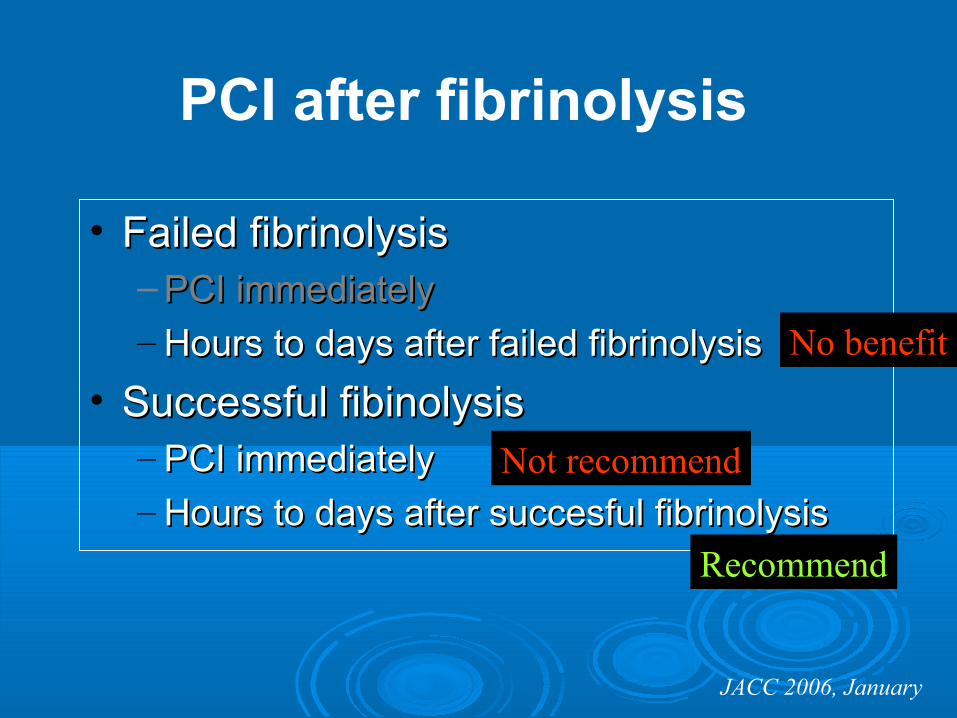
• Failed fibrinolysisFailed fibrinolysis– PCI immediatelyPCI immediately– Hours to days after failed fibrinolysisHours to days after failed fibrinolysis
• Successful fibinolysisSuccessful fibinolysis– PCI immediatelyPCI immediately– Hours to days after succesful fibrinolysisHours to days after succesful fibrinolysis
JACC 2006, January
Not recommend
No benefit
Recommend
PCI after fibrinolysis

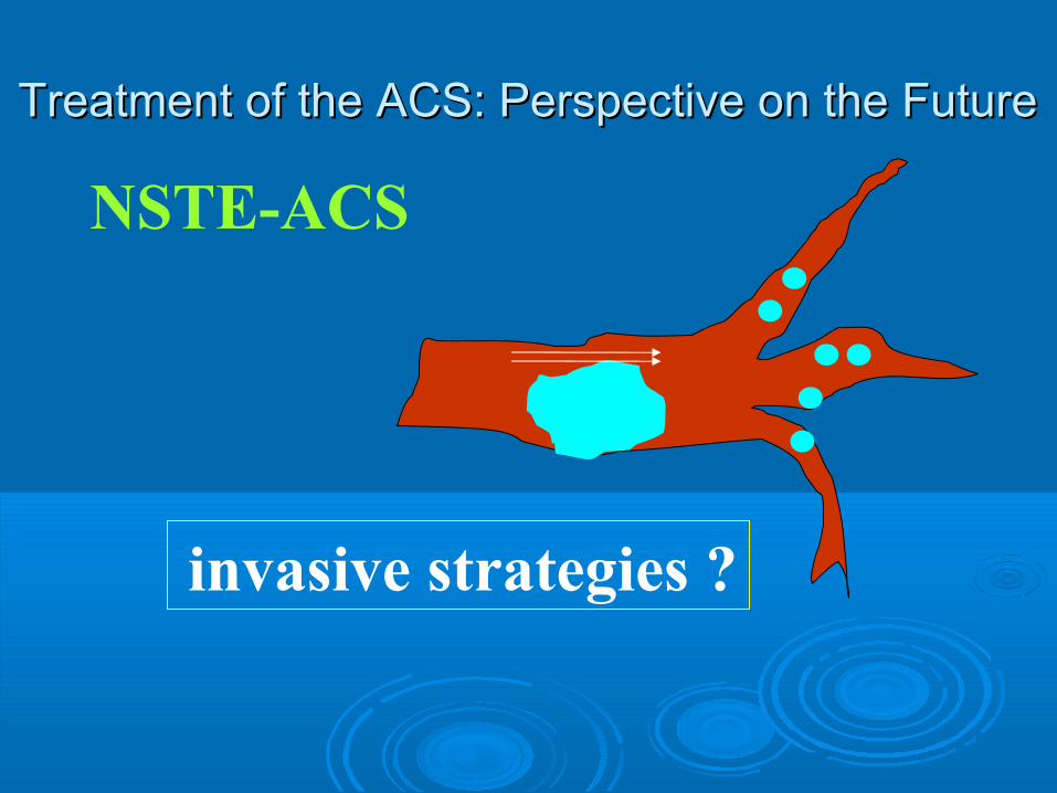
Treatment of the ACS: Perspective on the FutureTreatment of the ACS: Perspective on the Future
NSTE-ACS
invasive strategies ?
European Heart Journal (2002) 23, 1809–1840doi:10.1053/euhj.2002.3385, available online at http://www.idealibrary.com
Task Force Report
Management of acute coronary syndromes in patients presenting without persistent ST-segment elevation
The Task Force on the Management of Acute Coronary Syndromes of the European Society of Cardiology*
Michel E. Bertrand, Chair, Maarten L. Simoons, Keith A. A. Fox, Lars C. Wallentin,Christian W. Hamm, Eugene McFadden, Pim J. De Feyter,Giuseppe Specchia, Witold Ruzyllo
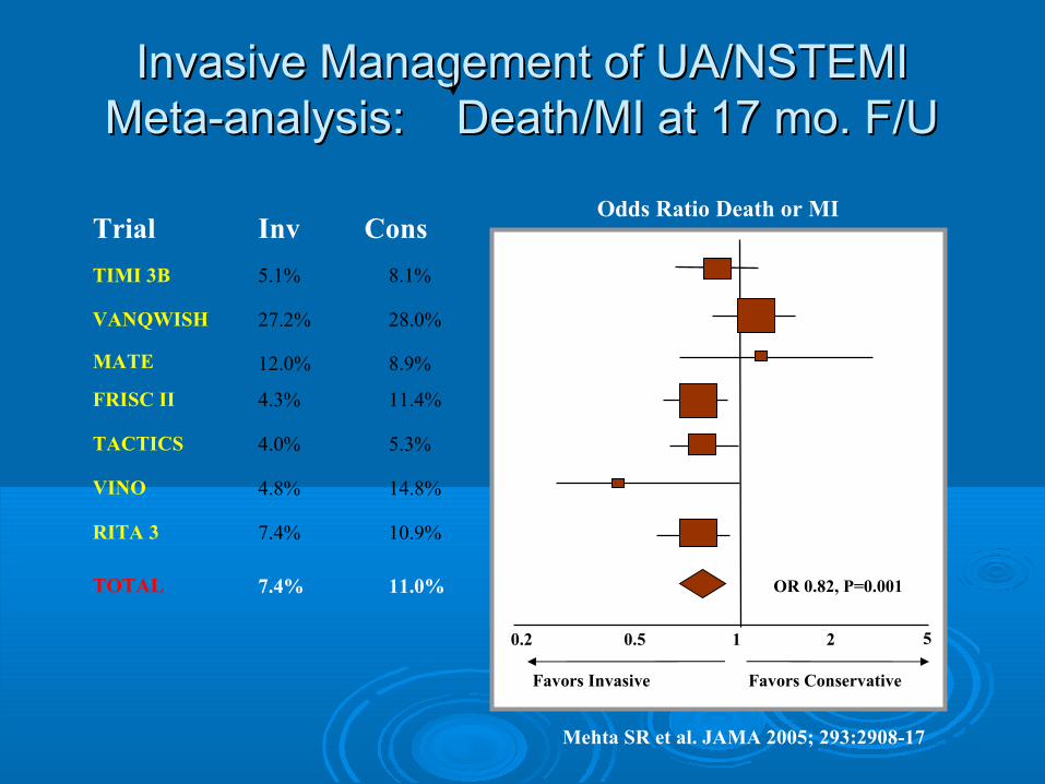
Invasive Management of UA/NSTEMIInvasive Management of UA/NSTEMIMeta-analysis: Death/MI at 17 mo. F/UMeta-analysis: Death/MI at 17 mo. F/U
Mehta SR et al. JAMA 2005; 293:2908-17
0.2 10.5 2 5
Favors Invasive Favors Conservative
OR 0.82, P=0.001
Odds Ratio Death or MITrial Inv Cons
TIMI 3B 5.1% 8.1%
VANQWISH 27.2% 28.0%
MATE 12.0% 8.9%
FRISC II 4.3% 11.4%
TACTICS 4.0% 5.3%
VINO 4.8% 14.8%
RITA 3 7.4% 10.9%
TOTAL 7.4% 11.0%
NSTEACS

Risk Scores in ACS-NSTERisk Scores in ACS-NSTE
TIMI (0-7) Age > 65 years 3 risk factors for CADUse of ASA (last 7 days) Known CAD (stenosis > 50%) > 1 episode rest angina in < 24hST-segment deviationElevated cardiac markers
PURSUIT (0-18)
GRACE (0-258)
8 (11)9 (12)
11 (13)12 (14)
Age, separate points for enrolment diagnosis - Decade [UA (MI)]
MaleFemale
50607080
Sex
10
No angina or CCS I/IICCS III/IV
Worst CCS-class in previous 6 weeks
02
Signs of heart failure
St-depression on presenting ECG
< 4040-4950-5960-6970-79≥80
Age (years)
01836557391
< 7070-8990-109110-149150-199> 200
Heart rate (bpm)
07
13233646
1111111
< 8080-99100-119120-139140-159160-199> 200
Systolic BP (mmHg)
6358473726110
Class IClass IIClass IIIClass IV
Killip class
0214364
Cardiac arrest at admissionElevated cardiac markesST-segment deviation
431530
1
2

• Recurrent ischemicRecurrent ischemic• Elevated troponin levelElevated troponin level• New ST segment depressionNew ST segment depression• CHF, new or worsening MRCHF, new or worsening MR• Depressed LV functionDepressed LV function• Hemodynamic instabilityHemodynamic instability• Sustained VTSustained VT• PCI in 6 monthsPCI in 6 months• Prior CABGPrior CABG
JACC 2006, January
Early invasive strategy in NSTE-ACS
European Heart Journal (2007) 28, 2525–2538 Expert consensus document
Universal definition of myocardial infarctionKristian Thygesen, Joseph S. Alpert and Harvey D. White on behalf of the Joint ESC/ACCF/AHA/WHF Task Force for the Redefinition of Myocardial Infarction
Task Force Members:Chairpersons: Kristian Thygesen (Denmark)*, Joseph S. Alpert (USA)*, Harvey D. White (New Zealand)*Biomarker Group: Allan S. Jaffe, Co-ordinator (USA), Fred S. Apple (USA), Marcello Galvani (Italy), Hugo A. Katus (Germany), L. Kristin Newby (USA), Jan Ravkilde (Denmark)ECG Group: Bernard Chaitman, Co-ordinator (USA), Peter M. Clemmensen (Denmark), Mikael Dellborg (Sweden), Hanoch Hod (Israel), Pekka Porela (Finland)Imaging Group: Richard Underwood, Co-ordinator (UK), Jeroen J. Bax (The Netherlands) George A. Beller (USA), Robert Bonow (USA), Ernst E. Van Der Wall (The Netherlands)Intervention Group: Jean-Pierre Bassand, Co-ordinator (France), William Wijns, Co-ordinator (Belgium), T. Bruce Ferguson (USA), Philippe G. Steg (France), Barry F. Uretsky (USA), David O. Williams (USA)Clinical Investigation Group: Paul W. Armstrong, Co-ordinator (Canada), Elliott M. Antman (USA), Keith A. Fox (UK), Christian W. Hamm (Germany), E. Magnus Ohman (USA), Maarten L. Simoons(The Netherlands)Global Perspective Group: Philip A. Poole-Wilson, Co-ordinator (UK), Enrique P. Gurfinkel (Argentina), ́Jose-Luis Lopez-Sendon (Spain), Prem Pais (India), Shanti Mendis‡ (Switzerland), Jun-Ren Zhu (China)́Implementation Group: Lars C. Wallentin Co-ordinator (Sweden), Francisco Fernandez-Aviles (Spain),Kim M. Fox (UK), Alexander N. Parkhomenko (Ukraine), Silvia G. Priori (Italy), Michal Tendera (Poland),Liisa-Maria Voipio-Pulkki (Finland)ESC Committee for Practice Guidelines: Alec Vahanian, Chair (France), A. John Camm (UK), Raffaele De Caterina (Italy),Veronica Dean (France), Kenneth Dickstein (Norway), Gerasimos Filippatos (Greece), Christian Funck-Brentano (France),Irene Hellemans (The Netherlands), Steen Dalby Kristensen (Denmark), Keith McGregor (France), Udo Sechtem (Germany), ́Sigmund Silber (Germany), Michal Tendera (Poland), Petr Widimsky (Czech Republic), Jose Luis Zamorano (Spain)Document Reviewers: Joao Morais, Review Co-ordinator (Portugal), Sorin Brener (USA), Robert Harrington (USA),David Morrow (USA), Udo Sechtem (Germany), Michael Lim (Singapore), Marco A. Martinez-Rios (Mexico), Steve Steinhubl(USA), Glen N. Levine (USA), W. Brian Gibler (USA), David Goff (USA), Marco Tubaro (Italy),Darek Dudek (Poland), Nawwar Al-Attar (France)
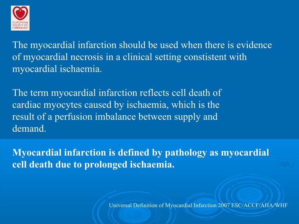
The myocardial infarction should be used when there is evidence of myocardial necrosis in a clinical setting constistent with myocardial ischaemia.
The term myocardial infarction reflects cell death ofcardiac myocytes caused by ischaemia, which is theresult of a perfusion imbalance between supply anddemand.
Myocardial infarction is defined by pathology as myocardialcell death due to prolonged ischaemia.
Universal Definition of Myocardial Infarction 2007 ESC/ACCF/AHA/WHF
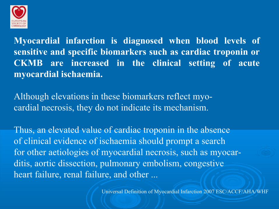
Myocardial infarction is diagnosed when blood levels of sensitive and specific biomarkers such as cardiac troponin or CKMB are increased in the clinical setting of acute myocardial ischaemia.
Although elevations in these biomarkers reflect myo-cardial necrosis, they do not indicate its mechanism.
Thus, an elevated value of cardiac troponin in the absenceof clinical evidence of ischaemia should prompt a searchfor other aetiologies of myocardial necrosis, such as myocar-ditis, aortic dissection, pulmonary embolism, congestiveheart failure, renal failure, and other ...
Universal Definition of Myocardial Infarction 2007 ESC/ACCF/AHA/WHF
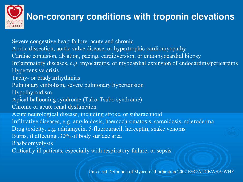
Severe congestive heart failure: acute and chronicAortic dissection, aortic valve disease, or hypertrophic cardiomyopathyCardiac contusion, ablation, pacing, cardioversion, or endomyocardial biopsyInflammatory diseases, e.g. myocarditis, or myocardial extension of endocarditis/pericarditisHypertensive crisisTachy- or bradyarrhythmiasPulmonary embolism, severe pulmonary hypertensionHypothyroidismApical ballooning syndrome (Tako-Tsubo syndrome)Chronic or acute renal dysfunctionAcute neurological disease, including stroke, or subarachnoidInfiltrative diseases, e.g. amyloidosis, haemochromatosis, sarcoidosis, sclerodermaDrug toxicity, e.g. adriamycin, 5-fluorouracil, herceptin, snake venomsBurns, if affecting .30% of body surface areaRhabdomyolysisCritically ill patients, especially with respiratory failure, or sepsis
Non-coronary conditions with troponin elevations
Universal Definition of Myocardial Infarction 2007 ESC/ACCF/AHA/WHF
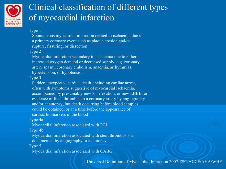
Clinical classification of different types of myocardial infarctionType 1 Spontaneous myocardial infarction related to ischaemia due to a primary coronary event such as plaque erosion and/or rupture, fissuring, or dissectionType 2 Myocardial infarction secondary to ischaemia due to either increased oxygen demand or decreased supply, e.g. coronary artery spasm, coronary embolism, anaemia, arrhythmias, hypertension, or hypotensionType 3 Sudden unexpected cardiac death, including cardiac arrest, often with symptoms suggestive of myocardial ischaemia, accompanied by presumably new ST elevation, or new LBBB, or evidence of fresh thrombus in a coronary artery by angiography and/or at autopsy, but death occurring before blood samples could be obtained, or at a time before the appearance of cardiac biomarkers in the bloodType 4a Myocardial infarction associated with PCIType 4b Myocardial infarction associated with stent thrombosis as documented by angiography or at autopsyType 5 Myocardial infarction associated with CABG Universal Definition of Myocardial Infarction 2007 ESC/ACCF/AHA/WHF

It should also be noted that the term myocardial infarctiondoes not include myocardial cell death associated withmechanical injury from coronary artery bypass grafting(CABG), for example ventricular venting, or manipulationof the heart; nor does it include myocardial necrosis dueto miscellaneous causes, e.g. renal failure, heart failure,cardioversion, electrophysiological ablation, sepsis, myocarditis, cardiac toxins, or infiltrative diseases.
Universal Definition of Myocardial Infarction 2007 ESC/ACCF/AHA/WHF
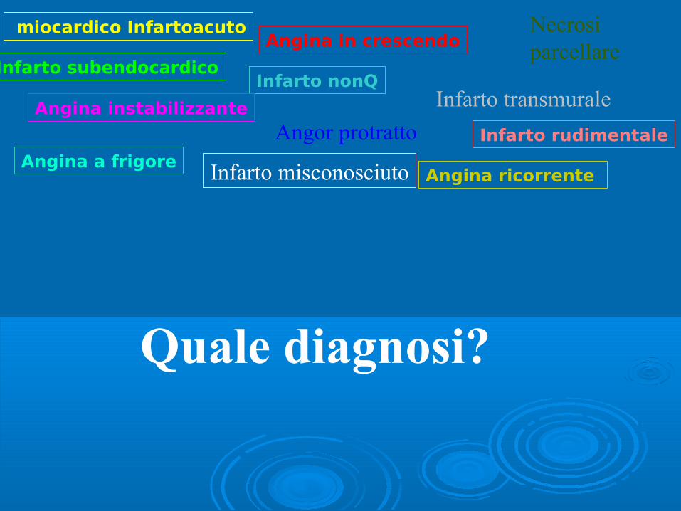
miocardico Infartoacuto
Infarto nonQ
Infarto misconosciuto
Angina instabilizzante
Infarto subendocardico
Angina a frigoreAngina ricorrente
Infarto rudimentale
Angina in crescendo
Infarto transmurale
Necrosi parcellare
Angor protratto
Quale diagnosi?
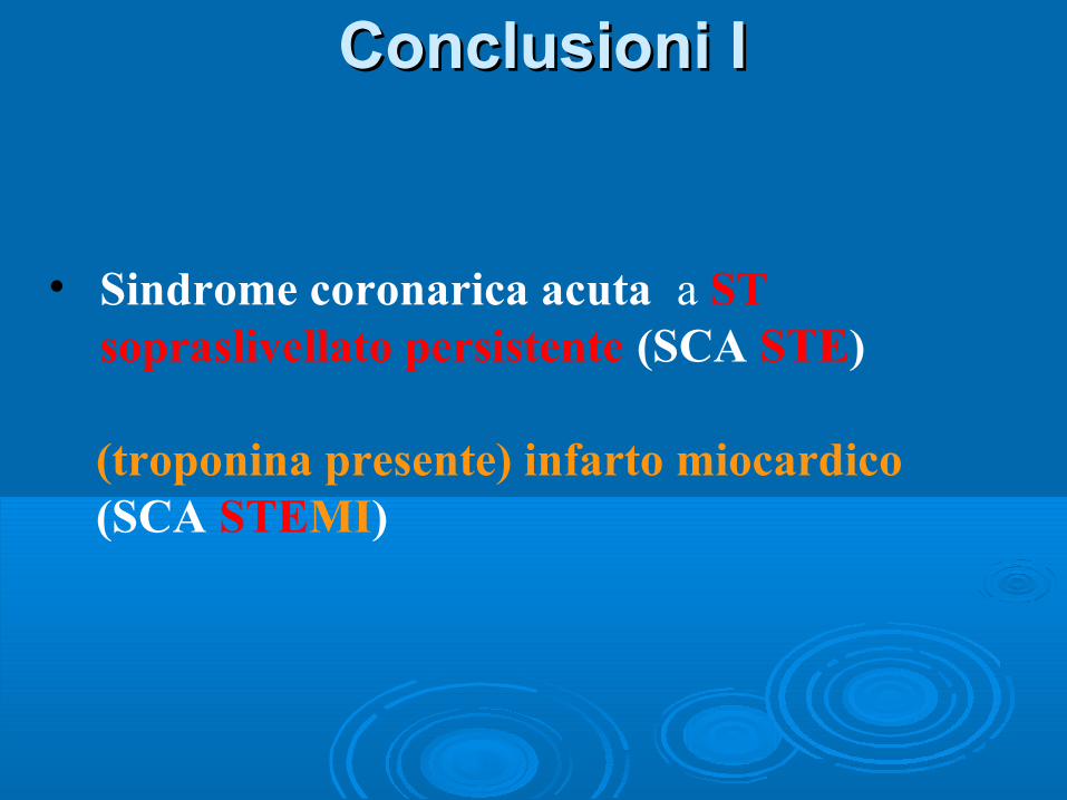
Conclusioni IConclusioni I
• Sindrome coronarica acuta a ST sopraslivellato persistente (SCA STE)
(troponina presente) infarto miocardico (SCA STEMI)
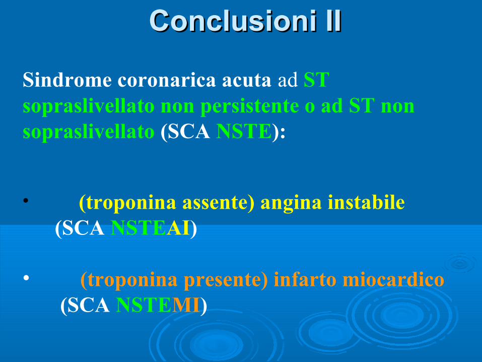
Conclusioni IIConclusioni II
Sindrome coronarica acuta ad STsopraslivellato non persistente o ad ST nonsopraslivellato (SCA NSTE):
• (troponina assente) angina instabile (SCA NSTEAI) • (troponina presente) infarto miocardico (SCA NSTEMI)

Grazie per l'attenzione