Embed Size (px)
Citation preview

Infective Endocarditis
• Febrile illness• Persistent bacteremia• Characteristic lesion of microbial infection
of the endothelial surface of the heart
– Variable in size– Amorphous mass of fibrin & platelets– Abundant organisms– Few inflammatory cells
The vegetation


Infective Endocarditis
• Acute – Toxic presentation– Progressive valve destruction & metastatic infection developing
in days to weeks– Most commonly caused by S. aureus
• Subacute– Mild toxicity– Presentation over weeks to months– Rarely leads to metastatic infection– Most commonly S. viridans or enterococcus

Infective Endocarditis
• Case rate may vary between 2-3 cases /100,000 to as high as 15-30/100,000 depending on incidence of i.v. drug abuse and age of the population– 55-75% of patients with native valve endocarditis (NVE) have
underlying valve abnormalities• MVP
• Rheumatic
• Congenital
• ASH or:
• i.v. drug abuse

Infective Endocarditis
• Case rates– 7-25% of cases involve prosthetic valves– 25-45% of cases predisposing condition can
not be identified
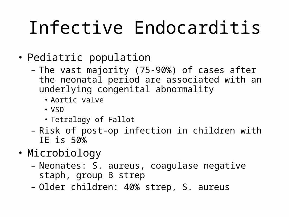
Infective Endocarditis
• Pediatric population– The vast majority (75-90%) of cases after the
neonatal period are associated with an underlying congenital abnormality
• Aortic valve• VSD• Tetralogy of Fallot
– Risk of post-op infection in children with IE is 50%
• Microbiology– Neonates: S. aureus, coagulase negative staph,
group B strep– Older children: 40% strep, S. aureus

Infective Endocarditis
• Adult population– MVP – prominent predisposing factor
• High prevalence in population– 2-4%– 20% in young women
• Accounts for 7 – 30% NVE in cases not related to drug abuse or nosocomial infection
– Relative risk in MVP ~3.5 – 8.2, largely confined to patients with murmur, but also increased in men and patients >45 years old
• MVP with murmur – incidence IE 52/100/000 pt. years• MVP w/o murmur – incidence IE 4.6/100,000 pt. years

Infective Endocarditis
• Adult population– Rheumatic Heart Disease
• 20 – 25% of cases of IE in 1970’s & 80’s• 7 – 18% of cases in recent reported series• Mitral site more common in women• Aortic site more common in men
– Congenital Heart Disease• 10 – 20% of cases in young adults• 8% of cases in older adults• PDA, VSD, bicuspid aortic valve (esp. in men>60)

Infective Endocarditis
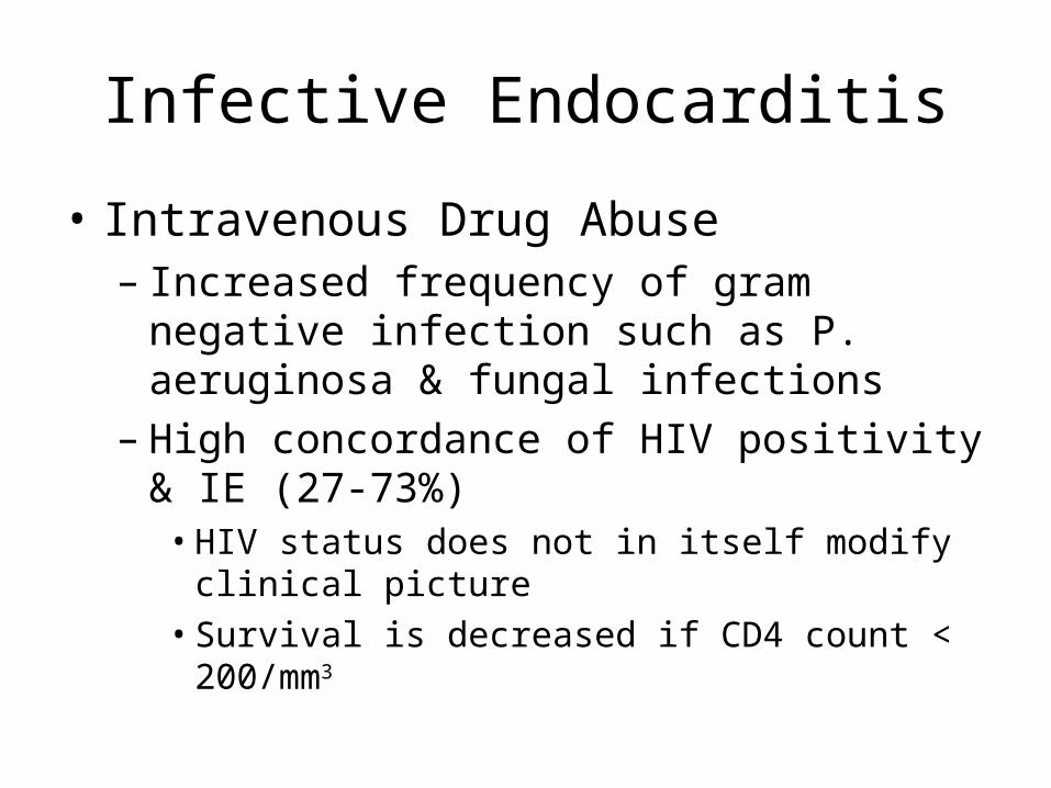
• Intravenous Drug Abuse– Risk is 2 – 5% per pt./year– Tendency to involve right-sided valves
• Distribution in clinical series– 46 – 78% tricuspid– 24 – 32% mitral– 8 – 19% aortic
– Underlying valve normal in 75 – 93%– S. aureus predominant organism (>50%, 60-
70% of tricuspid cases)
Infective Endocarditis
• Intravenous Drug Abuse– Increased frequency of gram negative
infection such as P. aeruginosa & fungal infections
– High concordance of HIV positivity & IE (27-73%)
• HIV status does not in itself modify clinical picture• Survival is decreased if CD4 count < 200/mm3
Infective Endocarditis
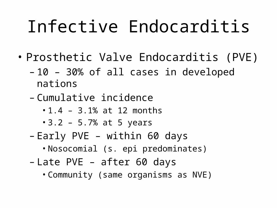
• Prosthetic Valve Endocarditis (PVE)– 10 – 30% of all cases in developed nations– Cumulative incidence
• 1.4 – 3.1% at 12 months• 3.2 – 5.7% at 5 years
– Early PVE – within 60 days• Nosocomial (s. epi predominates)
– Late PVE – after 60 days• Community (same organisms as NVE)

Infective Endocarditis
• Pathology– NVE infection is largely confined to leaflets– PVE infection commonly extends beyond
valve ring into annulus/periannular tissue• Ring abscesses• Septal abscesses• Fistulae• Prosthetic dehiscence
– Invasive infection more common in aortic position and if onset is early
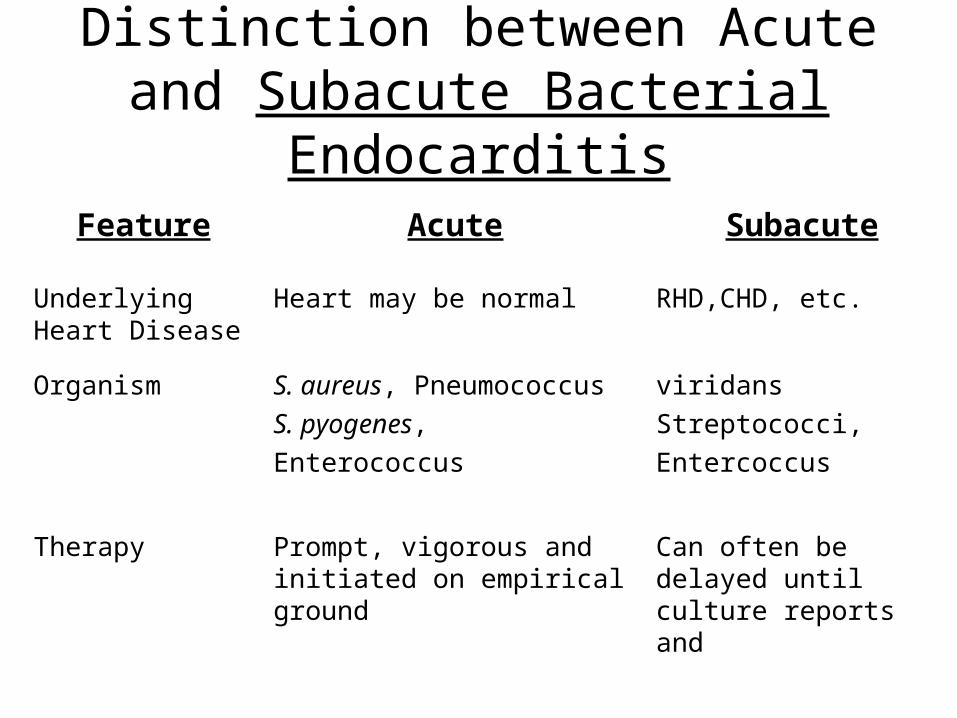
Distinction between Acute and Subacute Bacterial Endocarditis
Feature Acute Subacute
Underlying Heart Disease
Heart may be normal RHD,CHD, etc.
Organism S. aureus, Pneumococcus
S. pyogenes,
Enterococcus
viridans
Streptococci,
Entercoccus
Therapy Prompt, vigorous and initiated on empirical ground
Can often be delayed until culture reports and susceptibilities available
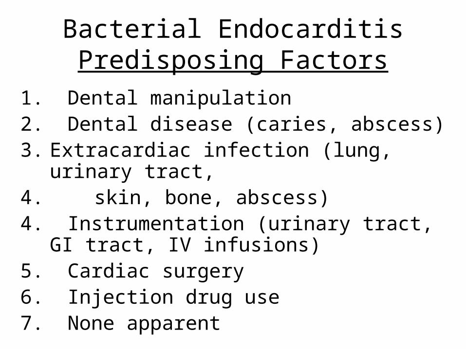
Bacterial EndocarditisPredisposing Factors
1. Dental manipulation2. Dental disease (caries, abscess)3. Extracardiac infection (lung, urinary tract,4. skin, bone, abscess)4. Instrumentation (urinary tract, GI tract, IV
infusions)5. Cardiac surgery6. Injection drug use7. None apparent

Bacterial EndocarditisClinical Features
1. Fever. Antibiotics, salicylates, steroids, severe CHF, uremia may mask temperature elevations.
2. Murmurs3. Petechiae and cutaneous manifestations. Roth spots
conjunctival and mucosal petechiae, splinter hemorrhages, Osler nodes and Janway lesions.
4. Splenomegaly 5. Embolism. Septic or sterile. CNS, spleen, lung, retinal
vessels, coronary artery, large vessels.6. Renal disease, infarction. Multiple abscesses.
Glomerulonephritis and uremia7. CHF8. General. Weight loss, anorexia, debilitation, loss of libido.

Bacterial Endocarditis Laboratory Features
1. Anemia
2. Most commonly elevated WBC
3. ESR elevated, ↓ C′ in patients with glomerulonephritis
4. Microscopic hematuria
5. Bacteremia. Persistent.≥ 3, ≤ 5 blood cultures. Aerobic and anaerobic. Different sites.

Bacterial EndocarditisTherapy and Prophylaxis
1. Prolonged; high dosages; use of bactericidal drugs
2. Serum antibiotic levels and MBC of the organism
3. Viridans streptococci, Enterococcus, S. aureus, S. pneumoniae, S. pyogenes
4. Institution of therapy on empirical grounds5. Proven negative blood cultures on Abx6. Prophylaxis: dental extraction, GU.

Infective Endocarditis
• Majority of cases caused by streptococcus, staphylococcus, enterococcus, or fastidious gram negative cocco-bacillary forms
• Gram negative organisms– P. aeruginosa most common– HACEK - slow growing, fastidious organisms that
may need 3 weeks to grow out of culture• Haemophilus sp.• Actinobacillus• Cardiobacterium• Eikenella• Kingella

Pathophysiology
• Embolization• Clinically evident 11 – 43% of patients• Pathologically present 45 – 65%• High risk for embolization
» Large > 10 mm vegetation» Hypermobile vegetation» Mitral vegetations (esp. anterior leaflet)
• Pulmonary (septic) – 65 – 75% of i.v. drug abusers with tricuspid IE

Clinical Features
• Interval between index bacteremia & onset of sx’s usually < 2 weeks
• May be substantially longer in early PVE
• Fever most common sign• May be absent in elderly/debilitated pt.
• Murmur present in 80 – 85%• Generally indication of underlying lesion• Frequently absent in tricuspid IE
• Changing murmur

Classical Peripheral Manifestations
• Less common today
• Not seen in tricuspid endocarditis
• Petechiae most common

Janeway Lesions

Splinter Hemorrhage


Subconjunctival Hemorrhages

Roth’s Spots

Clinical Features
• Systemic emboli• Incidence decreases with effective anti-microbial Rx
• Neurological sequelae• Embolic stroke 15 – 20% of patients• Mycotic aneurysm• Cerebritis
• CHF• Due to mechanical disruption• High mortality without surgical intervention
• Renal insufficiency• Immune complex mediated• Impaired hemodynamics/drug toxicity
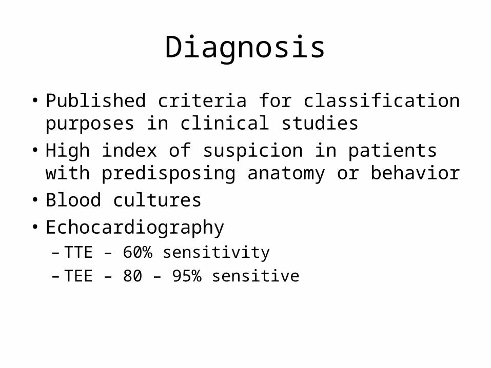
Diagnosis
• Published criteria for classification purposes in clinical studies
• High index of suspicion in patients with predisposing anatomy or behavior
• Blood cultures
• Echocardiography– TTE – 60% sensitivity– TEE – 80 – 95% sensitive

Goals of Therapy
Eradicate infection
Definitively treat sequelae of destructive intra-cardiac and extra-cardiac lesions

Antibiotic Therapy
• Treatment tailored to etiologic agent– Important to note MIC/MBC relationship for
each causative organism and the antibiotic used
– High serum concentration necessary to penetrate avascular vegetation
– ID CONSULT EVERY TIME
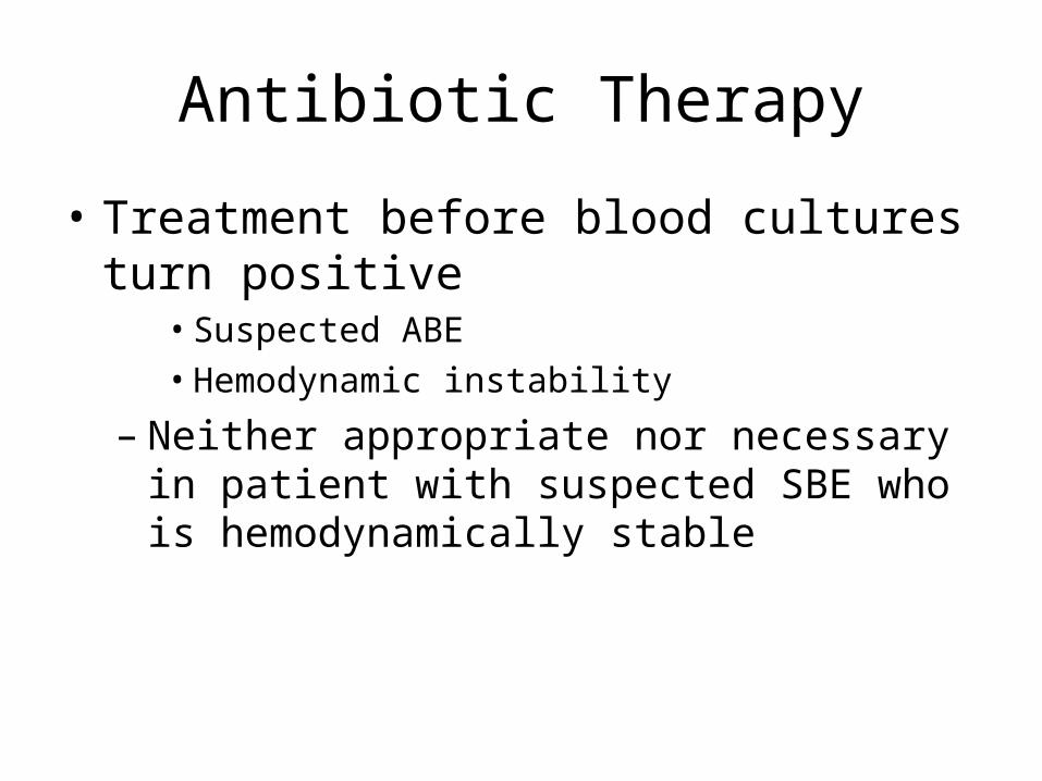
Antibiotic Therapy
• Treatment before blood cultures turn positive
• Suspected ABE• Hemodynamic instability
– Neither appropriate nor necessary in patient with suspected SBE who is hemodynamically stable

Antibiotic Therapy
• Effective antimicrobial treatment should lead to defervescence within 7 – 10 days– Persistent fever in:
• IE due to staph, pseudomonas, culture negative• IE with microvascular complications/major emboli• Intracardiac/extracardiac septic complications• Drug reaction

Surgical Treatment of Intra-Cardiac Complications
• NYHA Class III/IV CHF due to valve dysfunction– Surgical mortality – 20-40%– Medical mortality – 50-90%
• Unstable prosthetic valve– Surgical mortality – 15-55%– Medical mortality – near 100% at 6 months
• Uncontrolled infection

Surgical Treatment of Intra-Cardiac Complications
• Difficult to cure:– Fungal endocarditis– Brucella
• S. aureus PVE with any intra-cardiac complication
• Relapse of PVE after optimal therapy

Surgical Treatment of Intra-Cardiac Complications
• Relative indications– Perivalvular extension of infection– Poorly responsive S. aureus NVE– Relapse of NVE– Culture negative NVE/PVE with persistent fever (> 10
days)– Large (> 10mm) or hypermobile vegetation– Endocarditis due to highly resistant enterococcus– Embolism despite therapy

Prevention
• Prophylactic regimen targeted against likely organism– Strep. viridans – oral, respiratory, eosphogeal – Enterococcus – genitourinary, gastrointestinal– S. aureus – infected skin, mucosal surfaces

Prevention – the procedure
• Dental procedures known to produce bleeding
• Tonsillectomy • Surgery involving GI,
respiratory mucosa• Esophageal dilation• ERCP for obstruction
• Gallbladder surgery• Cystoscopy, urethral
dilation• Urethral catheter if
infection present• Urinary tract surgery,
including prostate• I&D of infected tissue

Prevention – the underlying lesion
• High risk lesions– Prosthetic valves
– Prior IE
– Cyanotic congenital heart disease
– PDA
– AR, AS, MR,MS with MR
– VSD
– Coarctation
– Surgical systemic-pulmonary shunts
• Intermediate risk– MVP with murmur– Pure MS– Tricuspid disease– Pulmonary stenosis– ASH– Bicuspid Ao valve with no
hemodynamic significance
Lesions at highest risk

Prevention – the underlying lesion
• Low/no risk– MVP without murmur– Trivial valvular regurg.– Isolated ASD– Implanted device
(pacer, ICD)– CAD– CABG


ChemoprophylaxisAdult Prophylaxis: Dental, Oral, Respiratory, Esophageal
Standard Regimen
Amoxicillin 2g PO 1h before procedure or Ampicillin 2g IM/IV 30m before procedure
Penicillin Allergic Clindamycin
600 mg PO 1h before procedure or 600 mg IV 30m before
Cephalexin OR Cefadroxil 2g PO 1 hour before Cefazolin 1.0g IM/IV 30 min before procedure Azithromycin or Clarithromycin 500mg PO 1h before

Adult Genitourinary or Gastrointestinal Procedures High Risk Patients
Standard Regimen Before procedure (30 minutes):
Ampicillin 2g IV/IM AND Gentamicin 1.5 mg/kg (MAX 120 mg) IM/IV
After procedure (6 hours later) Ampicillin 1g IM/IV OR Amoxicillin 1g PO
Penicillin Allergic Complete infusion 30 minutes before procedure
Vancomycin 1g IV over 1-2h AND Gentamicin 1.5 mg/kg IV/IM (MAX 120 mg)
Moderate Risk Patients Standard Regimen
Amoxicillin 2g PO 1h before OR Ampicillin 2g IM/IV 30m before
Penicillin Allergic Vancomycin 1g IV over 1-2h, complete 30m before


![InvestigationofBacteremiadueto ...Aeromonas sp. and other bacterial species [13], the present report is the first to describe a comparative study on Aer-omonas bacteremia and bacteremia](https://img.pdfslide.tips/doc/110x75/6128da66bf259c143c71095b/investigationofbacteremiadueto-aeromonas-sp-and-other-bacterial-species-13.jpg)