Embed Size (px)
Citation preview
Khám đồng tử
PGS.TS Cao Phi Phong
2017
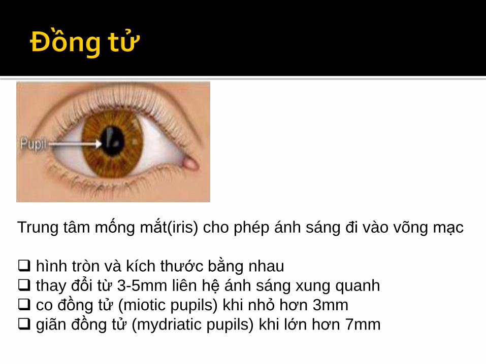
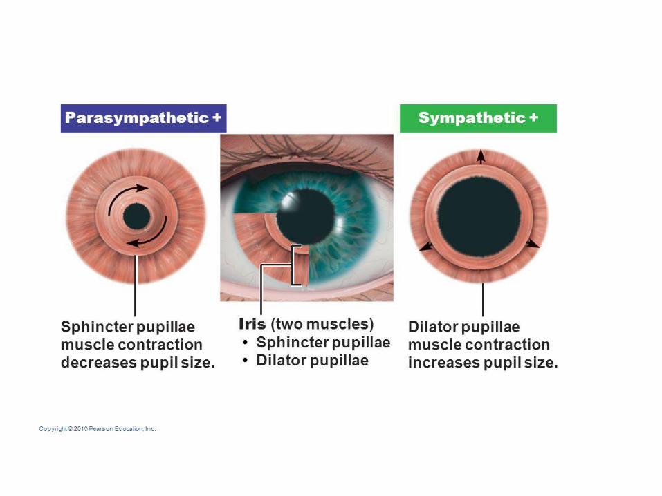
Trung tâm mống mắt(iris) cho phép ánh sáng đi vào võng mạc
hình tròn và kích thước bằng nhau
thay đổi từ 3-5mm liên hệ ánh sáng xung quanh
co đồng tử (miotic pupils) khi nhỏ hơn 3mm
giãn đồng tử (mydriatic pupils) khi lớn hơn 7mm
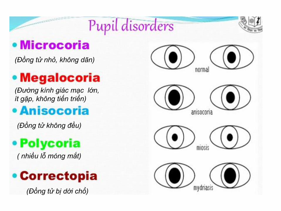
( nhiều lỗ móng mắt)
(Đồng tử bị dời chổ)
(Đường kính giác mạc lớn,
ít gặp, không tiến triển)
(Đồng tử nhỏ, không dãn)
(Đồng tử không đều)
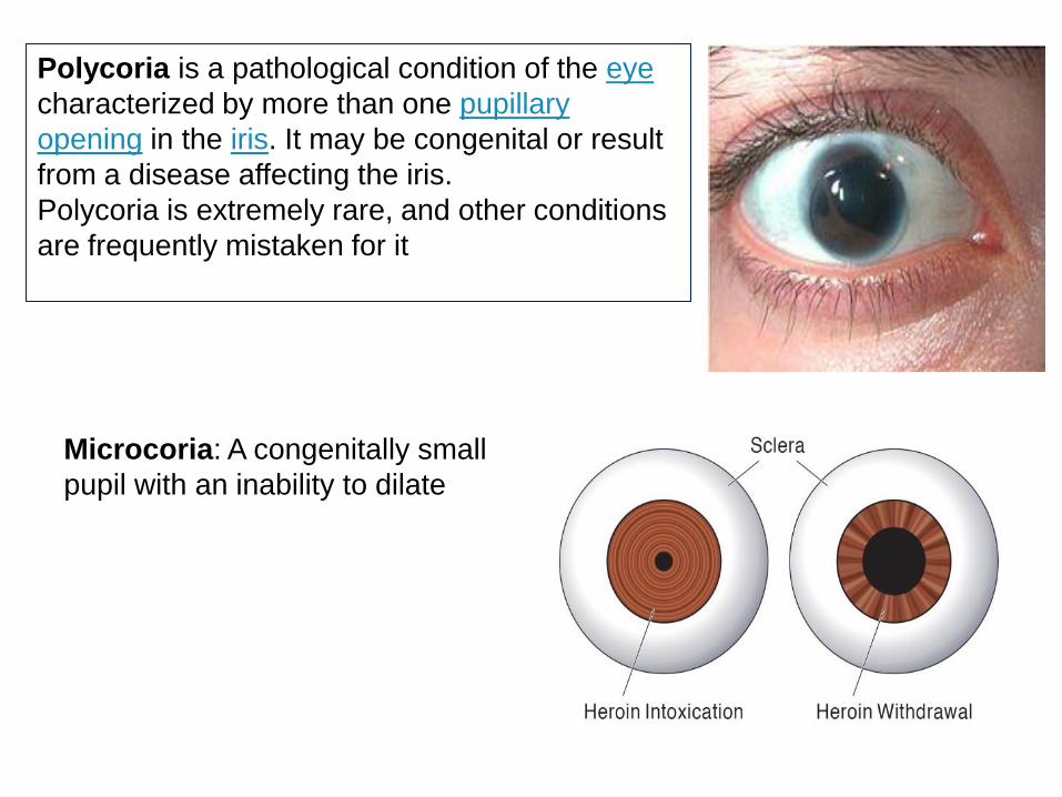
Polycoria is a pathological condition of the eye
characterized by more than one pupillary
opening in the iris. It may be congenital or result
from a disease affecting the iris.
Polycoria is extremely rare, and other conditions
are frequently mistaken for it
Microcoria: A congenitally small
pupil with an inability to dilate
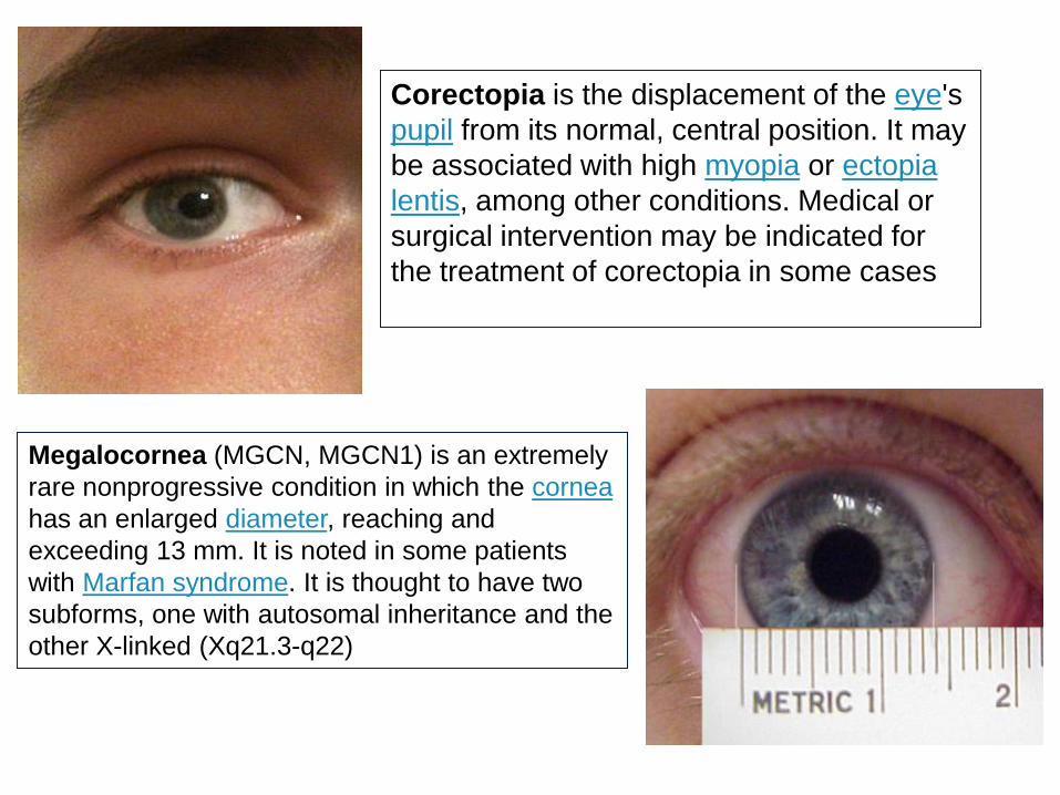
Corectopia is the displacement of the eye's
pupil from its normal, central position. It may
be associated with high myopia or ectopia
lentis, among other conditions. Medical or
surgical intervention may be indicated for
the treatment of corectopia in some cases
Megalocornea (MGCN, MGCN1) is an extremely
rare nonprogressive condition in which the cornea
has an enlarged diameter, reaching and
exceeding 13 mm. It is noted in some patients
with Marfan syndrome. It is thought to have two
subforms, one with autosomal inheritance and the
other X-linked (Xq21.3-q22)
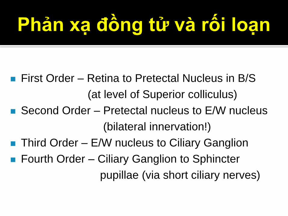
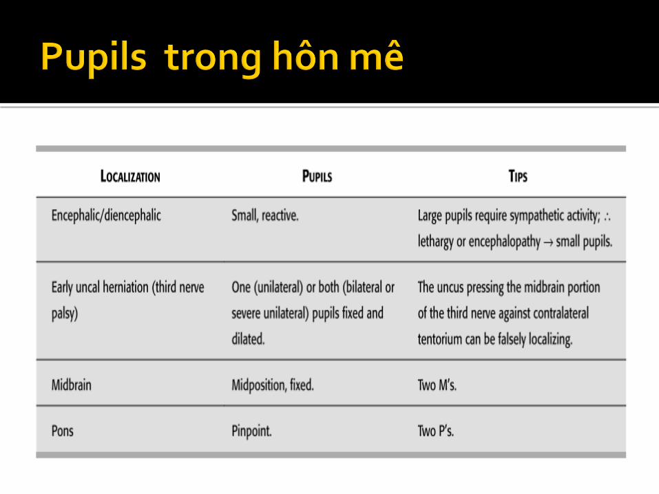
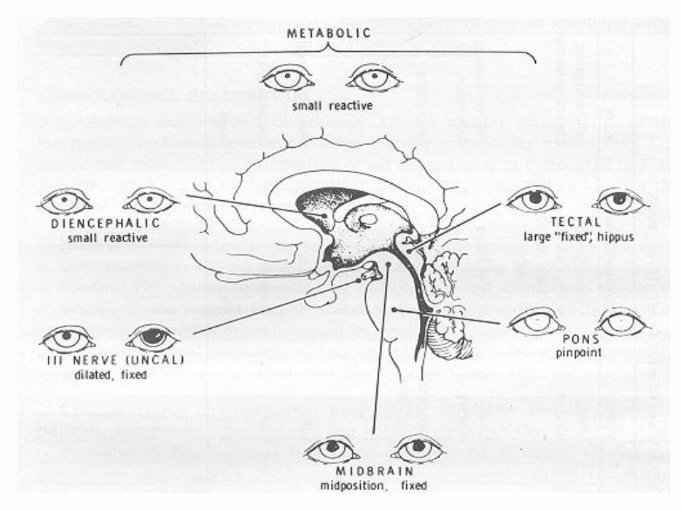
First Order – Retina to Pretectal Nucleus in B/S
(at level of Superior colliculus)
Second Order – Pretectal nucleus to E/W nucleus
(bilateral innervation!)
Third Order – E/W nucleus to Ciliary Ganglion
Fourth Order – Ciliary Ganglion to Sphincter
pupillae (via short ciliary nerves)
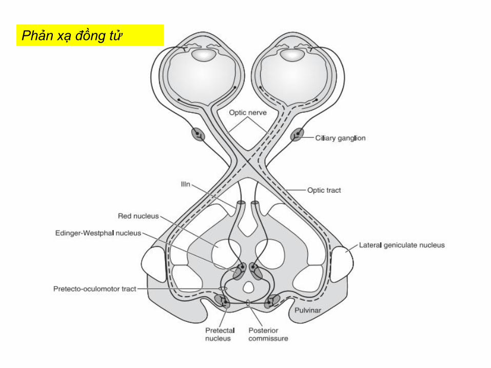
Phản xạ đồng tử
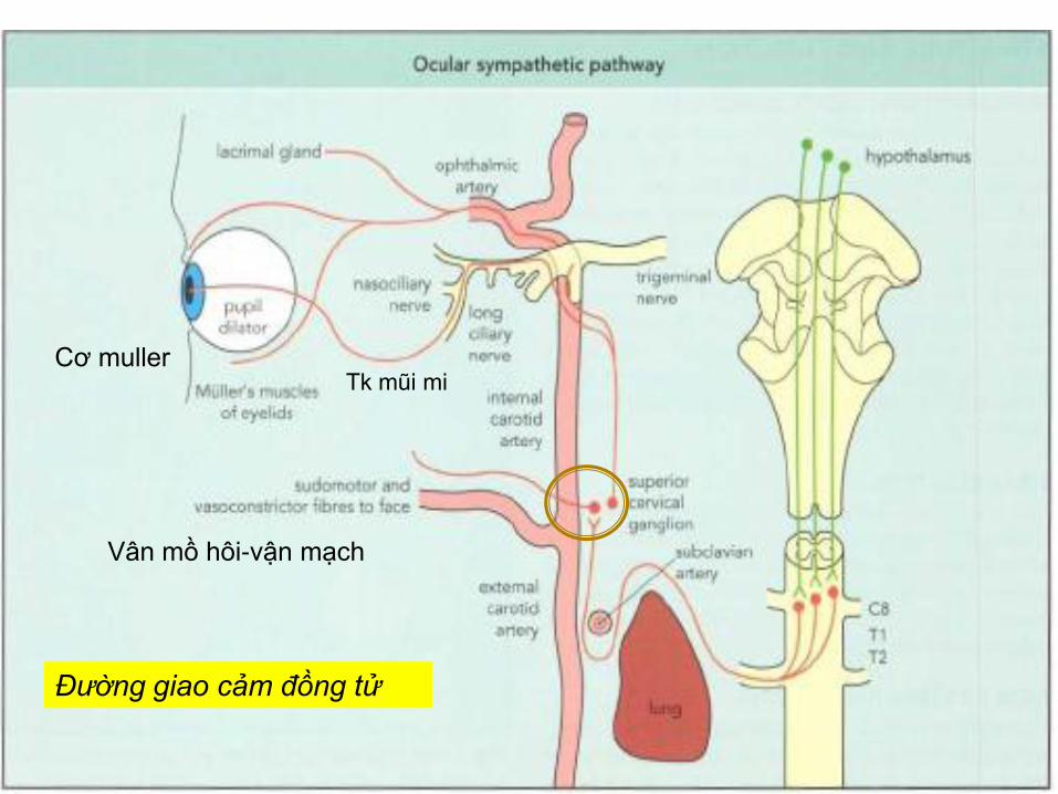
Đường giao cảm đồng tử
Tk mũi mi
Vân mồ hôi-vận mạch
Cơ muller
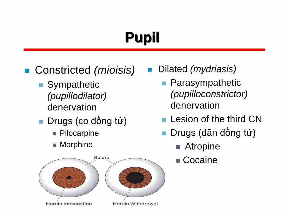
Pupil
Constricted (mioisis)
Sympathetic
(pupillodilator)
denervation
Drugs (co đồng tử)
Pilocarpine
Morphine
Dilated (mydriasis)
Parasympathetic
(pupilloconstrictor)
denervation
Lesion of the third CN
Drugs (dãn đồng tử)
Atropine
Cocaine
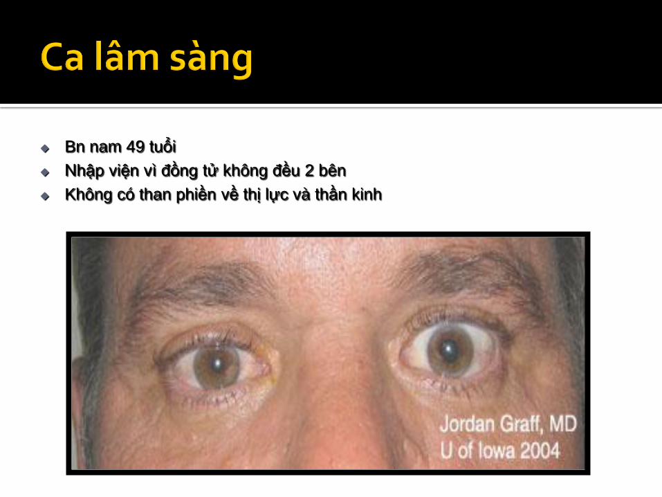
Bn nam 49 tuổi
Nhập viện vì đồng tử không đều 2 bên
Không có than phiền về thị lực và thần kinh
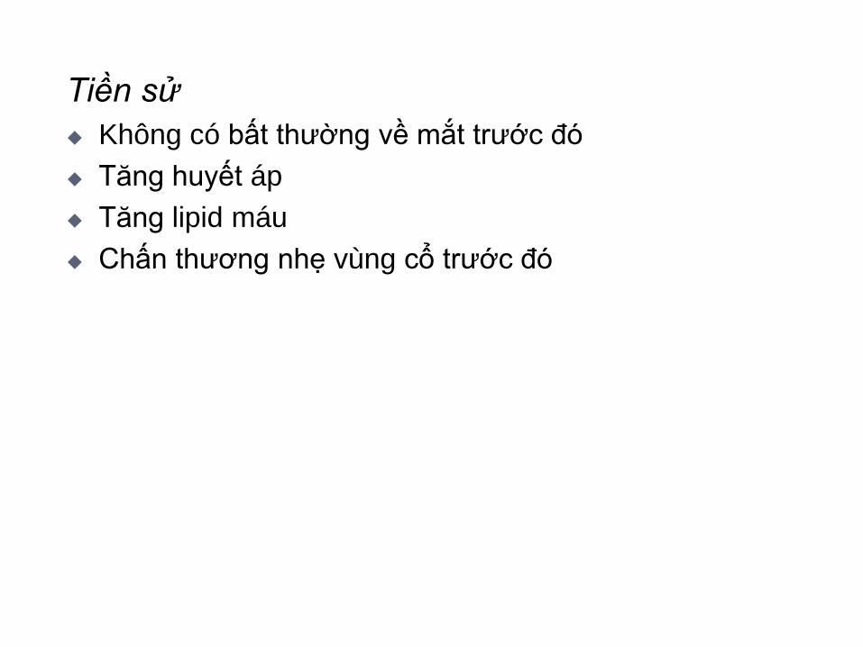
Tiền sử
Không có bất thường về mắt trước đó
Tăng huyết áp
Tăng lipid máu
Chấn thương nhẹ vùng cổ trước đó
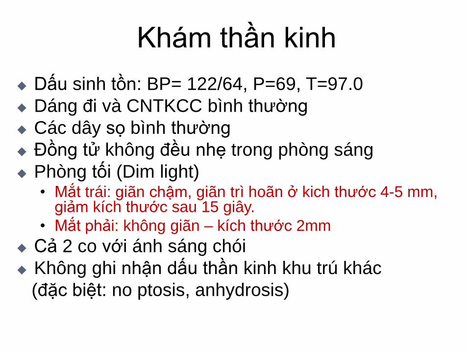
Khám thần kinh
Dấu sinh tồn: BP= 122/64, P=69, T=97.0
Dáng đi và CNTKCC bình thường
Các dây sọ bình thường
Đồng tử không đều nhẹ trong phòng sáng
Phòng tối (Dim light) • Mắt trái: giãn chậm, giãn trì hoãn ở kich thước 4-5 mm,
giảm kích thước sau 15 giây.
• Mắt phải: không giãn – kích thước 2mm
Cả 2 co với ánh sáng chói
Không ghi nhận dấu thần kinh khu trú khác
(đặc biệt: no ptosis, anhydrosis)

Chẩn đoán: Đồng tử không đều
Tiếp cận chẩn đoán ?
Khám hệ thống đồng tử
First step: xác định đồng tử có đáp ứng ánh sáng
Second step: so sánh kích thước đồng tử
Third step: thực hiện “swinging flashlight test”
(when there is an interocular difference of 0.5 mm
or more in pupillary diameter), (khi có sự khác biệt đường
kinh giữa 2 mắt là 0,5mm hay hơn)
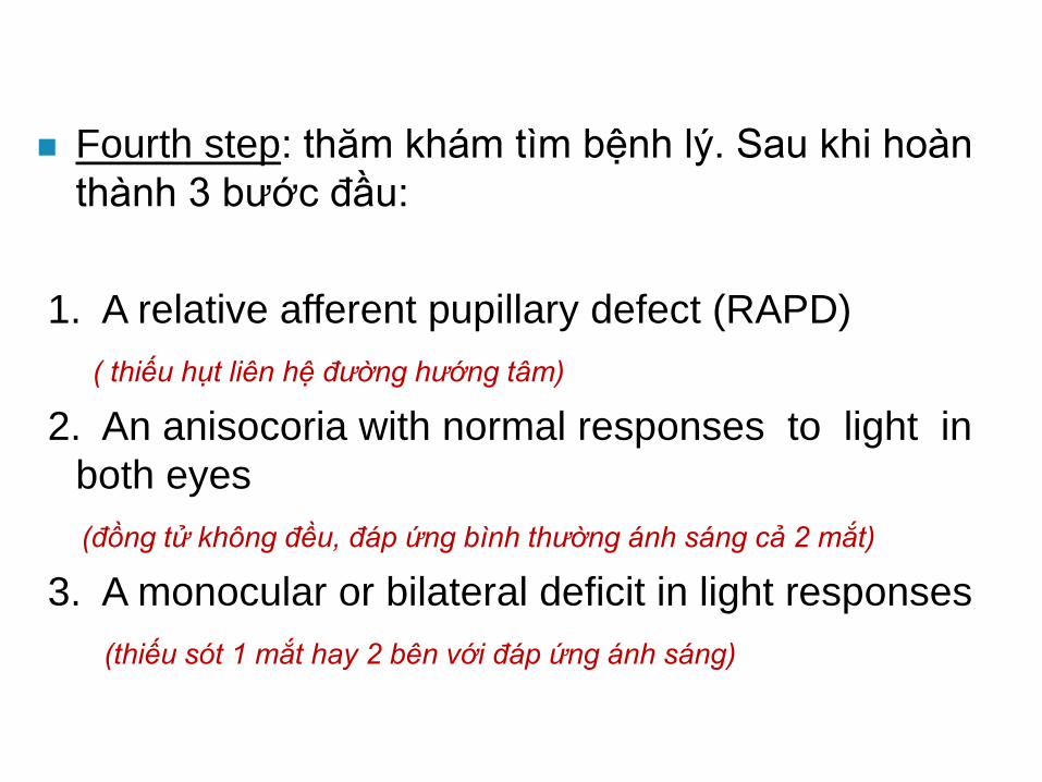
Fourth step: thăm khám tìm bệnh lý. Sau khi hoàn
thành 3 bước đầu:
1. A relative afferent pupillary defect (RAPD)
( thiếu hụt liên hệ đường hướng tâm)
2. An anisocoria with normal responses to light in
both eyes
(đồng tử không đều, đáp ứng bình thường ánh sáng cả 2 mắt)
3. A monocular or bilateral deficit in light responses
(thiếu sót 1 mắt hay 2 bên với đáp ứng ánh sáng)
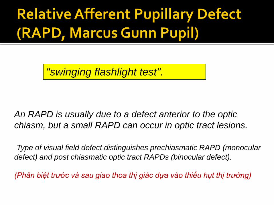
"swinging flashlight test".
An RAPD is usually due to a defect anterior to the optic
chiasm, but a small RAPD can occur in optic tract lesions.
Type of visual field defect distinguishes prechiasmatic RAPD (monocular
defect) and post chiasmatic optic tract RAPDs (binocular defect).
(Phân biệt trước và sau giao thoa thị giác dựa vào thiếu hụt thị trường)
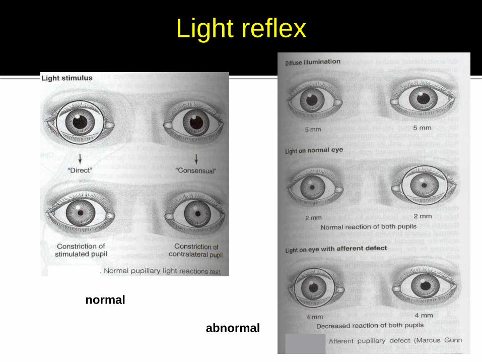
Light reflex
normal
abnormal
(Co 2 bên vừa phải)
(Nhanh, co nhiều hơn 2 bên)
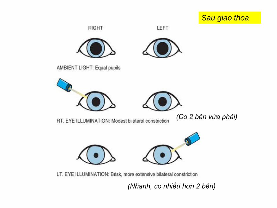
Sau giao thoa
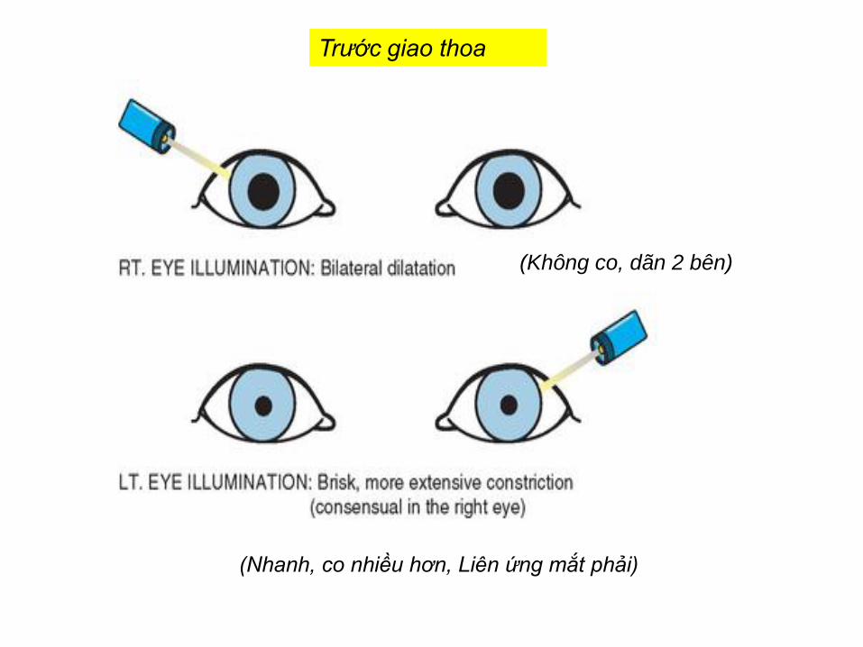
(Không co, dãn 2 bên)
(Nhanh, co nhiều hơn, Liên ứng mắt phải)
Trước giao thoa
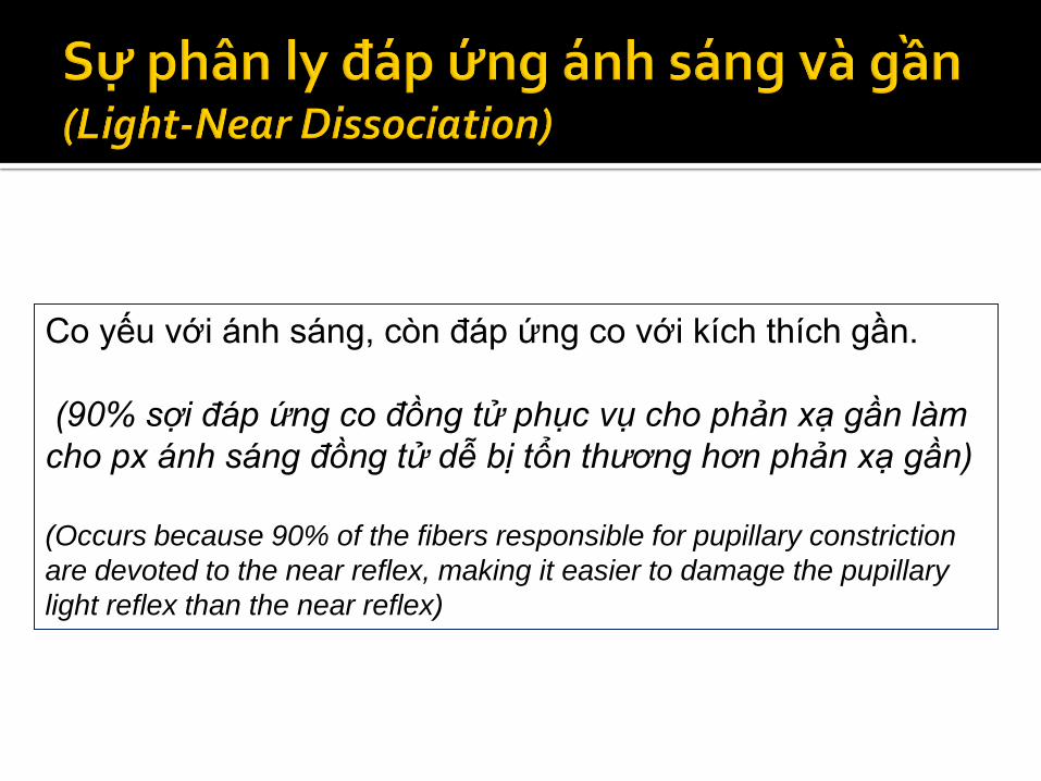
Co yếu với ánh sáng, còn đáp ứng co với kích thích gần.
(90% sợi đáp ứng co đồng tử phục vụ cho phản xạ gần làm
cho px ánh sáng đồng tử dễ bị tổn thương hơn phản xạ gần)
(Occurs because 90% of the fibers responsible for pupillary constriction
are devoted to the near reflex, making it easier to damage the pupillary
light reflex than the near reflex)
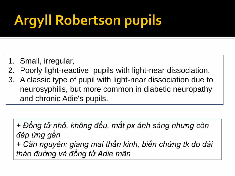
1. Small, irregular,
2. Poorly light-reactive pupils with light-near dissociation.
3. A classic type of pupil with light-near dissociation due to
neurosyphilis, but more common in diabetic neuropathy
and chronic Adie's pupils.
+ Đồng tử nhỏ, không đều, mất px ánh sáng nhưng còn
đáp ứng gần
+ Căn nguyên: giang mai thần kinh, biến chứng tk do đái
tháo đường và đồng tử Adie mãn
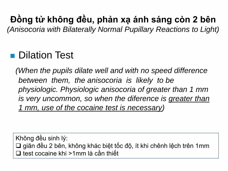
Đồng tử không đều, phản xạ ánh sáng còn 2 bên (Anisocoria with Bilaterally Normal Pupillary Reactions to Light)
Dilation Test
(When the pupils dilate well and with no speed difference
between them, the anisocoria is likely to be
physiologic. Physiologic anisocoria of greater than 1 mm
is very uncommon, so when the diference is greater than
1 mm, use of the cocaine test is necessary)
Không đều sinh lý:
giãn đều 2 bên, không khác biệt tốc độ, ít khi chênh lệch trên 1mm
test cocaine khi >1mm là cần thiết
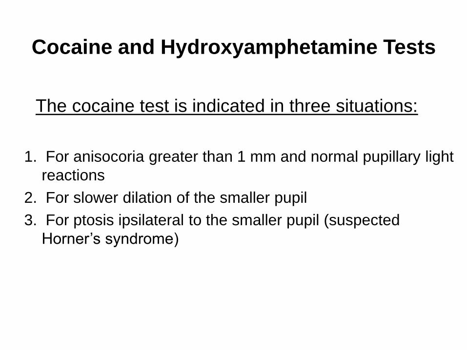
Cocaine and Hydroxyamphetamine Tests
The cocaine test is indicated in three situations:
1. For anisocoria greater than 1 mm and normal pupillary light
reactions
2. For slower dilation of the smaller pupil
3. For ptosis ipsilateral to the smaller pupil (suspected
Horner’s syndrome)
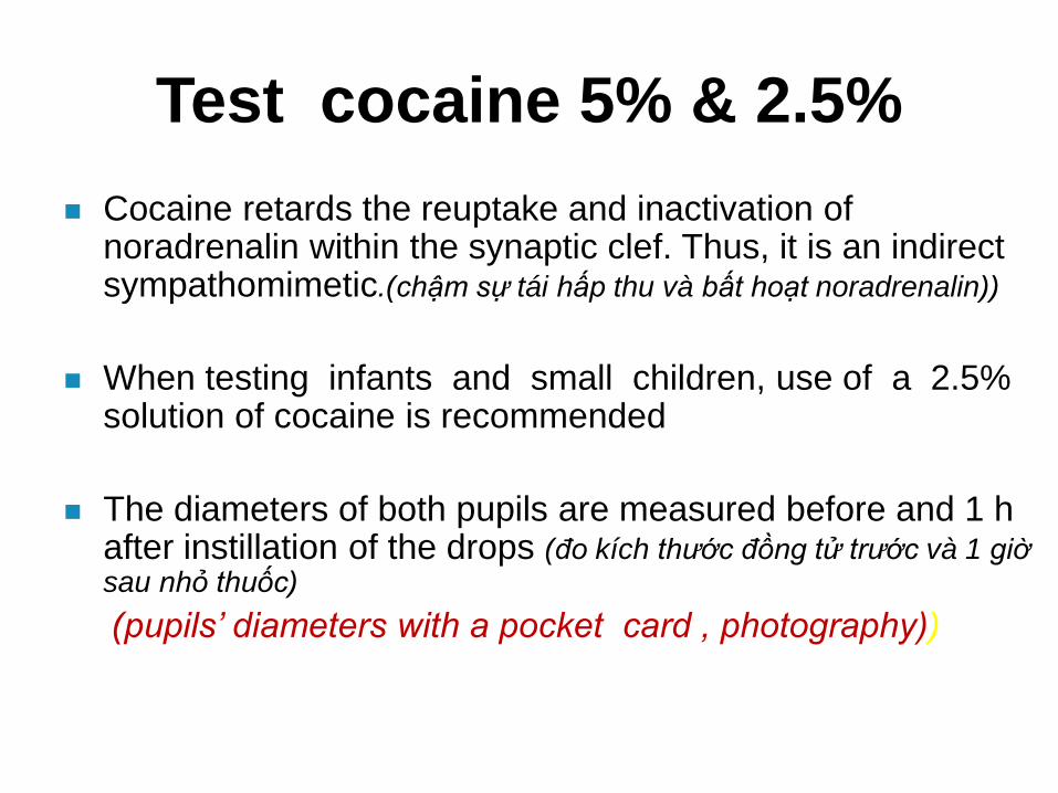
Test cocaine 5% & 2.5%
Cocaine retards the reuptake and inactivation of noradrenalin within the synaptic clef. Thus, it is an indirect sympathomimetic.(chậm sự tái hấp thu và bất hoạt noradrenalin))
When testing infants and small children, use of a 2.5% solution of cocaine is recommended
The diameters of both pupils are measured before and 1 h after instillation of the drops (đo kích thước đồng tử trước và 1 giờ sau nhỏ thuốc)
(pupils’ diameters with a pocket card , photography))
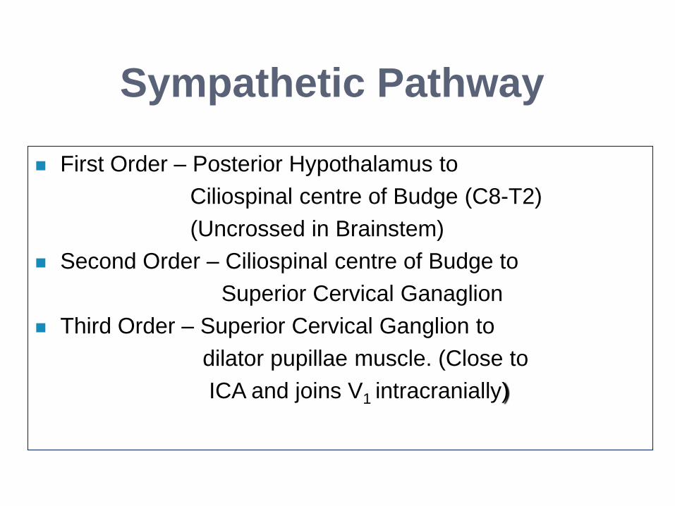
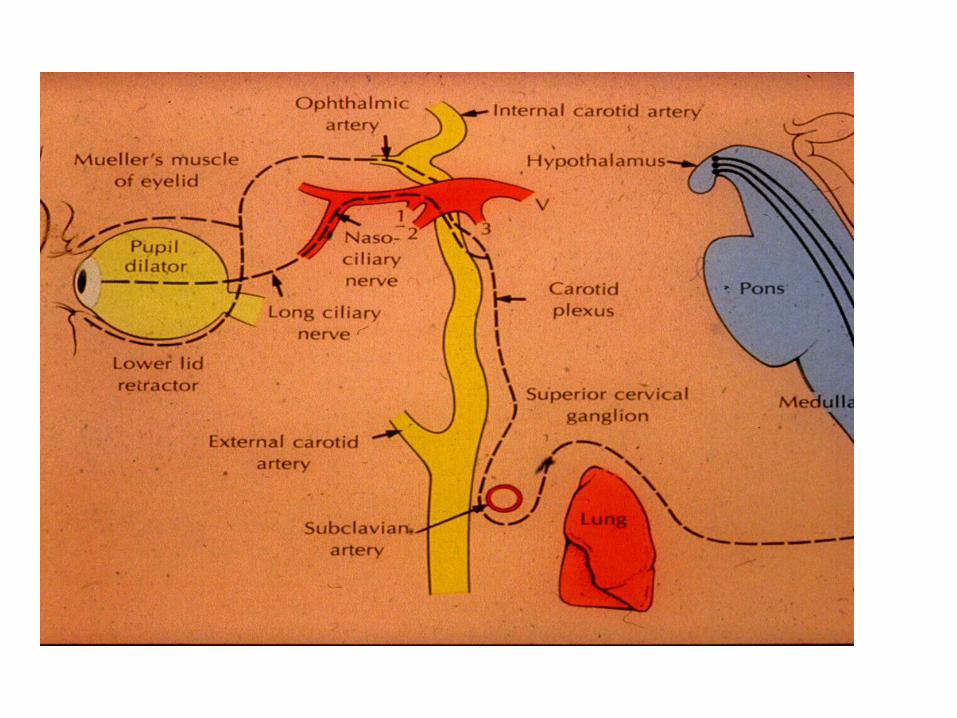
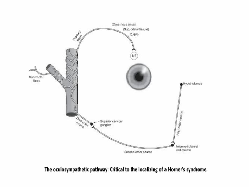
Sympathetic Pathway
First Order – Posterior Hypothalamus to
Ciliospinal centre of Budge (C8-T2)
(Uncrossed in Brainstem)
Second Order – Ciliospinal centre of Budge to
Superior Cervical Ganaglion
Third Order – Superior Cervical Ganglion to
dilator pupillae muscle. (Close to
ICA and joins V1 intracranially)
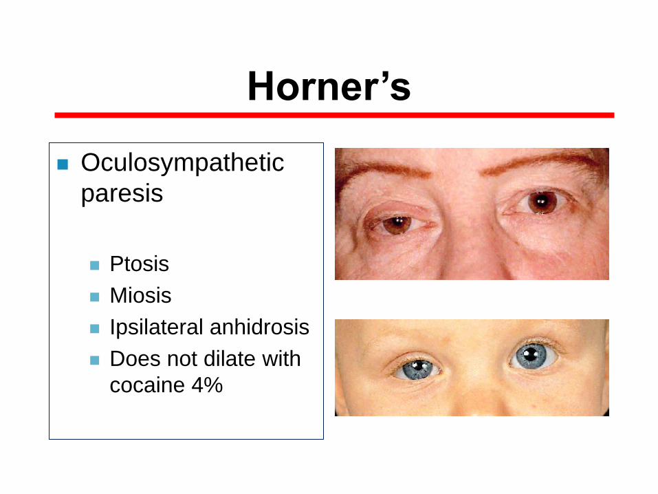
Horner’s
Oculosympathetic
paresis
Ptosis
Miosis
Ipsilateral anhidrosis
Does not dilate with
cocaine 4%
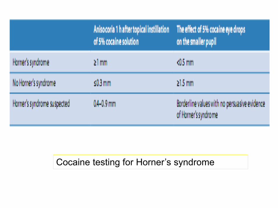
Cocaine testing for Horner’s syndrome
Test 1% hydroxyamphetamine
hay 2.5% tyramine
Stimulating the release of noradrenalin into the synaptic clef
at the terminus of the end neuron of the sympathetic chain
(kích thích phóng thích noradrenalin vào khe si-nap ở nơ-ron tận cùng)
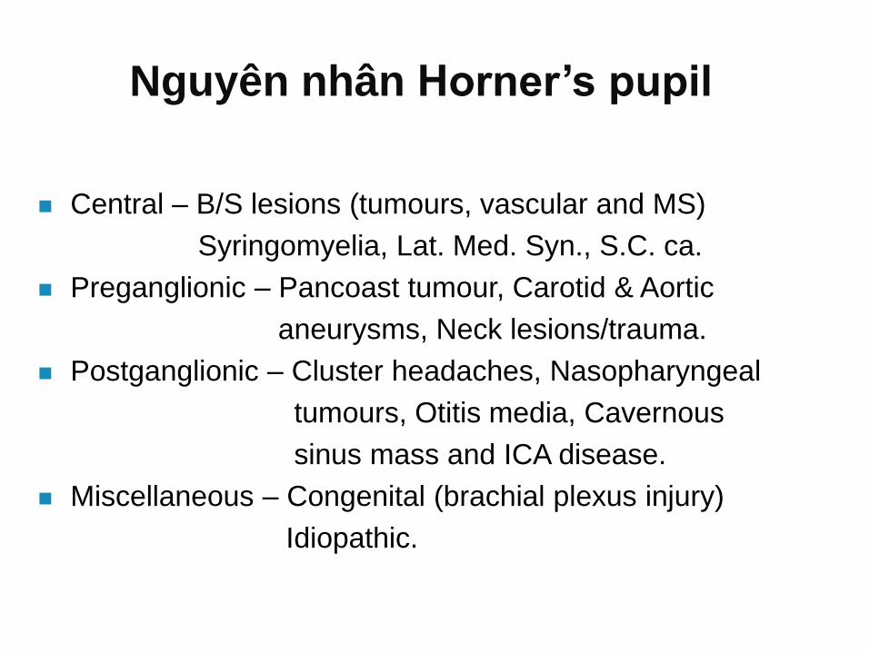
Nguyên nhân Horner’s pupil
Central – B/S lesions (tumours, vascular and MS)
Syringomyelia, Lat. Med. Syn., S.C. ca.
Preganglionic – Pancoast tumour, Carotid & Aortic
aneurysms, Neck lesions/trauma.
Postganglionic – Cluster headaches, Nasopharyngeal
tumours, Otitis media, Cavernous
sinus mass and ICA disease.
Miscellaneous – Congenital (brachial plexus injury)
Idiopathic.
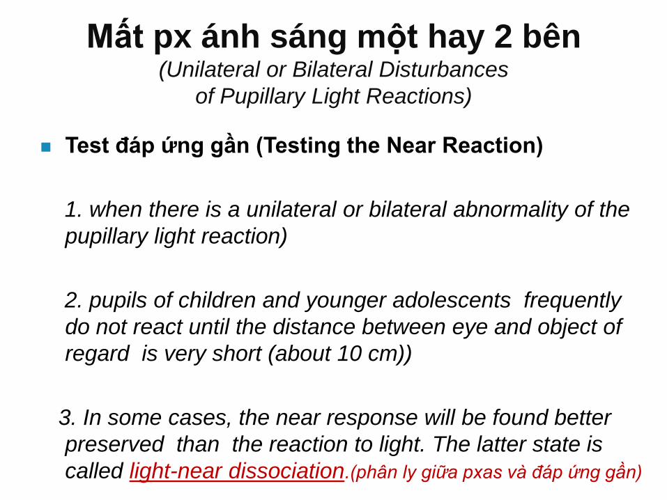
Mất px ánh sáng một hay 2 bên (Unilateral or Bilateral Disturbances
of Pupillary Light Reactions)
Test đáp ứng gần (Testing the Near Reaction)
1. when there is a unilateral or bilateral abnormality of the
pupillary light reaction)
2. pupils of children and younger adolescents frequently
do not react until the distance between eye and object of
regard is very short (about 10 cm))
3. In some cases, the near response will be found better
preserved than the reaction to light. The latter state is
called light-near dissociation.(phân ly giữa pxas và đáp ứng gần)
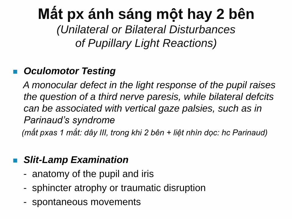
Mất px ánh sáng một hay 2 bên (Unilateral or Bilateral Disturbances
of Pupillary Light Reactions)
Oculomotor Testing
A monocular defect in the light response of the pupil raises
the question of a third nerve paresis, while bilateral defcits
can be associated with vertical gaze palsies, such as in
Parinaud’s syndrome
(mất pxas 1 mắt: dây III, trong khi 2 bên + liệt nhìn dọc: hc Parinaud)
Slit-Lamp Examination
- anatomy of the pupil and iris
- sphincter atrophy or traumatic disruption
- spontaneous movements
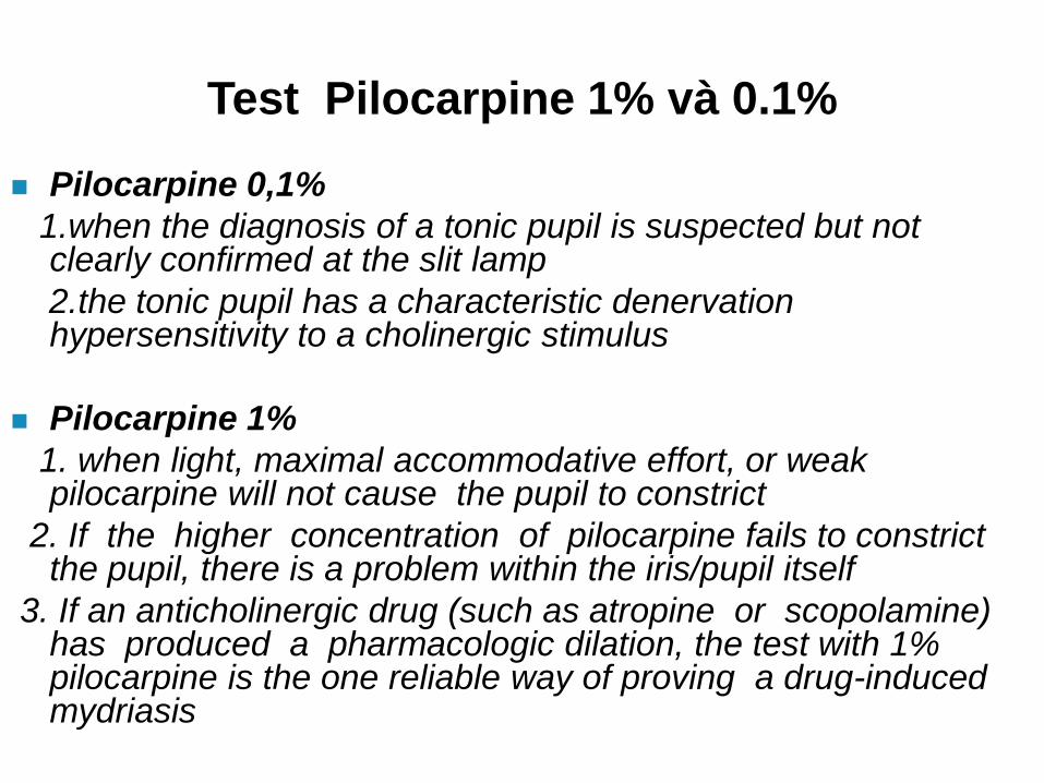
Test Pilocarpine 1% và 0.1%
Pilocarpine 0,1%
1.when the diagnosis of a tonic pupil is suspected but not clearly confirmed at the slit lamp
2.the tonic pupil has a characteristic denervation hypersensitivity to a cholinergic stimulus
Pilocarpine 1%
1. when light, maximal accommodative effort, or weak pilocarpine will not cause the pupil to constrict
2. If the higher concentration of pilocarpine fails to constrict the pupil, there is a problem within the iris/pupil itself
3. If an anticholinergic drug (such as atropine or scopolamine) has produced a pharmacologic dilation, the test with 1% pilocarpine is the one reliable way of proving a drug-induced mydriasis
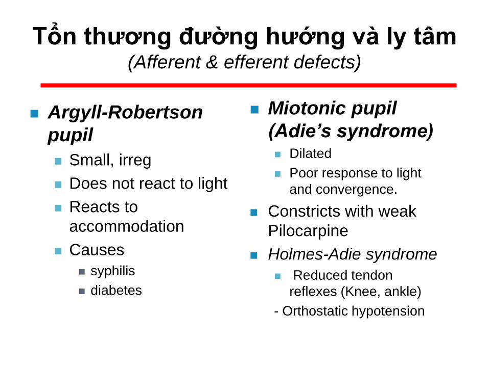
Argyll-Robertson
pupil
Small, irreg
Does not react to light
Reacts to
accommodation
Causes
syphilis
diabetes
Miotonic pupil
(Adie’s syndrome) Dilated
Poor response to light
and convergence.
Constricts with weak
Pilocarpine
Holmes-Adie syndrome
Reduced tendon
reflexes (Knee, ankle)
- Orthostatic hypotension
Tổn thương đường hướng và ly tâm (Afferent & efferent defects)

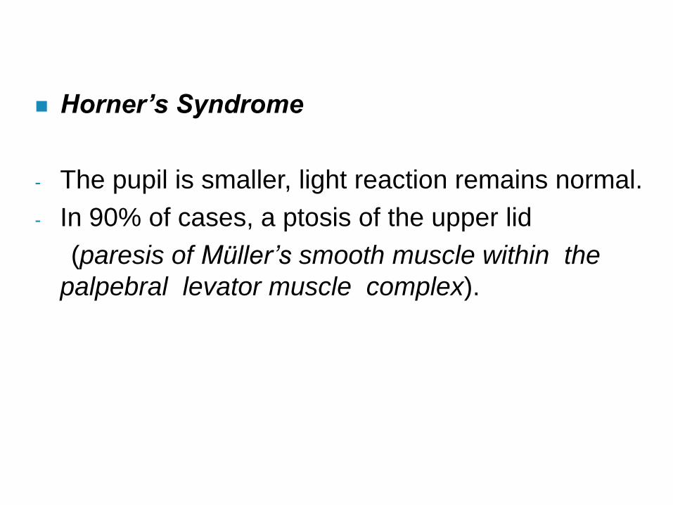
Horner’s Syndrome
- The pupil is smaller, light reaction remains normal.
- In 90% of cases, a ptosis of the upper lid
(paresis of Müller’s smooth muscle within the
palpebral levator muscle complex).
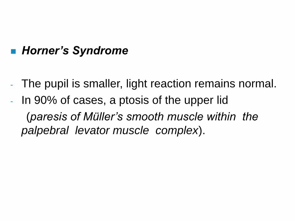
Horner’s Syndrome
- The pupil is smaller, light reaction remains normal.
- In 90% of cases, a ptosis of the upper lid
(paresis of Müller’s smooth muscle within the
palpebral levator muscle complex).
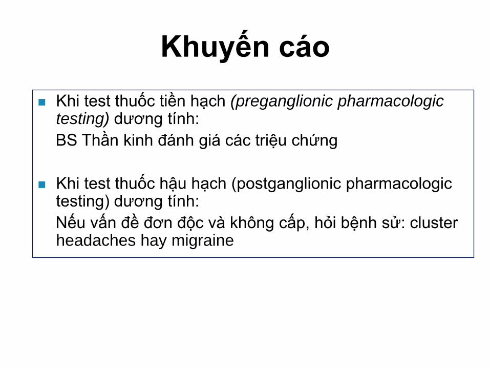
Khuyến cáo
Khi test thuốc tiền hạch (preganglionic pharmacologic testing) dương tính:
BS Thần kinh đánh giá các triệu chứng
Khi test thuốc hậu hạch (postganglionic pharmacologic testing) dương tính:
Nếu vấn đề đơn độc và không cấp, hỏi bệnh sử: cluster headaches hay migraine
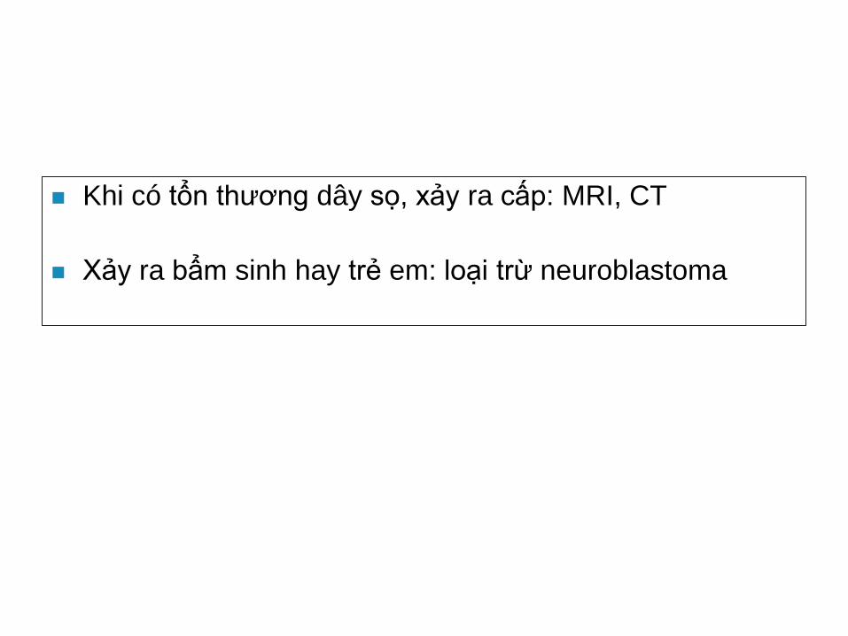
Khi có tổn thương dây sọ, xảy ra cấp: MRI, CT
Xảy ra bẩm sinh hay trẻ em: loại trừ neuroblastoma
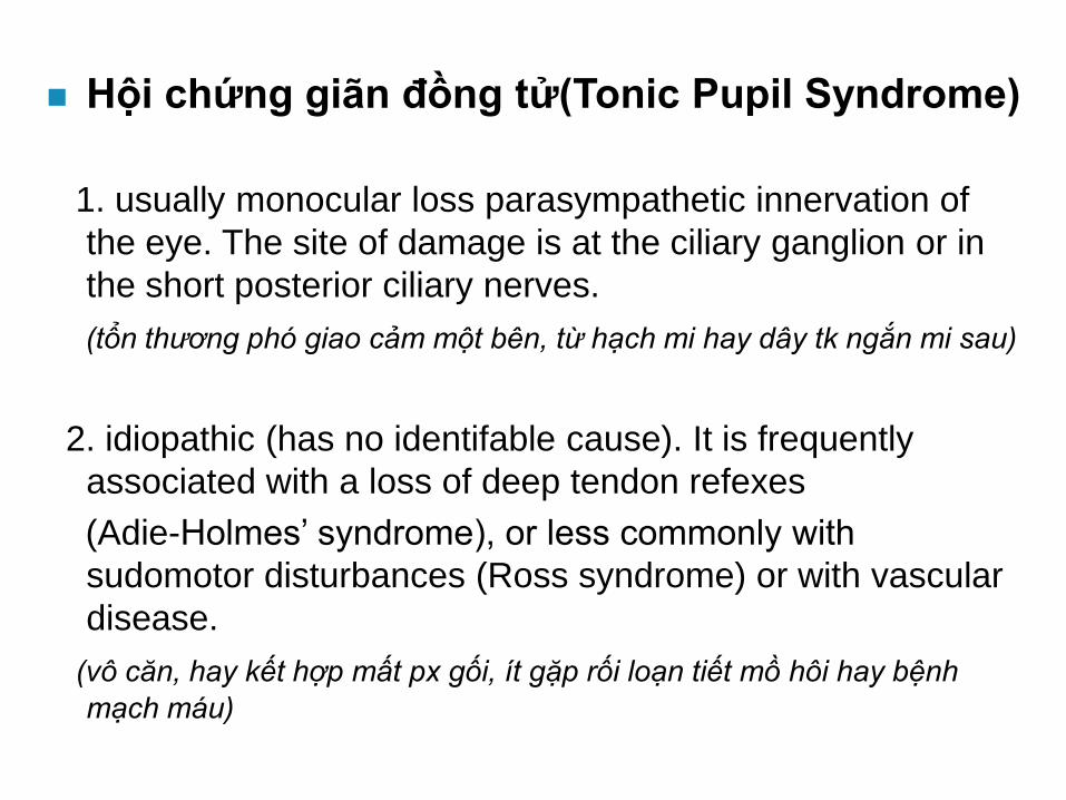
Hội chứng giãn đồng tử(Tonic Pupil Syndrome)
1. usually monocular loss parasympathetic innervation of
the eye. The site of damage is at the ciliary ganglion or in
the short posterior ciliary nerves.
(tổn thương phó giao cảm một bên, từ hạch mi hay dây tk ngắn mi sau)
2. idiopathic (has no identifable cause). It is frequently
associated with a loss of deep tendon refexes
(Adie-Holmes’ syndrome), or less commonly with
sudomotor disturbances (Ross syndrome) or with vascular
disease.
(vô căn, hay kết hợp mất px gối, ít gặp rối loạn tiết mồ hôi hay bệnh
mạch máu)
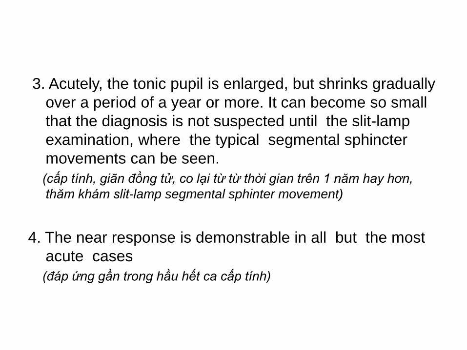
3. Acutely, the tonic pupil is enlarged, but shrinks gradually
over a period of a year or more. It can become so small
that the diagnosis is not suspected until the slit-lamp
examination, where the typical segmental sphincter
movements can be seen.
(cấp tính, giãn đồng tử, co lại từ từ thời gian trên 1 năm hay hơn,
thăm khám slit-lamp segmental sphinter movement)
4. The near response is demonstrable in all but the most
acute cases
(đáp ứng gần trong hầu hết ca cấp tính)
Nguyên nhân
Most commonly, the affected patients are women between 30 and 40 years of age
In a few cases, the cause can be established, or at least confidently suspected:
- after orbital trauma,(sau chấn thương hốc mắt)
- extensive panretinal photocoagulation (quang đông),
- outbreaks of varicella zoster, (siêu vi)
- orbital ischemia with active giant cell arteritis, (viêm đm đại bào)
- and only rarely associated with a malignancy and a suspected paraneoplastic syndrome.(cận ung thư)
testing of the erythrocyte sedimentation rate(VS)
or of C-reactive protein(CRP) levels, since giant
cell arteritis can present in this way
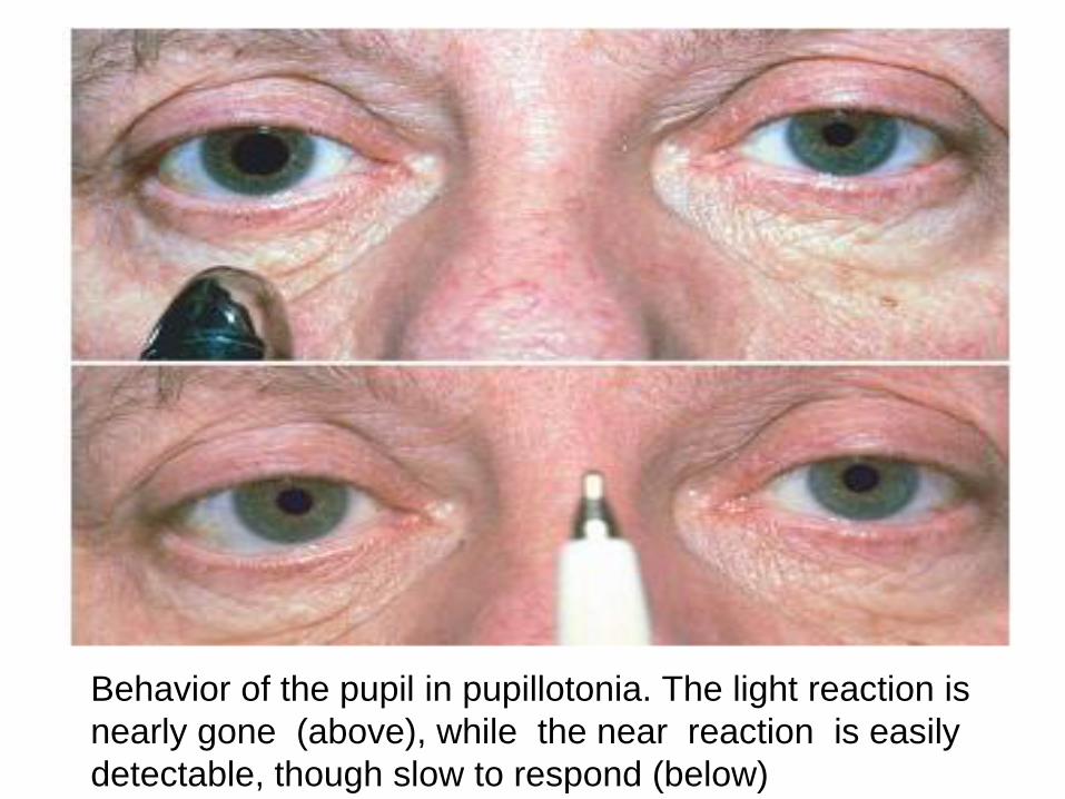
Behavior of the pupil in pupillotonia. The light reaction is
nearly gone (above), while the near reaction is easily
detectable, though slow to respond (below)
Liên hệ đồng tử trong liệt dây III
1. damage to the third cranial nerve at locations
between the oculomotor nucleus and the ciliary
ganglion.(tổn thương từ nhân dây III và hạch mi)
2. pupillary mydriasis caused by internal
ophthalmoplegia as part of a third nerve palsy
indicates a high probability of a compressive
mechanism, such as by a tumor or an aneurysm
(giãn đồng tử trong liệt dây III: chèn ép, u hay
phình mạch….)
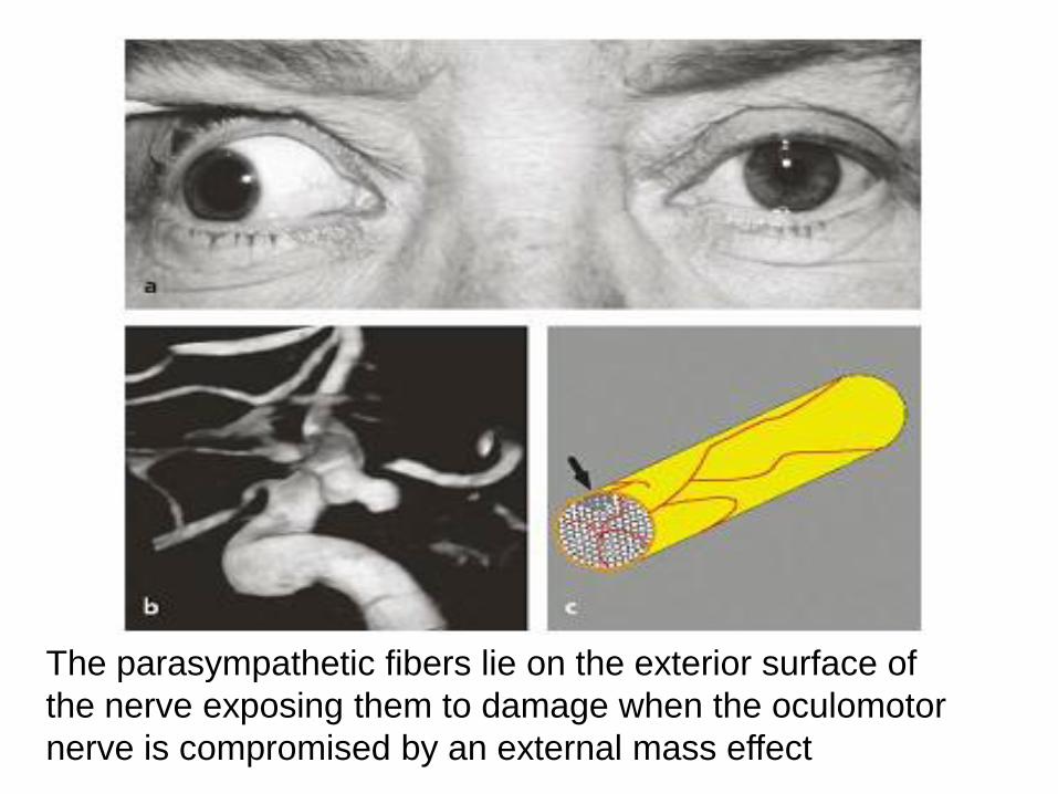
The parasympathetic fibers lie on the exterior surface of
the nerve exposing them to damage when the oculomotor
nerve is compromised by an external mass effect
Tổn thương vùng lưng não giữa- hc Parinaud
(Lesions of the Dorsal Midbrain: Parinaud’s Syndrome)
1. deficit in the light reactions of the pupil
2. retained accommodative miosis, and loss of connection to
the Edinger-Westphal nucleus and the final common
pathway of third nerve function
3. the clinical presentation of this syndrome includes loss of
upward saccadic movements, and convergence retraction
nystagmus.
mất pxas
phân ly đáp ứng ánh sáng-gần
mất saccade nhìn lên
convergence retraction nystagmus
Tổn thương mống mắt (Iris)
1. identifed at the slit lamp, can find even subtle tears in
the iris sphincter
2. An attack of angle closure glaucoma typically causes a
mid-dilated pupil that is unresponsive to a light stimulus
3. pharmacologic pupil can be confirmed by instillation of
1% pilocarpine, which will have no effect on the size of
the pupil.
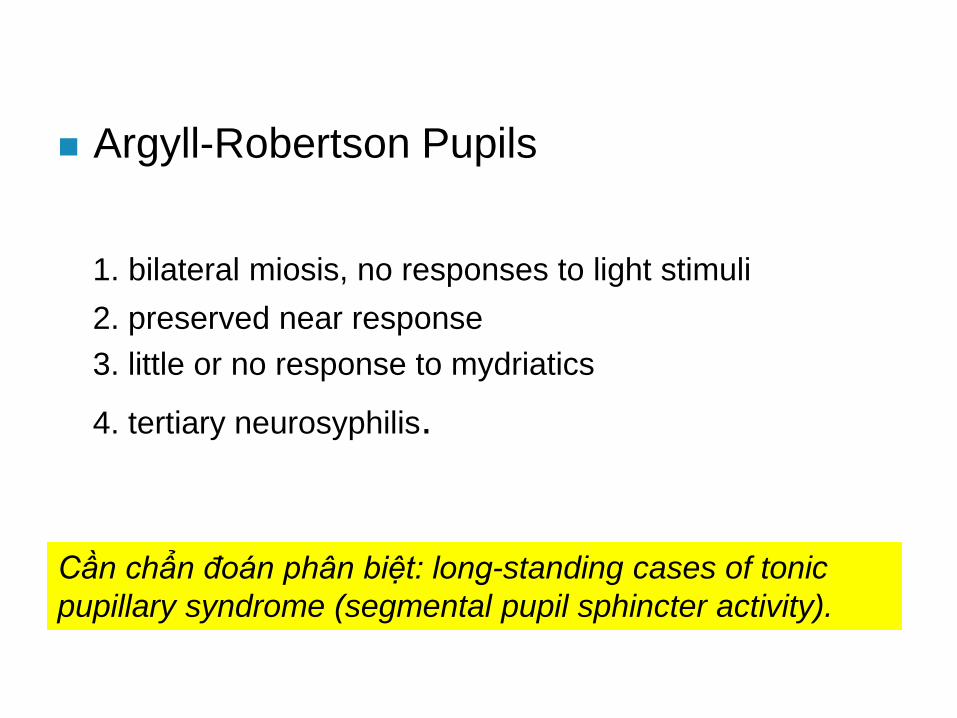
Argyll-Robertson Pupils
1. bilateral miosis, no responses to light stimuli
2. preserved near response
3. little or no response to mydriatics
4. tertiary neurosyphilis.
Cần chẩn đoán phân biệt: long-standing cases of tonic
pupillary syndrome (segmental pupil sphincter activity).
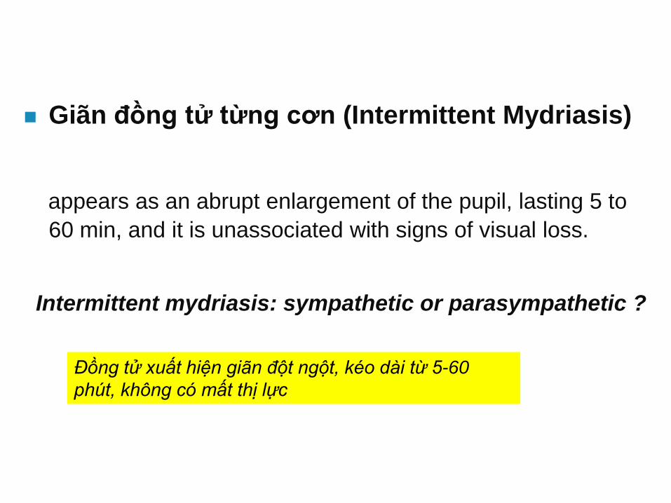
Giãn đồng tử từng cơn (Intermittent Mydriasis)
appears as an abrupt enlargement of the pupil, lasting 5 to
60 min, and it is unassociated with signs of visual loss.
Intermittent mydriasis: sympathetic or parasympathetic ?
Đồng tử xuất hiện giãn đột ngột, kéo dài từ 5-60
phút, không có mất thị lực
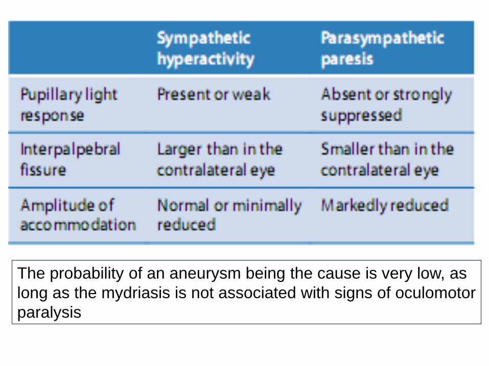
The probability of an aneurysm being the cause is very low, as
long as the mydriasis is not associated with signs of oculomotor
paralysis
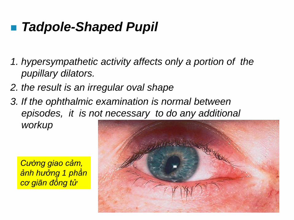
Tadpole-Shaped Pupil
1. hypersympathetic activity affects only a portion of the
pupillary dilators.
2. the result is an irregular oval shape
3. If the ophthalmic examination is normal between
episodes, it is not necessary to do any additional
workup
Cường giao cảm,
ảnh hưởng 1 phần
cơ giãn đồng tử
Paradoxical Pupils(đồng tư nghich ly)
1. response to a light (constriction in the dark) has been
associated with heredofamilial retinal dystrophies.
Co đồng tử bẩm sinh và giãn đồng tử dai dẳng
(Congenital Miosis and Persistent Mydriasis)
1. two anomalies of pupillary size are of clinical
importance, one is congenital miosis, and the other is a
persistent mydriasis
2. develops during the second or third decade of age
Đồng tử dao động(Oscillations of the Pupil)
1. illumination has continuous oscillatory movement
2. pupillary unrest
3. physiologic phenomenon.
4. In the past, and still used in current publications, this has
been called hippus (from the Greek word for “horse”)
Còn dùng từ hippus (tiếng Hy lạp: ngựa)
Chiếu sáng vào đồng tử dao động liên tục, không nghỉ
Hiện tượng sinh lý
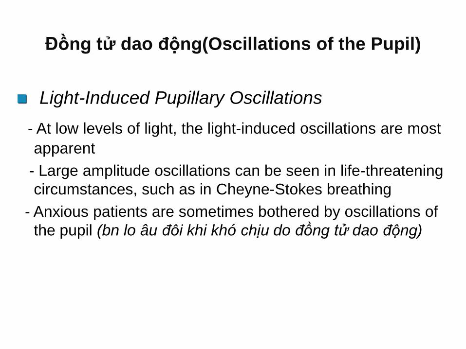
Đồng tử dao động(Oscillations of the Pupil)
Light-Induced Pupillary Oscillations
- At low levels of light, the light-induced oscillations are most
apparent
- Large amplitude oscillations can be seen in life-threatening
circumstances, such as in Cheyne-Stokes breathing
- Anxious patients are sometimes bothered by oscillations of
the pupil (bn lo âu đôi khi khó chịu do đồng tử dao động)
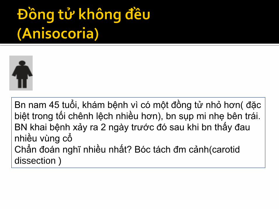
Bn nam 45 tuổi, khám bệnh vì có một đồng tử nhỏ hơn( đặc
biệt trong tối chênh lệch nhiều hơn), bn sụp mi nhẹ bên trái.
BN khai bệnh xảy ra 2 ngày trước đó sau khi bn thấy đau
nhiều vùng cổ
Chẩn đoán nghĩ nhiều nhất? Bóc tách đm cảnh(carotid
dissection )
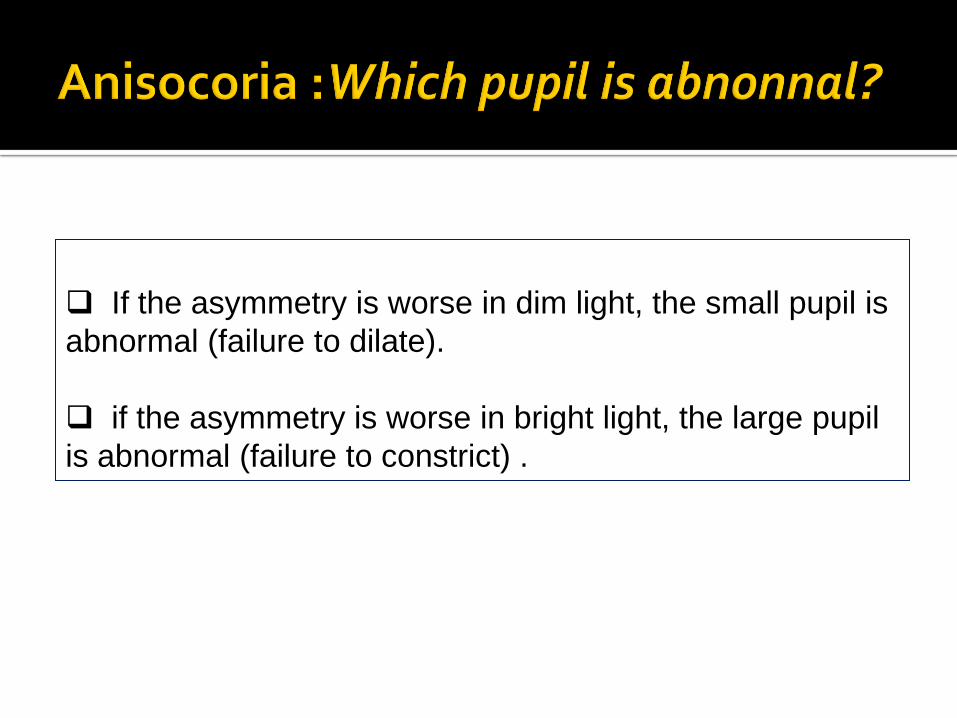
If the asymmetry is worse in dim light, the small pupil is
abnormal (failure to dilate).
if the asymmetry is worse in bright light, the large pupil
is abnormal (failure to constrict) .
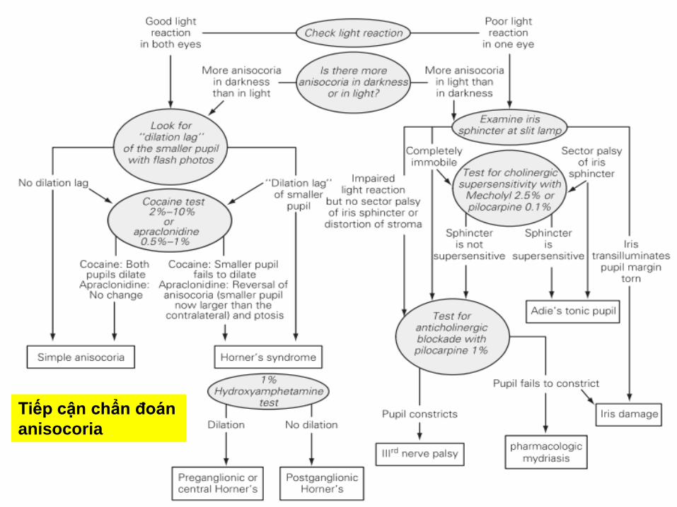
Tiếp cận chẩn đoán
anisocoria
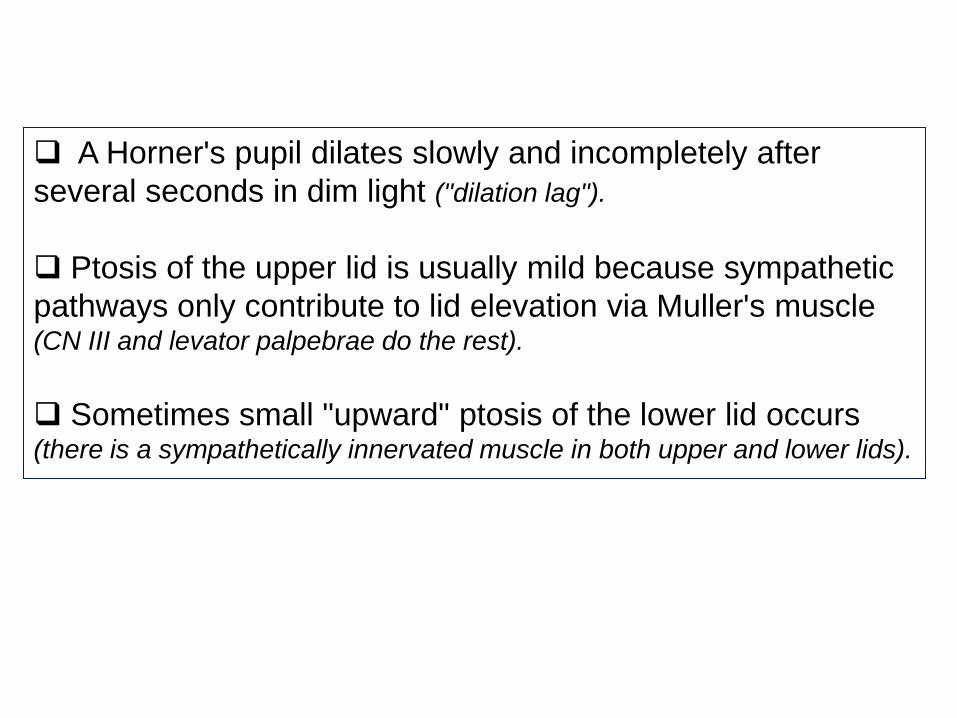
A Horner's pupil dilates slowly and incompletely after
several seconds in dim light ("dilation lag").
Ptosis of the upper lid is usually mild because sympathetic
pathways only contribute to lid elevation via Muller's muscle (CN III and levator palpebrae do the rest).
Sometimes small "upward" ptosis of the lower lid occurs (there is a sympathetically innervated muscle in both upper and lower lids).
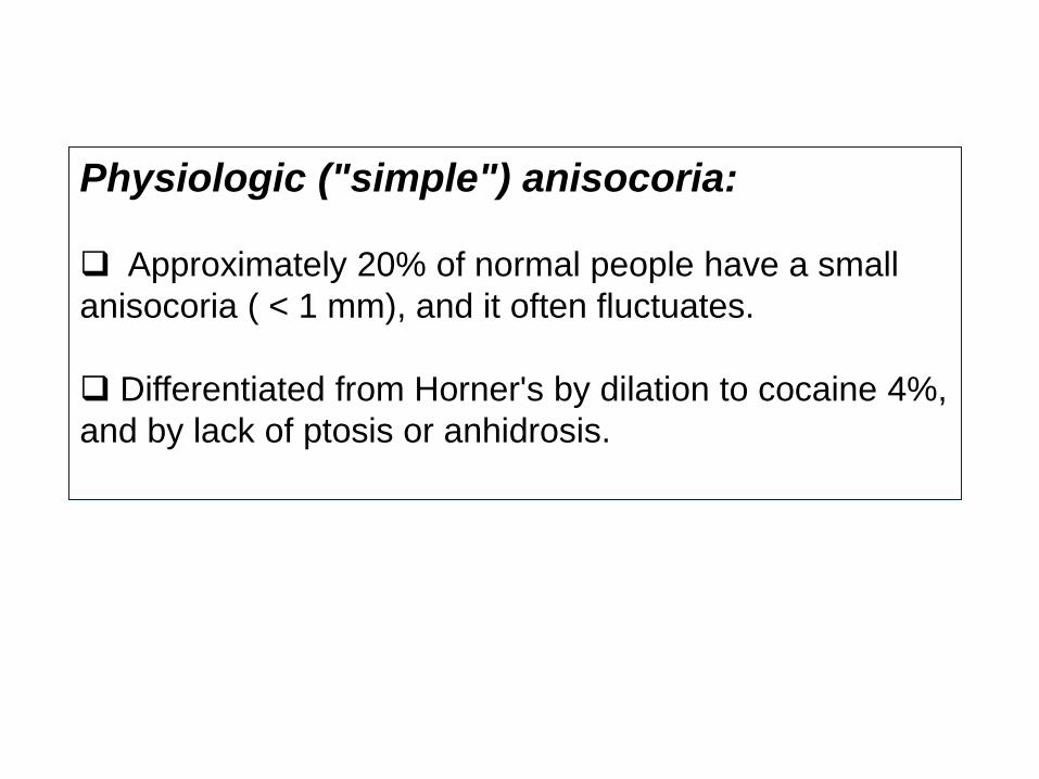
Physiologic ("simple") anisocoria:
Approximately 20% of normal people have a small
anisocoria ( < 1 mm), and it often fluctuates.
Differentiated from Horner's by dilation to cocaine 4%,
and by lack of ptosis or anhidrosis.
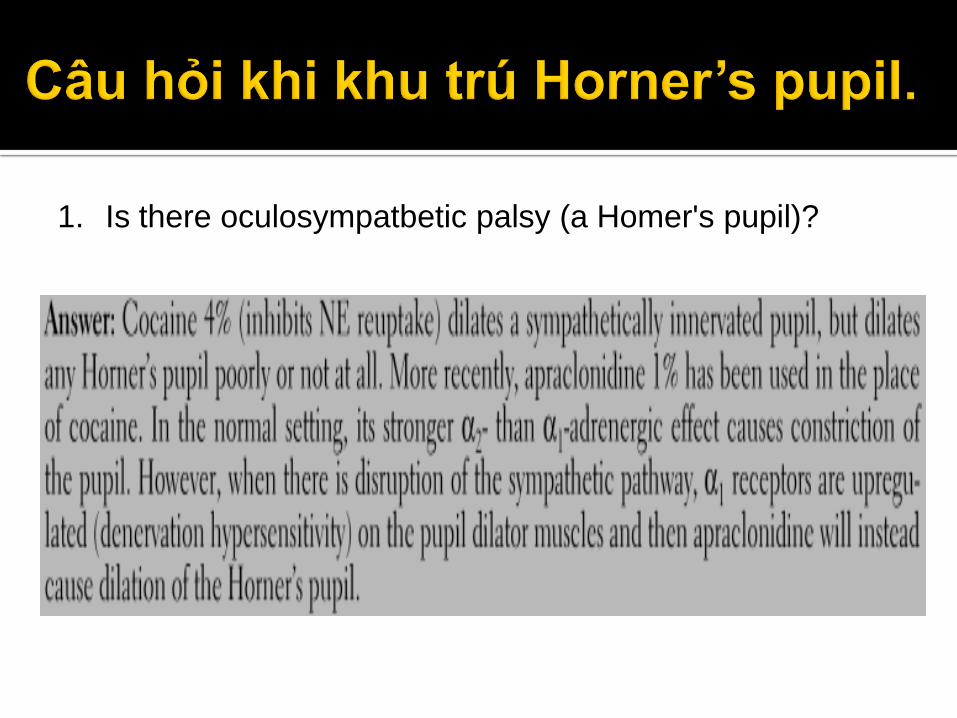
1. Is there oculosympatbetic palsy (a Homer's pupil)?
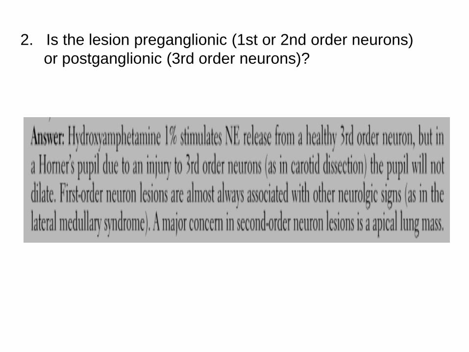
2. Is the lesion preganglionic (1st or 2nd order neurons)
or postganglionic (3rd order neurons)?
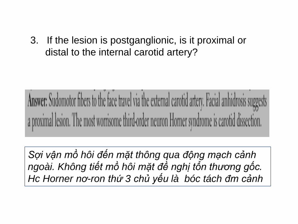
3. If the lesion is postganglionic, is it proximal or
distal to the internal carotid artery?
Sợi vận mồ hôi đến mặt thông qua động mạch cảnh
ngoài. Không tiết mồ hôi mặt đề nghị tổn thương gốc.
Hc Horner nơ-ron thứ 3 chủ yếu là bóc tách đm cảnh

Bn nữ 32 tuổi than phiền đồng tử giãn bên phải, bn ghi nhân
sau khi soi gương. BN không có triệu chứng gì khác. Khám
không có sụp mi, vận động mắt bình thường, đồng tử mất
đáp ứng với ánh sáng bên(p). Các test tại chổ tiếp theo?
Chẩn đoán nếu đồng tử co lai khi nhìn hội tụ.
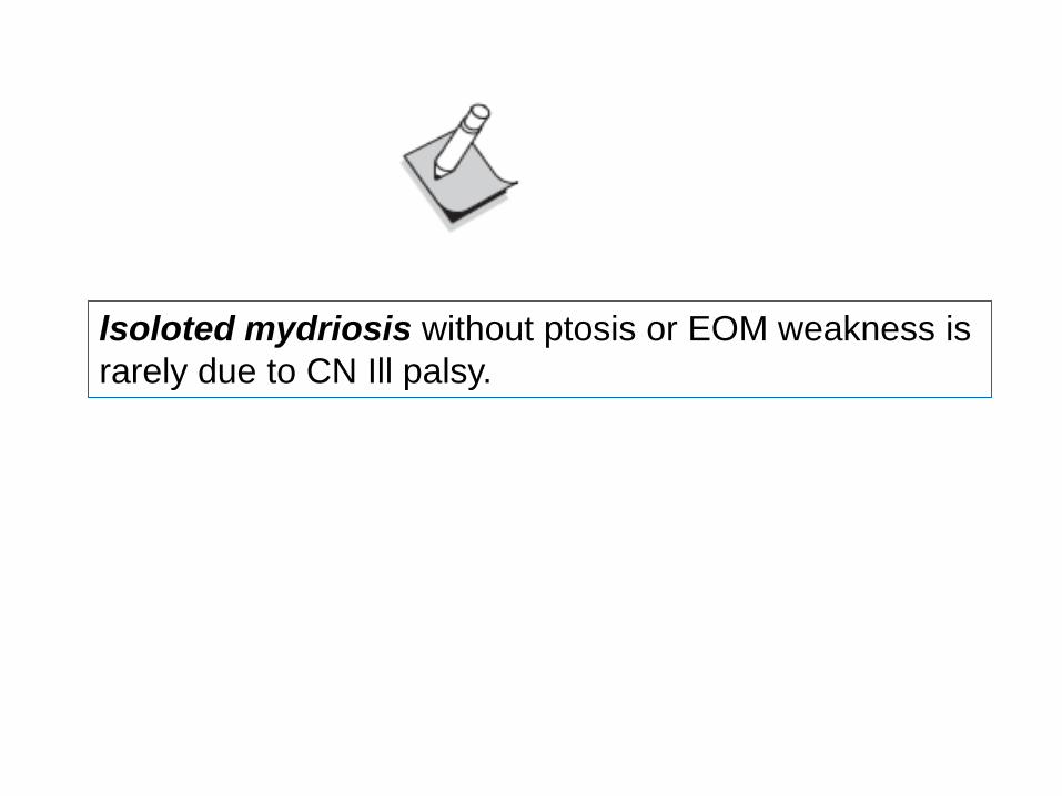
lsoloted mydriosis without ptosis or EOM weakness is
rarely due to CN Ill palsy.
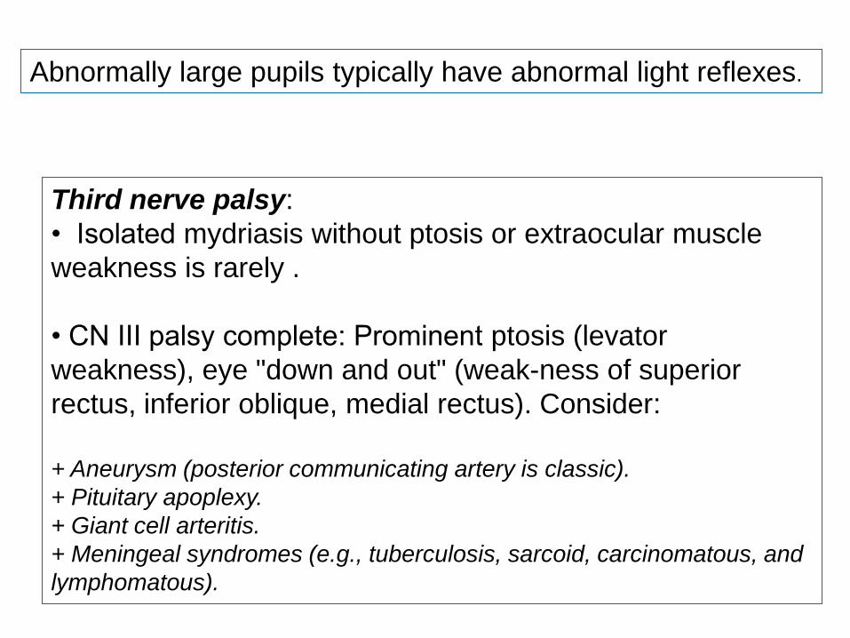
Abnormally large pupils typically have abnormal light reflexes.
Third nerve palsy:
• Isolated mydriasis without ptosis or extraocular muscle
weakness is rarely .
• CN III palsy complete: Prominent ptosis (levator
weakness), eye "down and out" (weak-ness of superior
rectus, inferior oblique, medial rectus). Consider:
+ Aneurysm (posterior communicating artery is classic).
+ Pituitary apoplexy.
+ Giant cell arteritis.
+ Meningeal syndromes (e.g., tuberculosis, sarcoid, carcinomatous, and
lymphomatous).
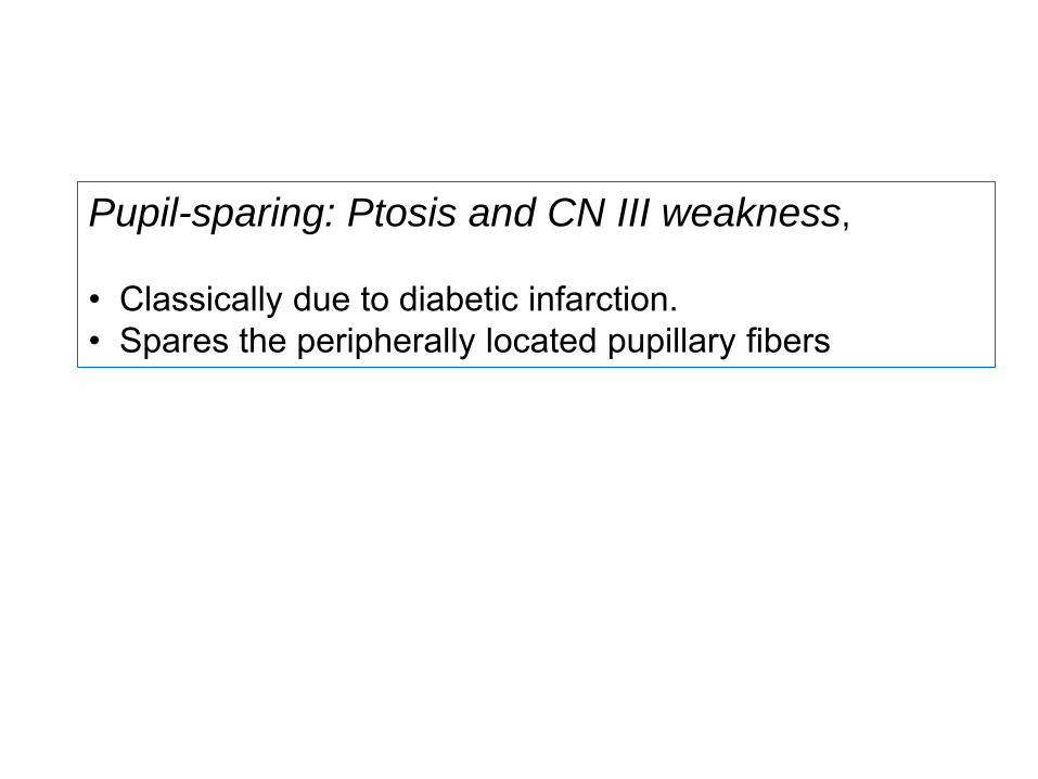
Pupil-sparing: Ptosis and CN III weakness,
• Classically due to diabetic infarction.
• Spares the peripherally located pupillary fibers
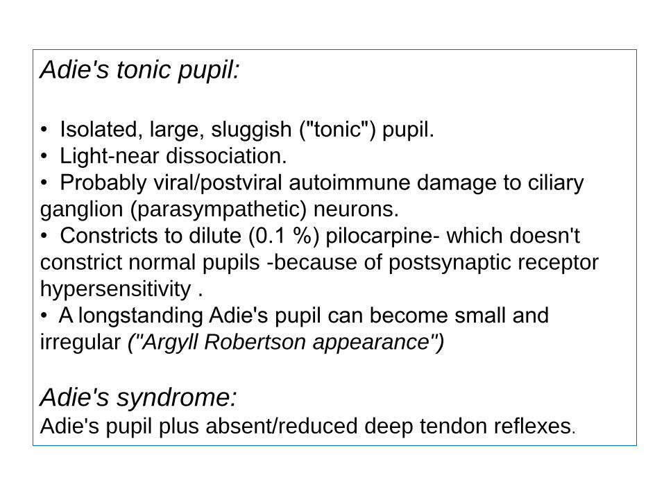
Adie's tonic pupil:
• Isolated, large, sluggish ("tonic") pupil.
• Light-near dissociation.
• Probably viral/postviral autoimmune damage to ciliary
ganglion (parasympathetic) neurons.
• Constricts to dilute (0.1 %) pilocarpine- which doesn't
constrict normal pupils -because of postsynaptic receptor
hypersensitivity .
• A longstanding Adie's pupil can become small and
irregular ("Argyll Robertson appearance")
Adie's syndrome: Adie's pupil plus absent/reduced deep tendon reflexes.
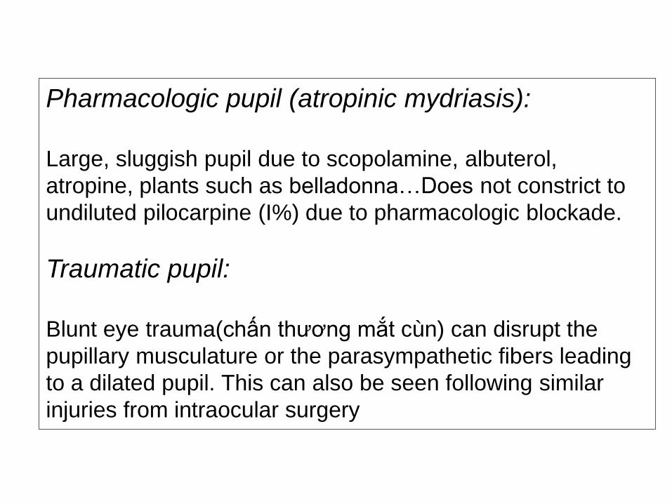
Pharmacologic pupil (atropinic mydriasis):
Large, sluggish pupil due to scopolamine, albuterol,
atropine, plants such as belladonna…Does not constrict to
undiluted pilocarpine (I%) due to pharmacologic blockade.
Traumatic pupil:
Blunt eye trauma(chấn thương mắt cùn) can disrupt the
pupillary musculature or the parasympathetic fibers leading
to a dilated pupil. This can also be seen following similar
injuries from intraocular surgery
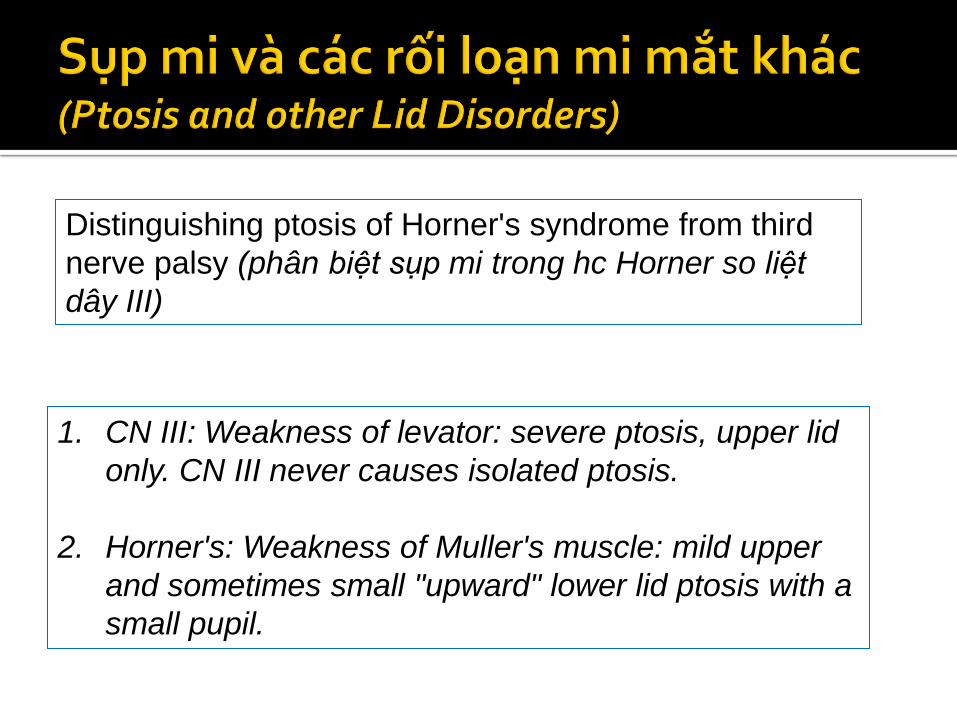
Distinguishing ptosis of Horner's syndrome from third
nerve palsy (phân biệt sụp mi trong hc Horner so liệt
dây III)
1. CN III: Weakness of levator: severe ptosis, upper lid
only. CN III never causes isolated ptosis.
2. Horner's: Weakness of Muller's muscle: mild upper
and sometimes small "upward" lower lid ptosis with a
small pupil.
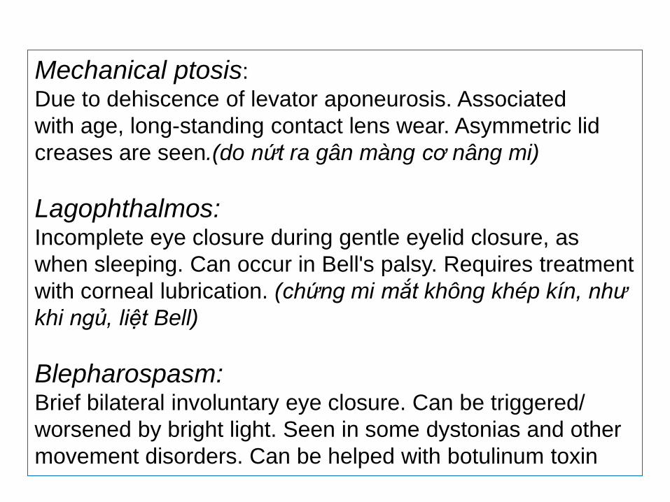
Mechanical ptosis:
Due to dehiscence of levator aponeurosis. Associated
with age, long-standing contact lens wear. Asymmetric lid
creases are seen.(do nứt ra gân màng cơ nâng mi)
Lagophthalmos: Incomplete eye closure during gentle eyelid closure, as
when sleeping. Can occur in Bell's palsy. Requires treatment
with corneal lubrication. (chứng mi mắt không khép kín, như
khi ngủ, liệt Bell)
Blepharospasm: Brief bilateral involuntary eye closure. Can be triggered/
worsened by bright light. Seen in some dystonias and other
movement disorders. Can be helped with botulinum toxin
Eyelid apraxia (thất điều mi mắt):
Most associated with blepharospasm, seen in other conditions
(Parkinson's, progressive supranuclear palsy). Difficulty opening the
eyelids, which appear only gently closed. Patients elevate eyebrows and
forehead in an attempt to open the eyes.
Lid lag(chậm trễ mi mắt):
While pursuing a visual target moving slowly from superior to inferior, the lid
will lag slightly behind its normal position (in normal patients the lid is
always at the limbus (đường biên) ready to protect the cornea). Associated
with thyroid eye disease
Lid twitch(giật mi mắt):
"Cogan's lid twitch" can be seen in myasthenia gravis. After looking
downward, when gaze returns to midposition the lid "jumps" higher before
settling into position. (Resting the levator allows brief return of nonnal
function in a myasthenic.)
Upgaze paralysis(liệt nhìn lên):
occurs with lesions of the posterior commisure or pretectal
area and is part of Parinaud's dorsal midbrain syndrome
(upgaze palsy),
+ Lid retraction,
+ Light-near dissociation,
+ Convergence-retraction nystagmus.
Downgaze paralysis(liệt nhìn xuống):
occurs with bilateral lesions in the rostral interstitial
nucleus of the MLF
Căn nguyên:
1. Vascular supply: Posterior thalamosubthalamic
paramedian branch of the PCA.
(nhánh sau cận đường giữa đồi hạ đồi)
2. Can occur in association with thalamic infarcts.
3. Up-and downgaze palsies seen in Whipple's disease,
progressive supranuclear palsy, diffuse upper brain
stem disorders.
Skew deviation (lệch nghiêng):
Vertical deviation of the eyes not caused by infranuclear
lesions (e.g., CN III or IV palsy).
Caused by lesions in the cerebellum, brain stem, and
vestibulo-ocular pathways.
Most common in lateral medullary syndromes in
which vestibular pathways are affected.
Ocular tilt reaction (OTR) (phản ứng nghiêng
nhãn cầu):
Normally when tilting the head, both eyes counter-roll in the
opposite direction (contralateral eye falls, ipsilateral eye
rises). (Bình thường khi nghiêng đầu, cả 2 mắt quay(lăn)cyclotorsion
ngược lại hướng đối bên, mắt đối bện rơi xuống, cùng bên nâng lên)
The OTR (abnormal) is a triad of spontaneous
(1) skew deviation,
(2) cyclotorsion of both eyes,
(3) paradoxical head tilt toward the lower eye.
Caused by damage anywhere along the vestibular
pathways/connections.

Cảm ơn sự theo dõi