Embed Size (px)
Citation preview
1
doi: 10.2169/internalmedicine.1220-18
Intern Med Advance Publication
http://internmed.jp
【 CASE REPORT 】
Miliary Lung Metastases from Genital Large CellNeuroendocrine Carcinomas
Akimasa Sekine 1, Makiko Satoh 2,3, Koji Okudela 4, Mai Matsumura 4, Yukio Morishita 5,
Yuko Minami 6, Kenji Hayashihara 6, Takefumi Saito 6, Tae Iwasawa 7 and Takashi Ogura 1
Abstract:We herein report two cases of miliary lung metastases from genital carcinoma in uterine cervix and en-
dometrium. Notably, these patients were unable to receive any anti-tumor chemotherapy due to rapid progres-
sion causing respiratory failure, and they ultimately died of disease progression within only a month after the
first visit to our hospitals. A postmortem examination confirmed the diagnosis of genital large-cell neuroen-
docrine carcinoma (LCNEC). Chest physicians should be aware of genital LCNEC with a dismal prognostic
entity as an important differential diagnosis of miliary lung metastases.
Key words: large-cell neuroendocrine carcinoma, miliary lung metastases, uterus, cervix, corpus
(Intern Med Advance Publication)(DOI: 10.2169/internalmedicine.1220-18)
Introduction
Miliary lung metastases are most frequently seen in lung
cancer (1, 2), although previous studies report that they can
rarely occur from malignancies of other organs, such as thy-
roid (3-6) and stomach (7). However, there are no current
reports of miliary lung metastases from genital carcinomas.
We herein report two cases of miliary lung metastases from
genital large-cell neuroendocrine carcinomas (LCNEC).
Case Presentation
Case 1
A 32-year-old never-smoking woman visited the previous
hospital due to persistent cough occurring for a month.
Chest radiography revealed multiple pulmonary nodules,
raising the suspicion of miliary tuberculosis (Fig. 1A). The
patient was referred to our hospital on the same day already
presenting with respiratory failure with a PaO2 of 46.8 Torr
on room air. Chest computed tomography (CT) showed tiny,
miliary nodules throughout the bilateral lungs (Fig. 1B). The
patient also had metastases to the liver, right kidney, adrenal
glands, and bones. The tumor markers were elevated with
CEA of 10.3 ng/mL, CYFRA of 264.0 ng/ml, and CA125
of 75 U/mL. Since her detailed medical history included a
one-month history of metrorrhagia, pelvic magnetic reso-
nance imaging (MRI) was performed. MRI revealed a swol-
len cervix of the uterus with an obscure boundary between
the normal myometrium and the tumor, which had invaded
the corpus uteri (Fig. 1C). The patient was then diagnosed
with stage 4 uterine cervical cancer.
Since the performance status was already 4 and the pa-
tient was suffering from severe dyspnea, it was not possible
to perform an invasive pathological evaluation or to adminis-
ter any anti-tumor agents. The patient ultimately died of dis-
ease progression 13 days after admission to our hospital.
A necropsy of the lungs and uterus was performed; both
specimens presented microscopic uniform, round-shaped
large tumor cells with moderate cytoplasm and nuclei of
granular chromatin pattern forming solid nests and cords
1Department of Respiratory medicine, Kanagawa Cardiovascular and Respiratory Center, Japan, 2Department of Obstetrics and Gynecology,
Graduate School of Medicine, Yokohama City University, Japan, 3Department of Obstetrics and Gynecology, Aomori City Hospital, Japan, 4De-
partment of Pathology, Graduate School of Medicine, Yokohama City University, Japan, 5Diagnostic Pathology Division, Tokyo Medical Univer-
sity Ibaraki Medical Center, Japan, 6Department of Respiratory medicine, National Hospital Organization, Ibarakihigashi National Hospital,
Japan and 7Department of Radiology, Kanagawa Cardiovascular and Respiratory Center, Japan
Received: March 20, 2018; Accepted: September 24, 2018; Advance Publication by J-STAGE: December 18, 2018
Correspondence to Dr. Akimasa Seine, [email protected]
Intern Med Advance Publication DOI: 10.2169/internalmedicine.1220-18
2
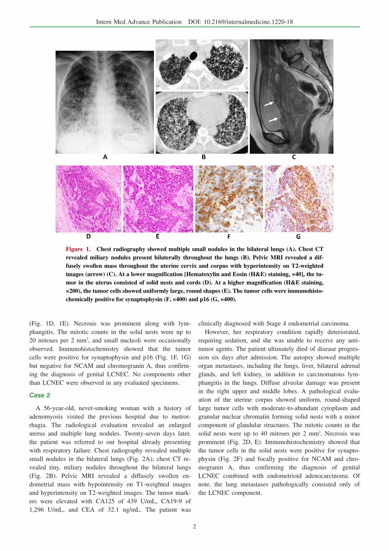
Figure 1. Chest radiography showed multiple small nodules in the bilateral lungs (A). Chest CT revealed miliary nodules present bilaterally throughout the lungs (B). Pelvic MRI revealed a dif-fusely swollen mass throughout the uterine cervix and corpus with hyperintensity on T2-weighted images (arrow) (C). At a lower magnification [Hematoxylin and Eosin (H&E) staining, ×40], the tu-mor in the uterus consisted of solid nests and cords (D). At a higher magnification (H&E staining, ×200), the tumor cells showed uniformly large, round shapes (E). The tumor cells were immunohisto-chemically positive for synaptophysin (F, ×400) and p16 (G, ×400).
(Fig. 1D, 1E). Necrosis was prominent along with lym-
phangitis. The mitotic counts in the solid nests were up to
20 mitoses per 2 mm2, and small nucleoli were occasionally
observed. Immunohistochemistry showed that the tumor
cells were positive for synaptophysin and p16 (Fig. 1F, 1G)
but negative for NCAM and chromogranin A, thus confirm-
ing the diagnosis of genital LCNEC. No components other
than LCNEC were observed in any evaluated specimens.
Case 2
A 56-year-old, never-smoking woman with a history of
adenomyosis visited the previous hospital due to metror-
rhagia. The radiological evaluation revealed an enlarged
uterus and multiple lung nodules. Twenty-seven days later,
the patient was referred to our hospital already presenting
with respiratory failure. Chest radiography revealed multiple
small nodules in the bilateral lungs (Fig. 2A); chest CT re-
vealed tiny, miliary nodules throughout the bilateral lungs
(Fig. 2B). Pelvic MRI revealed a diffusely swollen en-
dometrial mass with hypointensity on T1-weighted images
and hyperintensity on T2-weighted images. The tumor mark-
ers were elevated with CA125 of 439 U/mL, CA19-9 of
1,296 U/mL, and CEA of 32.1 ng/mL. The patient was
clinically diagnosed with Stage 4 endometrial carcinoma.
However, her respiratory condition rapidly deteriorated,
requiring sedation, and she was unable to receive any anti-
tumor agents. The patient ultimately died of disease progres-
sion six days after admission. The autopsy showed multiple
organ metastases, including the lungs, liver, bilateral adrenal
glands, and left kidney, in addition to carcinomatous lym-
phangitis in the lungs. Diffuse alveolar damage was present
in the right upper and middle lobes. A pathological evalu-
ation of the uterine corpus showed uniform, round-shaped
large tumor cells with moderate-to-abundant cytoplasm and
granular nuclear chromatin forming solid nests with a minor
component of glandular structures. The mitotic counts in the
solid nests were up to 40 mitoses per 2 mm2. Necrosis was
prominent (Fig. 2D, E). Immunohistochemistry showed that
the tumor cells in the solid nests were positive for synapto-
physin (Fig. 2F) and focally positive for NCAM and chro-
mogranin A, thus confirming the diagnosis of genital
LCNEC combined with endometrioid adenocarcinoma. Of
note, the lung metastases pathologically consisted only of
the LCNEC component.
Intern Med Advance Publication DOI: 10.2169/internalmedicine.1220-18
3
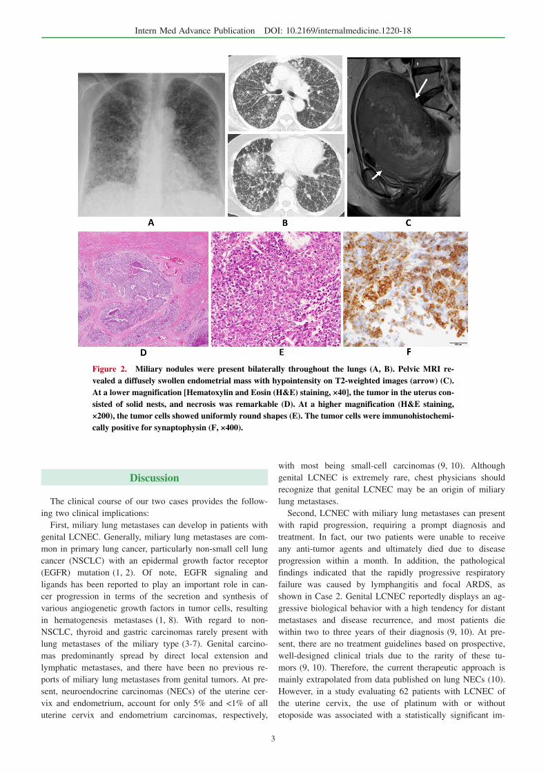
Figure 2. Miliary nodules were present bilaterally throughout the lungs (A, B). Pelvic MRI re-vealed a diffusely swollen endometrial mass with hypointensity on T2-weighted images (arrow) (C). At a lower magnification [Hematoxylin and Eosin (H&E) staining, ×40], the tumor in the uterus con-sisted of solid nests, and necrosis was remarkable (D). At a higher magnification (H&E staining, ×200), the tumor cells showed uniformly round shapes (E). The tumor cells were immunohistochemi-cally positive for synaptophysin (F, ×400).
Discussion
The clinical course of our two cases provides the follow-
ing two clinical implications:
First, miliary lung metastases can develop in patients with
genital LCNEC. Generally, miliary lung metastases are com-
mon in primary lung cancer, particularly non-small cell lung
cancer (NSCLC) with an epidermal growth factor receptor
(EGFR) mutation (1, 2). Of note, EGFR signaling and
ligands has been reported to play an important role in can-
cer progression in terms of the secretion and synthesis of
various angiogenetic growth factors in tumor cells, resulting
in hematogenesis metastases (1, 8). With regard to non-
NSCLC, thyroid and gastric carcinomas rarely present with
lung metastases of the miliary type (3-7). Genital carcino-
mas predominantly spread by direct local extension and
lymphatic metastases, and there have been no previous re-
ports of miliary lung metastases from genital tumors. At pre-
sent, neuroendocrine carcinomas (NECs) of the uterine cer-
vix and endometrium, account for only 5% and <1% of all
uterine cervix and endometrium carcinomas, respectively,
with most being small-cell carcinomas (9, 10). Although
genital LCNEC is extremely rare, chest physicians should
recognize that genital LCNEC may be an origin of miliary
lung metastases.
Second, LCNEC with miliary lung metastases can present
with rapid progression, requiring a prompt diagnosis and
treatment. In fact, our two patients were unable to receive
any anti-tumor agents and ultimately died due to disease
progression within a month. In addition, the pathological
findings indicated that the rapidly progressive respiratory
failure was caused by lymphangitis and focal ARDS, as
shown in Case 2. Genital LCNEC reportedly displays an ag-
gressive biological behavior with a high tendency for distant
metastases and disease recurrence, and most patients die
within two to three years of their diagnosis (9, 10). At pre-
sent, there are no treatment guidelines based on prospective,
well-designed clinical trials due to the rarity of these tu-
mors (9, 10). Therefore, the current therapeutic approach is
mainly extrapolated from data published on lung NECs (10).
However, in a study evaluating 62 patients with LCNEC of
the uterine cervix, the use of platinum with or without
etoposide was associated with a statistically significant im-
Intern Med Advance Publication DOI: 10.2169/internalmedicine.1220-18
4
provement in the overall survival compared with other regi-
mens (11). When interpreting our two cases in light of those
reports, we propose that although the prognosis for patients
with miliary lung metastases from genital LCNEC is poor, it
can potentially be improved through treatment with platinum
agents. Therefore, taking a detailed medical history empha-
sizing symptoms such as metrorrhagia is crucial for achiev-
ing a prompt diagnosis and treatment, particularly in women
with miliary lung metastases. In addition, a tumor biopsy of
the uterus or lung should be immediately performed, as the
disease progresses very rapidly once respiratory failure de-
velops.
In conclusion, we encountered two cases of miliary lung
metastases from genital LCNEC. Although rare, since this
condition can progress rapidly, chest physicians should be
aware of genital LCNEC as a potential origin of miliary
lung metastases in order to initiate aggressive treatment
promptly.
The authors state that they have no Conflict of Interest (COI).
References
1. Togashi Y, Masago K, Kubo T, et al. Association of diffuse, ran-
dom pulmonary metastases, including miliary metastases, with epi-
dermal growth factor receptor mutations in lung adenocarcinoma.
Cancer 117: 819-825, 2011.
2. Wu SG, Hu FC, Chang YL, et al. Frequent EGFR Mutations in
NSCLC Presenting with Miliary Intrapulmonary Carcinomatosis.
Eur Respir J 41: 417-424, 2012.
3. Vahidova D, Persaud R, Kirsch C, et al. Radiology quiz case 1.
Papillary thyroid carcinoma with miliary lung metastases. Arch
Otolaryngol Head Neck Surg 130: 1442-1446, 2004.
4. Chariot P, Feliz A, Monnet I. Miliary opacities diagnosed as lung
metastases of a thyroid carcinoma after 13 years of stability. Chest
104: 981-982, 1993.
5. Scott JX, Gnananayagam JE, Sundaravalli EK, et al. Unusual
cause for miliary lung mottling in a child. Indian J Chest Dis Al-
lied Sci 46: 291-293, 2004.
6. Vermeer-Mens JC, Goemaere NN, Kuenen-Boumeester V, et al.
Childhood papillary thyroid carcinoma with miliary pulmonary
metastases. J Clin Oncol 24: 5788-5789, 2006.
7. Ku YH, Lee KW, Kim JH, et al. Metastatic gastric carcinoma in a
19-year-old man. J Clin Oncol 25: 5026-5028, 2007.
8. Mitsudomi T, Yatabe Y. Epidermal growth factor receptor in rela-
tion to tumor development: EGFR gene and cancer. FEBS J 277:
301-308, 2010.
9. Kobayashi A, Yahata T, Nanjo S, et al. Rapidly progressing large-
cell neuroendocrine carcinoma arising from the uterine corpus: A
case report and review of the literature. Mol Clin Oncol 6: 881-
885, 2017.
10. Gadducci A, Carinelli S, Aletti G. Neuroendrocrine tumors of the
uterine cervix: A therapeutic challenge for gynecologic oncolo-
gists. Gynecol Oncol 144: 637-646, 2017.
11. Embry JR, Kelly MG, Post MD, et al. Large cell neuroendocrine
carcinoma of the cervix: prognostic factors and survival advantage
with platinum chemotherapy. Gynecol Oncol 120: 444-448, 2011.
The Internal Medicine is an Open Access journal distributed under the Creative
Commons Attribution-NonCommercial-NoDerivatives 4.0 International License. To
view the details of this license, please visit (https://creativecommons.org/licenses/
by-nc-nd/4.0/).
Ⓒ The Japanese Society of Internal Medicine
Intern Med Advance Publication