Embed Size (px)
Citation preview

Multiscale imaging of colitis in miceusing confocal laser endomicroscopy,light-sheet fluorescence microscopy,and magnetic resonance imaging
Tianmeng LiHui HuiChaoen HuHe MaXin YangJie Tian
Tianmeng Li, Hui Hui, Chaoen Hu, He Ma, Xin Yang, Jie Tian, “Multiscale imaging of colitis in miceusing confocal laser endomicroscopy, light-sheet fluorescence microscopy, and magnetic resonanceimaging,” J. Biomed. Opt. 24(1), 016003 (2019), doi: 10.1117/1.JBO.24.1.016003.
Downloaded From: https://www.spiedigitallibrary.org/journals/Journal-of-Biomedical-Optics on 24 Nov 2021Terms of Use: https://www.spiedigitallibrary.org/terms-of-use

Multiscale imaging of colitis in mice using confocallaser endomicroscopy, light-sheet fluorescencemicroscopy, and magnetic resonance imaging
Tianmeng Li,a,b,c,† Hui Hui,b,c,d,† Chaoen Hu,b,c He Ma,a,* Xin Yang,b,c,* and Jie Tianb,c,d,*aNortheastern University, Sino-Dutch Biomedical and Information Engineering School, Shenyang, ChinabChinese Academy of Sciences, Institute of Automation, CAS Key Laboratory of Molecular Imaging, Beijing, ChinacInstitute of Automation, Beijing Key Laboratory of Molecular Imaging, Beijing, ChinadUniversity of Chinese Academy of Sciences, Beijing, China
Abstract. The objective of our study is to develop a multimodality approach by combining magnetic resonanceimaging (MRI) and optical imaging methods to assess acute murine colitis at the macro- and microscopic level.In vivoMRI is used to measure the cross-sectional areas of colons at the macroscopic level. Dual-color confocallaser endomicroscopy (CLE) allows in vivo examination of the fluorescently labeled epithelial cells and micro-vessels in the mucosa with a spatial resolution of ∼1.4 μm during ongoing endoscopy. To further validate thestructural changes of the colons in three-dimensions, ex vivo light-sheet fluorescence microscopy (LSFM) isapplied for in-toto imaging of cleared colon sections. MRI, LSFM, and CLE findings are significantly correlatedwith histological scoring (p < 0.01) and the inflammation-associated activity index (p < 0.01). Our multimodalityimaging technique permits visualization of mucosa in colitis at different scales, which can enhance our under-standing of the pathogenesis of inflammatory bowel diseases. © The Authors. Published by SPIE under a Creative CommonsAttribution 4.0 Unported License. Distribution or reproduction of this work in whole or in part requires full attribution of the original publication, including itsDOI. [DOI: 10.1117/1.JBO.24.1.016003]
Keywords: multimodality imaging; confocal laser endomicroscopy; light-sheet fluorescence microscopy; magnetic resonance imag-ing; inflammatory bowel disease.
Paper 180551RR received Sep. 30, 2018; accepted for publication Jan. 15, 2019; published online Jan. 30, 2019.
1 IntroductionInflammatory bowel diseases (IBDs) are chronic inflammatorydiseases with intestinal immune disorders, including Crohn’sdisease (CD) and ulcerative colitis (UC).1 Patients with IBDsshow an increased risk for developing colorectal cancer (CRC),one of the leading causes of death worldwide.2 In the last de-cade, new imaging techniques have been developed for earlydetection of inflammatory lesions, which has resulted in a signifi-cant reduction in the incidences of CRC.3–6 However, the etiologyof IBDs remains unclear due to its heterogeneous disorders withvarious appearances. In the previous study, the inflammation ofUC has been mainly observed in the mucosal and submucosallayers; however, in CD, the inflammation has been scatteredand may be found in all the layers of colon.7 Therefore, toenhance our understanding of the pathogenesis of IBDs, powerfulmultimodality imaging techniques that can assess the colon at themacro- and microscopic level should be developed.
Macroscopic imaging techniques, such as magnetic reso-nance imaging (MRI), are widely used to evaluate the luminalalternations of colitis.8,9 MRI is preferable because it does nothave the ionizing radiation emitted by computed tomography(CT) enterography.10 However, conventional MRI has too lowa resolution to detect mucosal changes at the cellular level. Animaging method, confocal laser endomicroscopy (CLE), pro-vides over 10-fold higher resolution than MRI for in vivotracking of fluorescently labeled epithelial cells and microvessels
of the mucosa.3 In a CLE system, thousands of thin fibers areincorporated in a probe, in which each fiber works as a point scan-ner and pinhole used in a conventional confocal microscope.11 Incontrast to standard white-light endoscopy, the CLE system hasreal-time imaging of the mucosal alterations via the administrationof fluorescent contrast agents to stain cells and vessels in themucosa.12 The lateral resolution of CLE for mouse applicationscan reach up to 1.4 μm with 10-μm optical sectioning and afield of view (FOV) diameter from 240 to 600 μm.
Although CLE provides accurate assessment of mucosa,whole colon section imaging in three-dimensions (3-D) isstill challenging due to its small FOV. Advanced imaging tech-niques, such as light-sheet fluorescence microscopy (LSFM)(also known as ultramicroscopy), allow for in-toto imaging ofthe fluorescently labeled transparent sample.13–16 Candeoet al.17 have developed a virtual unfolding of LSFM datasetfor quantitative analysis of the murine intestine. Gabanyiet al.18 have used intravital multiphoton microscopy to monitorcell dynamics in different layers of the intestinal wall of liveanimals, and a 3-D view of macrophage distribution in thesmall intestine has been visualized by light-sheet microscopy.The aim of this study was to compare and correlate MRI,CLE, and LSFM to analyze their potential as multimodalitytools to assess a chemically induced experimental model ofIBDs at the macro- and microscopic level.
2 Materials and Methods
2.1 Induction of Colitis Model
All animal experiments were approved in accordance with theguidelines of the Institutional Animal Care and Use Committee
*Address all correspondence to He Ma, E-mail: [email protected];Xin Yang, E-mail: [email protected]; Jie Tian, E-mail: [email protected]
†These authors contributed equally to this work.
Journal of Biomedical Optics 016003-1 January 2019 • Vol. 24(1)
Journal of Biomedical Optics 24(1), 016003 (January 2019)
Downloaded From: https://www.spiedigitallibrary.org/journals/Journal-of-Biomedical-Optics on 24 Nov 2021Terms of Use: https://www.spiedigitallibrary.org/terms-of-use

(IACUC) of Peking University, and all procedures were per-formed in accordance with the approved guidelines of IACUCof Peking University. Male C57BL/6 mice (n ¼ 15), six weeksold (Beijing Vital River Laboratory Animal Technology Co.,Ltd.), were kept under standard housing conditions providingwater and food ad libitum. To induce colitis, mice received 3.5%weight/volume dextran sodium sulfate (DSS, 36 to 50 kDa, MPBiomedicals) in their drinking water for three days (n ¼ 5) andsix days (n ¼ 5). Five mice were not treated with DSS and wereused as a control group. DSS-induced model development wasmonitored with weight loss, diarrhea, and stool bleeding.19–21
Prior to MRI and CLE, mice were fasted for 12 h.
2.2 Magnetic Resonance Imaging Assessment
MRI was performed on a 1-Tesla permanent magnet small ani-mal scanner (M3TM Aspect Imaging, Israel). To assess the ana-tomical changes in the mice with IBDs, we used the T1-weighted sequence (TR: 500 ms, TE: 12 ms, 195 μm in planeresolution, flip angle: 90 deg, acquisition matrix: 154 × 154,number of averages: 7, slice thickness: 1 mm, and duration:10 min and 5 s). MRI was started at day zero and repeatedon day three and six after DSS induction. For MRI, micewere anesthetized with 1.5% isoflurane. Animals were kepton a water tube heating pad to keep the body temperatureconstant. Measurement of the diameter and thickness ofthe colons was performed in the ImageJ package FIJI (version1.51).22
2.3 Confocal Laser Endomicroscopy Examination
Prior to CLE (CellVizio Dual Band, Mauna Kea Technologies,France) examination, the mice colon vasculature was stained viaintravenous injection with 100 μL 2% weight/volume Evansblue (MedChemExpress). Subsequently, 100 μL of 0.05%weight/volume acriflavine (Sigma-Aldrich) was administeredtopically for mucosa cell staining.23 Approximately 15 to 20 minafter administering the dyes, CLE was performed by placinga fiber optic probe against the distal colon mucosa of eachmouse. The CellVizio laser was used for confocal imaging;it generates excitation at both 488 and 660 nm and couplesa dual-laser beam into the probe with 2.6-mm tip diameter,1.4-μm lateral resolution, 10-μm optical sectioning, and a60-μm work distance. A CellVizio Mosaic Toolbox was usedto form a bigger FOV by following the probe’s track. We quan-tified the vessel length, area, and diameter with the CellVizioVessel Detection Module. The fiber optic probe cleaning pro-cedure was followed in accordance with the manufacturer’sinstructions.
2.4 Fixation and Clearing of Colon Samples
After CLE imaging, the mice were deeply anesthetized andeuthanized. Mice colons were removed aseptically, then washedwith phosphate-buffered saline (PBS), and fixed with 4% buf-fered formalin for 24 h at 4°C in the dark. Fixed colons weredehydrated in methanol (Beijing Chemical Works, China) rang-ing from 25% to 100% (in PBS) for 3 h and left in 100% meth-anol for 24 h at 25°C. Then, colons were cleared with benzylalcohol and benzyl benzoate (BABB at a 1:2 volume ratio) sol-ution over 24 h at 25°C.24
2.5 Light-Sheet Fluorescence Microscopy of MiceColons
The cleared colons were scanned with a commercial LSFM(LaVision BioTec, Germany). We combined a magnificationof 2× with a 2× objective lens (Mv PLAPO 2VC; Olympus)covered with a 6-mm working distance dipping cap. We useda supercontinuum white-light laser (SuperK EXTREME80 MHz VIS with wavelength from 400 to 2400 nm; NKTPhotonics, Cologne, Germany) as a laser source. The filterswere set as 470∕40 nm excitation and 525∕50 nm emissionfor acriflavine and 640∕30 nm excitation and 690∕50 nm emis-sion for Evans blue for the detection of cell morphology andvessels in the samples. The step size was set to 5 μm and a totalrange of up to 2 mm for colon transversal scanning. The mea-surements were performed with 385-ms exposure times perslice, and a total imaging time of ∼6 min per colon sample.Imaris software (Bitplane, Oxford Instruments Company) wasused to generate 3-D reconstructions of the tagged image fileformat images of the colons.
2.6 Histological Validations
To validate CLE and LSFM findings, mice colon sections werecollected and preserved in 4% buffered formalin for histologicstudy. The 4-μm coronal sections of paraffin-embedded mousecolons were cut and stained with hematoxylin and eosin (H&E)for histological scoring.21
2.7 Statistical Analysis
Data were represented as mean� standard deviation. Statisticalanalysis was performed in SPSS (IBM, version 23). The resultsof the two groups were compared using two-tail student’st-tests. Correlations were analyzed using Pearson’s correlationcoefficient (Pearson Product Moment Correlation) and signifi-cances were tested using two-tail student’s t-tests. Thedifferences with a p-value <0.05 were considered statisticallysignificant. Here, * denotes p < 0.05, ** denotes p < 0.01,and *** denotes p < 0.001.
3 ResultsOur multimodality imaging procedure of murine colitis modelswas shown in Fig. 1(a). As expected, the DSS-induced grouphad higher weight loss after two days than the control group[Fig. 1(b)]. Loose and bloody stools occurred three days afteradministration of DSS to mice. The inflammation-associatedactivity score was significantly higher in DSS-induced groupcompared to controls without DSS colitis21 [Fig. 1(c)].
3.1 In Vivo Monitoring of Luminal Changes by MRI
To monitor a transversal colon area for DSS-induced mice, MRIwas performed at three and six days after DSS water feeding.25
A significant increased colon lumen area was observed in DSS-induced mice, as measured by T1-weighted imaging [Figs. 2(a)–2(c)]. The cross-colon area was 1.27� 0.61 mm2 in healthymice and 3.0� 0.61 mm2 (p < 0.01) and 4.05� 0.7 mm2
(p < 0.05) in mice with DSS colitis on day three and six, respec-tively [Fig. 2(d)].
Journal of Biomedical Optics 016003-2 January 2019 • Vol. 24(1)
Li et al.: Multiscale imaging of colitis in mice using confocal laser endomicroscopy. . .
Downloaded From: https://www.spiedigitallibrary.org/journals/Journal-of-Biomedical-Optics on 24 Nov 2021Terms of Use: https://www.spiedigitallibrary.org/terms-of-use

Fig. 2 T1-weighted MRI of luminal changes: (a) the colon transversal area (white arrows) for healthymice, (b) for three days after DSS induction, and (c) for six days after DSS induction.(d) Quantification of the colon cross-sectional area for the T1-weighted images (scale bars represent2 mm).
Fig. 1 (a) Schematic illustration of the DSS-induced model and the imaging procedure; (b) comparison ofmice body weight loss and (c) inflammation-associated activity index score analysis.
Journal of Biomedical Optics 016003-3 January 2019 • Vol. 24(1)
Li et al.: Multiscale imaging of colitis in mice using confocal laser endomicroscopy. . .
Downloaded From: https://www.spiedigitallibrary.org/journals/Journal-of-Biomedical-Optics on 24 Nov 2021Terms of Use: https://www.spiedigitallibrary.org/terms-of-use
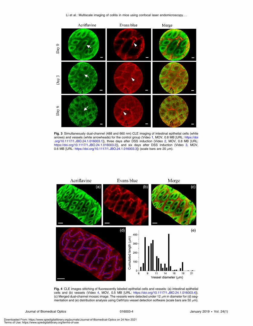
Fig. 3 Simultaneously dual-channel (488 and 660 nm) CLE imaging of intestinal epithelial cells (whitearrows) and vessels (white arrowheads) for the control group (Video 1, MOV, 0.8 MB [URL: https://doi.org/10.1117/1.JBO.24.1.016003.1]), three days after DSS induction (Video 2, MOV, 0.8 MB [URL:https://doi.org/10.1117/1.JBO.24.1.016003.2]), and six days after DSS induction (Video 3, MOV,0.6 MB [URL: https://doi.org/10.1117/1.JBO.24.1.016003.3]) (scale bars are 20 μm).
Fig. 4 CLE images stitching of fluorescently labeled epithelial cells and vessels: (a) intestinal epithelialcells and (b) vessels (Video 4, MOV, 0.5 MB [URL: https://doi.org/10.1117/1.JBO.24.1.016003.4]).(c) Merged dual-channel mosaic image. The vessels were detected under 12 μm in diameter for (d) seg-mentation and (e) distribution analysis using CellVizio vessel detection software (scale bars are 50 μm).
Journal of Biomedical Optics 016003-4 January 2019 • Vol. 24(1)
Li et al.: Multiscale imaging of colitis in mice using confocal laser endomicroscopy. . .
Downloaded From: https://www.spiedigitallibrary.org/journals/Journal-of-Biomedical-Optics on 24 Nov 2021Terms of Use: https://www.spiedigitallibrary.org/terms-of-use

3.2 In Vivo Monitoring of Mucosal Inflammation byCLE
CLE gives high-resolution in vivo imaging of fluorescentlystained cells and vessels throughout disease progression. Weused the recently developed dual-channel CLE imaging systemfor monitoring intestinal epithelial cells and microvessels ofcolon mucosa.26 The 488-nm laser with a fiber optic probe0.35 mm in diameter allowed for visualization of the acrifla-vine-stained colonic epithelial cells and crypt architectures, asshown in the first column of Fig. 3. Healthy mice had intactcolonic epithelial cells and crypts (Video 1). In DSS-colitismice, we found severe cell damage and loss of crypt structure(Videos 2 and 3). The 660-nm laser excitation allowed for visu-alization of the mucosal vasculature architecture, blood flow,and vasculature leakage (the second column of Fig. 3). Themerged results of the two-channel images are also shown inthe third column of Fig. 3.
To extend the FOV of CLE, the fiber probe’s trajectory wasfollowed and the images were stitched using the mosaic tech-nology in CellVizio [Figs. 4(a)–4(c), Video 4]. In addition,we quantitatively analyzed vessels using a vessel detection
software package, which allowed for segmentation analysisof vessels, and the cumulated length, area, and diameter ofthe acquired images. The vessel detection analysis was <12 μmin diameter for healthy mice [Fig. 4(d)], in which the mean ves-sel diameter was 11.1 μm and the total vessel length was2550 μm.
3.3 In-Toto 3-D Visualization of Colon VesselArchitecture and Morphology by LSFM
LSFM has ∼10 times higher resolution than MRI. After MRIand CLE imaging, mice were euthanized, and the colonswere removed for tissue clearing [Fig. 5(a)] by adapting theBABB protocol.24 The colonic mucosa and microvasculaturewere assessed in 3-D LSFM [Fig. 5(b), Video 5]. LSFM datasetsshowed disrupted colon villus structures and severe loss ofmucosal architecture in three and six days DSS-inducted mice,respectively. We found that colon thickness and length were sig-nificantly different between healthy mice (n ¼ 5) and mice withDSS-induced colitis (n ¼ 5). After six days of DSS induction,we observed a significant increase in colon thickness in DSS-colitis mice [0.5� 0.05 mm versus 0.25� 0.05 mm, p < 0.01,
Fig. 5 LSFM imaging of fluorescently labeled epithelial cells and vessels. Illustration of the mouse colonbefore and after clearing by BABB protocol. (a) The distal part of a cleared colon was selected for LSFMimaging. (b) Cleared LSFM images of acriflavine- and Evans blue-stained intestinal epithelial cells (leftcolumn) and vessels (middle column) (Video 5, MOV, 2.9 MB [URL: https://doi.org/10.1117/1.JBO.24.1.016003.5]). Quantification of (c) colon thickness and (d) length. Scale bars are 1 cm in (a) and 50 μm in(b).
Journal of Biomedical Optics 016003-5 January 2019 • Vol. 24(1)
Li et al.: Multiscale imaging of colitis in mice using confocal laser endomicroscopy. . .
Downloaded From: https://www.spiedigitallibrary.org/journals/Journal-of-Biomedical-Optics on 24 Nov 2021Terms of Use: https://www.spiedigitallibrary.org/terms-of-use

Fig. 5(c)]. Colon length was 85.75� 3.6 mm in healthy miceand 63.7� 8.5 mm (p < 0.05) in DSS-colitis mice.
3.4 Histological Evaluation of DSS-Induced Colitis
To characterize the development of DSS-induced colitis and val-idate MRI, CLE, and LSFM imaging results, histological alter-nations of healthy and DSS-induced mice after three and sixdays were assessed by H&E staining of paraffin-embeddedcolon sections. As DSS-induced mucosal damage progresses,the increasing ulceration and inflammation with the loss ofcrypt structure were observed [Figs. 6(a)–6(c)]. A histologicalscoring system based on epithelial damage and inflammatory
infiltrates was used to quantify the severity of the colitis21
[Fig. 6(d)].
3.5 Correlation of MRI, CLE, and LSFM
To correlate in vivo and ex vivo findings, we calculated the cor-relation coefficients of MRI (cross-sectional areas), LSFM(colonic thickness), and CLE (the crypt architecture, microvas-cular alteration and fluorescein leakage classification score)6
results with respect to weight loss, colon length, histologicalscore, and the inflammation-associated activity index fromhealthy and DSS-induced mice (after three and six days); thiscorrelation analysis is detailed in Table 1. The MRI resultshad a strong correlation with weight loss, and a significant cor-relation with the colitis activity index, colon length, and the his-tological score. The 3-D LSFM results were significantlycorrelated with the in vivo evaluation of weight loss and the dis-ease activity score. In addition, it was strongly correlated withcolon length and the histological score determined by post-mortem evaluation. The in vivomacroscopic imaging technique,MRI, was significantly correlated with the parameters represent-ing the development and severity of colitis, and microscopiclevel results, observed using the LSFM imaging method,were also significantly correlated with these parameters.
4 DiscussionThe development of optical multimodality imaging techniquescan improve IBD diagnosis.27,28 However, the analysis of coloninflammation is still challenging, mainly due to heterogeneousdistribution of lesions along the entire colon and in differentlayers of the colon. In addition, the functional and histologicalfeatures of the colon are variable at different scales, even withinnormal colon regions. In this study, we combined in vivo MRIand CLE with ex vivo LSFM of cleared colons to assess the
Fig. 6 H&E staining of paraffin-embedded transversal colon sections for histological changes analysis in(a) healthy mice, (b) DSS-induced mice after three days, and (c) DSS-induced mice after six days (scalebars are 100 μm). The black arrow represents the area of severe transmural inflammation with theloss of crypt structure. Black arrowheads indicate edematous submucosal inflammatory infiltrates.(d) Histological scores of the control group and DSS-induced groups (day three and six).
Table 1 Correlation of the mean values fromMRI (transversal areas),LSFM (colonic thickness) and CLE (the crypt architecture, microvas-cular alteration, and fluorescein leakage classification score) withbody weight loss, colon length, histological score, and the inflamma-tion-associated active index.
Correlationcoefficients
Bodyweightloss
Colonlength
Histologicalscore
Inflammation-associated
activity index
MRI −0.69** −0.85** 0.85** 0.88**
LSFM −0.8** −0.7** 0.82** 0.89**
CLE −0.58* −0.62* 0.79** 0.82**
Note: Significance was calculated using Pearson’s correlationcoefficient.*p < 0.05.**p < 0.01.
Journal of Biomedical Optics 016003-6 January 2019 • Vol. 24(1)
Li et al.: Multiscale imaging of colitis in mice using confocal laser endomicroscopy. . .
Downloaded From: https://www.spiedigitallibrary.org/journals/Journal-of-Biomedical-Optics on 24 Nov 2021Terms of Use: https://www.spiedigitallibrary.org/terms-of-use

development and severity of DSS-induced colitis at differentscales. First, noninvasive MRI was routinely performed to locatesuspicious inflammation areas in mouse models of colitis at themacroscopic scale. Then, minimal invasive CLE was used forobserving microscopic vascular architecture and cellular fea-tures alterations at these areas within mucosa. Finally, to over-come the limited FOVand imaging depth of CLE, we applied exvivo 3-D LSFM to image entire colon sections with cellular res-olution. All the findings from MRI, CLE, and LSFM were crossvalidated with correlation analysis, which demonstrated that thecorrelation of these three imaging modalities was consistent.
MRI is a versatile tool and it is a noninvasive measurement ofcolonic transversal areas at the macroscopic level for longi-tudinal study. In this study, using a permanent magnet small ani-mal MRI turned out to be feasible, safe, and low cost for routineexamination of colonic areas. Moreover, in vivo MRI resultswere significantly correlated with ex vivo colon length and his-tological score. However, the resolution of MRI in our study waslimited to ∼195 μm, which was unable to observe the cellularfeatures and microvessels architecture at the microscopic level.To overcome this limitation, CLE was employed for real-time,high-resolution visualization of the epithelial cell and microves-sels within the colon mucosa. For simultaneous monitoring ofcell features and vessel architecture using the CLE system, weapplied intravenous perfusion with acriflavine that tagged epi-thelial cells, followed by topical administration of Evans bluefor staining the vasculature of the colon mucosal layer. AfterCLE, colons were cleared using BABB, a well-developedprotocol.24 The high-resolution images of clearing colonacquired by LSFM were used for 3-D reconstruction of the flu-orescently tagged cells and blood vessel structure of the intactcolon section. Previous studies combined white-light endoscopywith CLE to obtain high-magnification images.29 In this study,we correlated in vivoMRI at the macroscopic level with CLE atthe microscopic level. In addition, we also correlated CLE intwo-dimensions and LSFM in 3-D. This approach can bridgethe gap between MRI and CLE at different levels, whichhave been separate domains in IBDs models, and it also allowsfor cross validation of in vivo CLE and ex vivo LSFM. Using thismultimodality approach, we visualized the colon mucosal layerat the cellular level and the vessel architecture level with singlevessel resolution. Furthermore, this multimodality approach notonly allows in vivo visualization of inflammation within mucosafor UC study but also enables ex vivo 3-D imaging of the colonwith cellular resolution for transmural investigation in CD,which can enhance our understanding of the pathogenesisof IBDs.
One of the main limitations of our study was CLE’s relativelysmall FOV. As described in Sec. 1, the FOV was limited to600 μm. One solution of this limitation is to stitch imagesacquired in a real-time scan to form a larger FOV using themosaic image software developed by CellVizio. Another disad-vantage was that clearing of the colon samples may haveresulted in decreased colon size due to dehydration. This draw-back may be corrected by quantifying the shrinkage of the colonvolume using CT imaging before and after clearing. The LSFMimages of cell and vessel parameters of cleared colons could becorrected by the volume reduction factor obtained by CT.
In conclusion, we demonstrated the feasibility of combiningin vivo MRI and CLE and ex vivo LSFM for assessing colonmorphology and dynamic changes of epithelial cells and vascu-lature in DSS-induced colitis models. This approach showed
multimodality imaging of mouse colons at different levels,which will improve the diagnostic precision of DSS colitis.This approach could also be applied to other experimental mod-els, such as trinitrobenzene sulfonic acid colitis and oxazolonecolitis. We envision that this multimodality imaging techniquewill improve the understanding of the pathogenesis, cell pro-gression, and microenvironment alternation of IBDs.
DisclosuresThe authors declare that this is an expanded and revised workbased on our previous SPIE proceedings, and there is no conflictof interests to declare.
AcknowledgmentsThis paper was supported by the National Key Research andDevelopment Program of China Nos. 2016YFC0103803,2017YFA0700401, and 2017YFA0205200; the NationalNatural Science Foundation of China under GrantNos. 81527805, 81671851, and 81227901; the StrategicPriority Research Program under Grant No. XDBS01030200,the Scientific Instrument R&D Programs under GrantNos. GJJSTD20170004 and YJKYYQ20170075, and theFrontier Science Key Research Program under GrantNo. QYZDJ-SSW-JSC005 from the Chinese Academy ofSciences; the Beijing Municipal Science and TechnologyCommission under Grant No. Z161100002616022. The authorswould like to acknowledge the instrumental and technical sup-port of multimodal biomedical imaging experimental platform,Institute of Automation, Chinese Academy of Sciences.
References1. S. Danese and C. Fiocchi, “Ulcerative colitis,” N. Engl. J. Med. 365(18),
1713–1725 (2011).2. L. J. Herrinton et al., “Incidence and mortality of colorectal adenocar-
cinoma in persons with inflammatory bowel disease from 1998 to2010,” Gastroenterology 143(2), 382–389 (2012).
3. L. Mielke et al., “Confocal laser endomicroscopy to monitor the colonicmucosa of mice,” J. Immunol. Methods 421(6), 81–88 (2015).
4. J. Phan et al., “Time to diagnostic colonoscopy and colonoscopic find-ings after positive fit in an ethnically diverse cohort of us veterans,”Gastroenterology 152(5), S538 (2017).
5. Y. Wu et al., “Inflammatory bowel disease: MR- and SPECT/CT-basedmacrophage imaging for monitoring and evaluating disease activity inexperimental mouse model—pilot study,” Radiology 271(2), 400–407(2014).
6. C. Q. Li et al., “Classification of inflammation activity in ulcerative col-itis by confocal laser endomicroscopy,” Am. J. Gastroenterol. 105(6),1391–1396 (2010).
7. I. Ordas et al., “Ulcerative colitis,” Lancet 380(9853), 1606–1619(2012).
8. J. Rimola et al., “Magnetic resonance imaging for evaluation of Crohn’sdisease: validation of parameters of severity and quantitative index ofactivity,” Inflammatory Bowel Dis. 17(8), 1759–1768 (2011).
9. J. Walldorf et al., “In-vivo monitoring of acute DSS-colitis using colon-oscopy, high resolution ultrasound and bench-top magnetic resonanceimaging in mice,” Eur. Radiol. 25(10), 2984–2991 (2015).
10. G. Masselli and G. Gualdi, “CT and MR enterography in evaluatingsmall bowel diseases: when to use which modality?” Abdom.Imaging 38(2), 249–259 (2013).
11. D. N. Rasmussen et al., “Confocal laser endomicroscopy in inflamma-tory bowel disease—a systematic review,” J. Crohn’s Colitis 9(12),1152–1159 (2015).
12. G. D. De Palma, “Confocal laser endomicroscopy in the ‘in vivo’ his-tological diagnosis of the gastrointestinal tract,”World J. Gastroenterol.15(46), 5770–5775 (2009).
Journal of Biomedical Optics 016003-7 January 2019 • Vol. 24(1)
Li et al.: Multiscale imaging of colitis in mice using confocal laser endomicroscopy. . .
Downloaded From: https://www.spiedigitallibrary.org/journals/Journal-of-Biomedical-Optics on 24 Nov 2021Terms of Use: https://www.spiedigitallibrary.org/terms-of-use

13. A. Arranz et al., “Helical optical projection tomography,” Opt. Express21(22), 25912–25925 (2013).
14. D. Dong et al., “Vertically scanned laser sheet microscopy,” J. Biomed.Opt. 19(10), 106001 (2014).
15. X. Liang et al., “Stripe artifact elimination based on nonsubsampledcontourlet transform for light sheet fluorescence microscopy,”J. Biomed. Opt. 21(10), 106005 (2016).
16. T. Li et al., “Hybrid of two-photon microscopy and optical multimodal-ity imaging for multi-scale imaging of small animals,” SPIE BiOS10499, 104991R (2018).
17. A. Candeo et al., “Virtual unfolding of light sheet fluorescence micros-copy dataset for quantitative analysis of the mouse intestine,” J. Biomed.Opt. 21(5), 056001 (2016).
18. I. Gabanyi et al., “Neuro-immune interactions drive tissue programmingin intestinal macrophages,” Cell 164(3), 378–391 (2016).
19. S. Melgar, A. Karlsson, and E. Michaëlsson, “Acute colitis induced bydextran sulfate sodium progresses to chronicity in C57BL/6 but not inBALB/c mice: correlation between symptoms and inflammation,” Am.J. Physiol. Gastrointest. Liver Physiol. 288(6), G1328 (2005).
20. N. Taghipour et al., “An experimental model of colitis induced by dex-tran sulfate sodium from acute progresses to chronicity in C57BL/6:correlation between conditions of mice and the environment,”Gastroenterol. Hepatol. Bed Bench 9(1), 45–52 (2016).
21. S. Wirtz et al., “Chemically induced mouse models of acute and chronicintestinal inflammation,” Nat. Protoc. 12(7), 1295–1309 (2017).
22. C. A. Schneider, W. S. Rasband, and K. W. Eliceiri, “NIH image toImageJ: 25 years of image analysis,” Nat. Methods 9(7), 671–675(2012).
23. M. J. Waldner et al., “Confocal laser endomicroscopy and narrow-bandimaging-aided endoscopy for in vivo imaging of colitis and colon cancerin mice,” Nat. Protoc. 6(9), 1471–1481 (2011).
24. J. Ahnfelt-Ronne et al., “An improved method for three-dimensionalreconstruction of protein expression patterns in intact mouse andchicken embryos and organs,” J. Histochem. Cytochem. 55(9), 925–930 (2007).
25. A. Bianchi et al., “Noninvasive longitudinal study of a magnetic reso-nance imaging biomarker for the quantification of colon inflammationin a mouse model of colitis,” Inflammatory Bowel Dis. 22(6), 1286–1295 (2016).
26. G. Vargas et al., “Topical injury evaluation of the murine colorectalmucosa using confocal endomicrosopy: a valuable method for assessingmucosal injuries associated with risk of pathogen transmission,”J. Microsc. 264(2), 227–237 (2016).
27. R. Atreya et al., “In vivo imaging using fluorescent antibodies to tumornecrosis factor predicts therapeutic response in Crohn’s disease,” Nat.Med. 20(3), 313–318 (2014).
28. E. Stanley, H. K. Moriarty, and C. G. Cronin, “Advanced multimodalityimaging of inflammatory bowel disease in 2015: an update,” World J.Radiol. 8(6), 571–580 (2016).
29. S. Coda and A. V. Thillainayagam, “State of the art in advanced endo-scopic imaging for the detection and evaluation of dysplasia and earlycancer of the gastrointestinal tract,” Clin. Exp. Gastroenterol. 7, 133–150 (2014).
Tianmeng Li is a second-year graduate student at the NortheasternUniversity in Shenyang, China. Her major is biomedical engineering.She received her bachelor’s degree in network engineering fromZaozhuangCollege, Shandong Province, China. Her current researchinterests include 3-D reconstruction and application of optical images.
Hui Hui is an associate professor at the Institute of Automation,Chinese Academy of Sciences, China. He received his BS and MSdegrees in mechanical engineering from Chang’an University,China, in 2003 and 2006, respectively, and his PhD in mechanicalengineering from the University of Franche-Comté, France, in2013. His current research interests include multimodal molecular im-aging and optical microscopy.
Chaoen Hu is a research assistant at the Institute of Automation,Chinese Academy of Sciences, China. He received his master’sdegree in control engineering major of Harbin University ofScience and Technology, China. His current research interestsinclude image processing and visualization.
He Ma is an associate professor, vice dean, master tutor at theInstitute of Biomedical and Information Engineering, NortheasternUniversity. He received his PhD in computer science and technologyfrom the Department of Computer Science, National University ofSingapore in 2014. His current research interests include lung cancerimagery and medical image process.
Xin Yang is a professor at the Institute of Automation, ChineseAcademy of Sciences, China. She received her PhD in precisioninstruments and optoelectronics engineering from Tianjin University,China, in 2000. Her current research interests include pattern recog-nition and medical image process.
Jie Tian is a professor at the Institute of Automation, ChineseAcademy of Sciences, China. He received his PhD in artificial intelli-gence from the Institute of Automation, Chinese Academy ofSciences, China, in 1992. His current research interests include medi-cal image process and molecular imaging.
Journal of Biomedical Optics 016003-8 January 2019 • Vol. 24(1)
Li et al.: Multiscale imaging of colitis in mice using confocal laser endomicroscopy. . .
Downloaded From: https://www.spiedigitallibrary.org/journals/Journal-of-Biomedical-Optics on 24 Nov 2021Terms of Use: https://www.spiedigitallibrary.org/terms-of-use


![Microscopia Confocal Tem [Recuperado]](https://img.pdfslide.tips/doc/110x75/563dbbbb550346aa9aafc610/microscopia-confocal-tem-recuperado.jpg)