Embed Size (px)
Citation preview

Nuclear Magnetic Resonance Studies of Hemoglobin Chesapeake: An alp2 Mutant?
Karen J . Wiechelman, Samuel Charache, and Chien Ho*
ABSTRACT: Proton nuclear magnetic resonance studies have been used to compare the deoxy and ligand-bound forms of hemoglobins A and Chesapeake (cy92FG4 Arg +Leu). Partial saturation studies have also allowed us to study the equilibrium binding of oxygen and carbon monox- ide to H b Chesapeake. The hyperfine shifted resonances of the heme protons of hemoglobins Chesapeake and A differ, indicating that the deoxy quaternary structure of hemoglo- bin Chesapeake has been altered in some way. The ring- current shifted proton resonances of both HbCO and Hb02
I t has been shown that hemoglobins with mutations at the aI IJ~ subunit interface exhibit decreased heme-heme inter- actions (Perutz and Lehmann, 1968) indicating that the structural integrity of this region of the hemoglobin (Hb)’ inolecule is critical for cooperative interactions in normal Hb A. X-Ray crystallographic studies have shown that the transition from the deoxy to the oxy quaternary structure results in a drastic change in the cyl$2 interface. On oxygen- ation the two subunits shift by 13’ relative to each other re- sulting in the breaking of the hydrogen bond between tyro- sine-a42(C7) and aspartic acid-/399(Gl) which helps stabi- lize the deoxy structure, and the formation of a new hydro- gen bond between aspartate-a94(GI ) and aspdragine- B102(G4) in the oxy quaternar) structure (Perutz, 1970: Perutz and Ten Eyck, 1971). The deoxy to oxy quaternary structural change results in a slight loosening of the N , I J , subunit interface but the two subunits do not shift relative to each other (Perutz and Ten Eyck. 1971). The alp’? in- terface is both smaller and smoother than the c y ) @ ) subunit interface and it is constructed so that the subunits can easi- ly slide past one another. I t is closely connected to the heme groups so that any changes in the a1/31 subunit interface would be expected to affect the heme environment (Perutz. 1969). Thus studies of hemoglobins with mutations at the iu,d? interface have proved to be invaluable in the study of structure-function relationships in hemoglobin.
Hb Chesapeake is an CY chain mutant with a leucine resi- due replacing the normal arginine residue at ( ~ 9 2 ( FG4). This substitution results in a higher than normal oxygen af-
t F-rom the Department of Biophysics and Microbiologq and the De- partment of Biochemistrj, Facultj of Arts and Sciences, Lniversit) of Pittsburgh. Pittsburgh, Pennsylvania 15260 (K.J .W. and C.H.) . and the Hematology Division. Department of Medicine, Johns Hopkins Lniversit) School of Medicine. Baltimore, Marqland 2 I205 (S .C. ) . K r w i l w f June 4, 1974. Supported by research grants from the Sa t ion - 31 Institutes of Health (HL-10383; HL-02799; RR-00292). National Science Foundation (GB-37096X), and the American Heart Associa- r i o n . Inc. ’ Abbreviations used are: ,Hb, hemoglobin; DPG, 2.3-diphosphoglq- cerate: I HP. inositol hexaphosphate; HbCO. carbonmonoxyhemoglo- bin; HbOz, oxj hemoglobin: Tris. tris(hydroxymeth)l)aminomethane: Bis-Tris. 2.2-bis(hydroxyethyl)-2.2’.2”-nitrilotrimethanol.
Chesapeake are identical with those of the corresponding ligand-bound forms of H b A suggesting that the tertiary structures of the cy and /3 heme pockets in liganded H b Che- sapeake are very similar to those of liganded H b A. Partial C O and 0 2 binding studies show that in H b Chesapeake the hemoglobin-ligand reaction is both ligand and phosphate specific. These studies also show that the cy and /3 chains within the H b Chesapeake tetramer are functionally more equivalent in their reactions with ligands than are the cy and B chains of H b A.
finity, a normal Bohr effect, and a reduced cooperativity which is reflected in the Hill coefficient (n) of 1 .3 (Nagel et al., 1967) as compared to a value of 2.9 for H b A (Antonini et al., 1964). In the presence of organic phosphates, such as 2,3-diphosphoglycerate (DPG) or inositol hexaphosphate (IHP), the oxygen affinity of H b Chesapeake is reduced and the cooperativity is increased (Imai, 1974).
Proton nuclear magnetic resonance (nmr) studies have shown that there are three prominent hyperfine (or contact) shifted resonances in deoxy H b A (Davis et al.. 1971: Ogawa and Shulman, 1972). Unpaired electrons of the paramagnetic iron interact with protons of the porphyrin ring and/or with the amino acid protons that are positioned sufficiently close to the iron atom to give resonances a t -17.6, -12.0, and -7.9 ppm from the residual H D O reso- nance (Davis et al., 1971). Nmr studies of various mutant hemoglobins have shown that the resonance a t - 17.6 ppm can be assigned to a /3 chain proton while the resonances a t -12.0 and -7.9 ppm are due to protons on the N chain (Davis et al . , 1971; Lindstrom et al., 1972a). These hyper- fine shifted resonances return to their normal positions bur- ied in the aromatic or aliphatic regions of the H b spectrum when ligands bind to the hemes making them diamagnetic.
The diamagnetic forms of hemoglobin (HbCO and HbO2) contain ring-current shifted proton resonances that are due to protons of amino acids that are positioned closely enough to the porphyrin ring to be affected by the local magnetic fields which are produced by the delocalized 7r
electrons in the heme groups (McDonald and Phillips, 1967: McDonald et al., 1969; Shulman et al., 1970). These ring- current shifted resonances have been shown to be very sen- sitive to changes in the tertiary structure around the ligand binding sites in HbCO (Lindstrom et al . , 1972b; Lindstrom and Ho, 1973).
In this communication we report the results of nmr stud- ies of the hyperfine and ring-current shifted proton reso- nances of H b Chesapeake.
Experimental Section Materials. H b Chesapeake was isolated and. purified by
chromatography on DEAE-Sephadex (A 50 Pharmacia)
N M R S T U D I E S O F H B C H E S A P E A K E
using the isolation and purification procedure of Huisman and Dozy (1965). H b A was isolated either from the H b Chesapeake hemolysate or was prepared by standard meth- ods from fresh whole blood samples obtained from the local blood bank (Davis et al., 1971). Hemoglobin was freed from phosphates by passing it through a column of Sepha- dex (3-25 (Pharmacia) equilibrated with 0.01 M Tris-HCI buffer containing 0.1 M NaCl a t pH 7.5 (Berman et a/., 1971). To reduce the intense proton resonance of aqueous samples, H 2 0 was exchanged with deuterium oxide (Merck, Sharp, and Dohme of Canada, Ltd.) by repeated dilution with D 2 0 and subsequent concentration through an Amicon UM-20E membrane. DPG was purchased from Calbiochem as the pentacyclohexylammonium salt, con- verted to the acid form with Bio-Rad AG50W-X8 cation exchange resin, and titrated to pH 7 with NaOH. I H P pur- chased from Sigma as the sodium salt was dissolved in D 2 0 and titrated to pD 7 with HC1. Both the DPG and I H P so- lutions were then repeatedly lyophilized and redissolved in D20. After the final lyophilization the residue was dissolved in DzO and the phosphate concentration of the solutions was determined (Bartlett, 1950). 2,2-Bis(hydroxyethyl)- 2,2',2"-nitrilotrimethanol (Bis-Tris) purchased from Al- drich was dissolved in DzO and titrated to pD 7 with HCI. The pD of all solutions was determined by adding 0.4 pH unit (Glasoe and Long, 1960) to the meter reading of a Ra- diometer Model 4 pH meter equipped with a Beckman 39030 combination electrode. Oxyhemoglobin was prepared from HbCO by replacing the C O with oxygen by flushing oxygen over the H b solution contained in a round-bottomed flask attached to a rotatory evaporator in an ice-water bath under a Sylvania 150-W flood lamp. Oxygen was removed by flushing the solution with prepurified nitrogen. The he- moglobin concentrations for our nmr studies varied from 9 to 14%.
For the oxygen or carbon monoxide saturation studies, partially liganded samples were prepared by one of two methods. When the study was carried out in 5-mm nmr sample tubes, appropriate amounts of deoxy H b were mixed with either HbCO or HbO2 to give partially liganded sam- ples. However, in the case of the oxygen saturation study in the presence of IHP, the experiments were carried out in the presence of the methemoglobin reductase system of Hayashi er al. (1973) with NADPH substituted for NADP. All of the materials used in the reductase system were purchased from Sigma. Appropriate amounts of air were injected into 5-mm tubes containing deoxy H b Chesa- peake and the sample was equilibrated for at least 30 min before nmr spectra were taken. In the IO-" sample tubes partially liganded samples were obtained by injecting small amounts of oxygen or carbon monoxide gas with a Hamil- ton microsyringe through a rubber stopper in the side arm of the tube. The samples were allowed to equilibrate for a t least 1.5 hr before nmr spectra were taken. The degree of ligand saturation was measured by monitoring the decrease in the deoxy peak a t 757 nm with either a Cary 14 or Zeiss PMQ I1 spectrophotometer. For the 5-mm sample tubes the absorbance was determined directly through the nmr tubes held in the light path of a specially designed holder. A piece of flat glass tubing (2-3 mm in optical path length) was fused to the top of the IO-" nmr tubes to serve as a cuvet.
Methods. Nmr spectra were obtained with 5-mm sample tubes on MPC-HF 250-MHz superconducting spectrometer (Dadok et al., 1970) and on a Bruker HFX 90-MHz spec- trometer with the IO-" sample tubes. Proton chemical
1 1 I , I 1 I - 22 -20 - ( 8 -16 -14 -12 -10 - 8 -6
PPM f r o m HDO
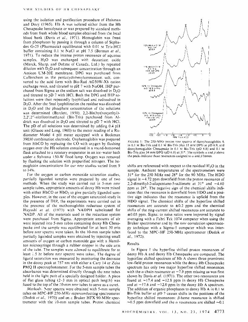
FIGURE 1: The 250-MHz proton nmr spectra of deoxyhemoglobin A In 0.1 M Brs-Tris and 0.1 M Bis-Tris plus 33 m u DPG at pD 6.9, and deoxyhemoglobin Chesapeake in 0.1 M Bis-Tris (pD 6.8) and 0.1 M Bis-Tris plus 34 mM DPG (pD 6.9) a t 31' The symbols 01 and p above the peaks indicate those resonances assigned to 01 and p hemes.
shifts are referenced with respect to the residual H2O in the sample. Ambient temperatures of the spectrometers were 31" for the 250 M H z and 28" for the 90 MHz. The H D O signal is -4.72 ppm downfield from the proton resonance of 2,2-dimethyl-2-silapentane-5-sulfonate a t 3 1 O and -4.80 ppm a t 28". The negative sign of the chemical shifts indi- cates that the resonance is downfield from H D O and a posi- tive sign indicates that the resonance is upfield from the HDO signal. The chemical shifts of the hyperfine shifted resonances are accurate to fO.2 ppm and the chemical shifts of the ring-current shifted resonances are accurate to f0 .05 ppm. Signal to noise ratios were improved by signal averaging with a Fabri Tek 1074 computer when using the Bruker spectrometer and by the nmr correlation spectrosco- py technique with a Sigma-5 computer which was inter- faced to the MPC-HF 250-MHz spectrometer (Dadok et al., 1972).
Results I n Figure I the hyperfine shifted proton resonances of
deoxy H b A and deoxy H b Chesapeake are compared. The hyperfine shifted spectrum of H b A shows three prominent low-field proton resonances while the deoxy H b Chesapeake spectrum has only two major hyperfine shifted resonances with the a chain resonance a t -7.9 ppm missing as was first shown by Davis et al. (1971). The other two resonances are found a t -17.4 and -12.8 ppm in deoxy H b Chesapeake and a t - 17.6 and - 12.0 ppm in the deoxy H b A spectrum. The addition of organic phosphates to deoxy H b A in 0.1 M Bis-Tris buffer a t pD 7 causes shifts in the positions of the hyperfine shifted resonances: P-heme resonance is shifted -0.5 ppm downfield and the a resonances are shifted -0.1
B I O C H E M I S T R Y . V O L . 1 3 , N O . 2 3 , 1 9 7 4 4773
W I E C H E L M A N . C H A R A C H E , A N D H O
1 1 1 1 1 1 1 , l ! , I l l ,
- 2 2 -20 -18 -16 -14 -12 -10 -8 -6 PPM f r o m HDO
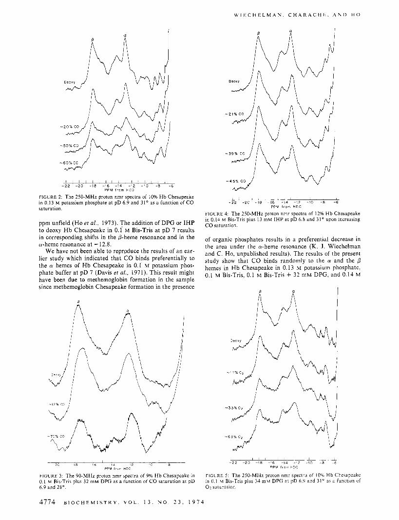
FIGURE 2: The 250-MHz proton nmr spectra of 10% Hb Chesapeake in 0.13 M potassium phosphate at pD 6.9 and 3 1 ' as a function of C O saturation
ppm unfield (Ho et al., 1973). The addition of DPG or I H P to deoxy H b Chesapeake in 0.1 M Bis-Tris a t pD 7 results in corresponding shifts in the 0-heme resonance and in the a-heme resonance at -12.8.
We have not been able to reproduce the results of an ear- lier study which indicated that C O binds preferentially to the a hemes of H b Chesapeake in 0.1 M potassium phos- phate buffer at pD 7 (Davis e? al., 1971). This result might have been due to methemoglobin formation in the sample since methemoglobin Chesapeake formation in the presence
1 8 1 ' 1 1 1
-72 -20 -18 -16 -14 -12 -10 -8 -6 _ _ ~~
PPM f r o m H D O
FIGURE 4: The 250-MHz proton nmr spectra of 12% Hb Chesapeake in 0.14 M Bis-Tris plus 13 mM IHP at pD 6.8 and 31' upon increasing C O saturation.
of organic phosphates results in a preferential decrease in the area under the a-heme resonance (K. J. Wiechelman and C. Ho, unpublished results). The results of the present study show that C O binds randomly to the a and the p hemes in H b Chesapeake in 0.13 M potassium phosphate, 0.1 M Bis-Tris, 0.1 M Bis-Tris + 32 mM DPG, and 0.14 M
I l l I l l - 2 2 -20 -18 -16 -14 -12 -10 - 8 -6
PPM f r o m H D O - 2 0 -18 -16 - ' 4 -12 -10 -8
PDM f r o m H D C
FIGURE 3: The 90-MHz proton nmr spectra of 9% Hb Chesapeake in 0.1 M Bis-Tris plus 32 mM DPG as a function of C O saturation at pD 6.9 and 28". 0 2 saturation.
F I G U R E 5 : The 250-MHz proton nmr spectra of 10% Hb Chesapeake in 0.1 M Bis-Tris plus 34 m M DPG at pD 6.9 and 3 1 " as a function of
4774 B I O C H E M I S T R Y , V O L . 1 3 , N O . 2 3 , 1 9 1 4

N M R S T U D I E S O F H B C H E S A P E A K E
a
l l l l 1 1 I I l I I I I I I I I - 2 2 -20 -18 -16 -14 -12 -10 -8 -6
PPM from HDO
FIGURE 6: The 250-MHz proton nmr spectra of 10% H b Chesapeake in 0.1 M Bis-Tris plus 9 mM I H P in the presence of methemoglobin re- ductase upon increasing 0 2 saturation a t pD 6.7 and 31'.
Bis-Tris + 13 mM IHP, all at pD 7 (Figures 2-4). If there is preferential ligand binding to either the cy or @ hemes in H b Chesapeake, one would observe a selective decrease in the intensity of either the cy or the @ chain resonances. On the contrary the area ratio of the cy resonance (at - -12.8 ppm) and the p resonance (at --17.6 ppm) remains essen- tially constant as a function of C O saturation (Figures 2- 4). This suggests that C O binds essentially randomly to the a and @ chains of H b Chesapeake. A detailed procedure for determining preferential binding of ligands to H b A by the nmr technique has been described by Ho and coworkers (Lindstrom and Ho, 1972; Johnson and Ho, 1974).
Figures 5 and 6 show the results of oxygen saturation studies for H b Chesapeake. When HB Chesapeake is in 0.1 M Bis-Tris buffer or in 0.1 M Bis-Tris + 34 mM DPG at pD 7, oxygen binds to the a and @ hemes essentially randomly as evidenced by the lack of a selective decrease in the area of either the a- or the @-heme resonance upon oxygenation (Figure 5). In the presence of 9 mM I H P oxygen binds to the cy hemes of H b Chesapeake in preference to the /3 hemes as shown by the preferential decrease of the a-heme reso- nance at --12.8 ppm as a function of oxygenation (Figure 6). In other words, the area ratio of the cy to @ resonances decreases upon oxygenation. Deoxy H b Chesapeake is un- stable in the presence of IHP and in the presence of oxygen significant amounts of methemoglobin were formed during the course of the experiment. The methemoglobin forma- tion could be prevented by the addition of the methemoglo- bin reductase system of Hayashi et al. (1973). The reduc- tase system in D20 will reduce a 10% solution of methemo- globin in about 45 min (when NADPH is introduced rather than being generated from NADP by glucose-6-phosphate dehydrogenase) so it can efficiently reduce the methemo- globin formed in the sample during the course of an nmr ex-
HbCO Chesapeake 0.1 M Bis-Tris
HbCO Chesapeake O.fM Bis-Tris
HbCO Chesapeake 0 1 M Bis-Tris
HbCO A 01 M Bis-Tris
HbCO A
3 0 m M DPG ' ' O 1 M Bis-Tris
I 1 I i 1 +5.0 + 5 5 +60 + 6 5 +70
PPM f rom HDO
FIGURE 7: The 250-MHz nmr spectra of the ring-current shifted pro- ton resonances of HbCO Chesapeake and HbCO A in the absence and presence of organic phosphates at 3 1 O . The assignments of the valine El 1 methyl groups of HbCO A are from Lindstrom et al. (1972b).
periment. This reductase system was used by Imai (1 974) to carry out oxygen equilibrium measurements on H b Chesapeake. The reductase system was also included in oxygen saturation experiments with H b A in the presence of 10 mM IHP and H b Chesapeake in the presence of 30 mM DPG. In both cases the results were the same as those ob- tained with samples which contained no reductase.
The ring-current shifted proton resonances of HbCO Chesapeake and HbOz Chesapeake in 0.1 M Bis-Tris, in the presence of 30 mM DPG, and in the presence of 10 mM IHP are very similar to the corresponding ring-current shifted resonances of HbCO A and HbO2 A (Figures 7 and 8). This suggests that the tertiary structures of the a- and @-heme pockets in liganded H b Chesapeake are very similar to those of liganded Hb A.
Discussion X-Ray crystallographic studies of deoxy H b Chesapeake
at 5.5-A resolution indicate that H b Chesapeake can as- sume a deoxy quaternary structure which is similar to that of deoxy H b A (Greer, 1971). The difference electron den- sity map of deoxy H b Chesapeake and deoxy H b A shows no significant changes other than the missing electron den- sity of the arginine residue at the mutation site. When air is introduced into the deoxy crystals, the crystals become highly disordered which indicates that a quaternary struc- tural change accompanies the deoxy to oxy transition in Hb Chesapeake (Greer, 1971). Crystals of oxy H b Chesapeake are isomorphous with oxy H b A crystals but the difference Fourier between the two hemoglobins shows that the overall conformations are not the same. The B, C, D, and G heli- cies of the same p chain which are in close proximity to the cy92(FG4) mutation site appear to have moved away from
B I O C H E M I S T R Y , V O L . 1 3 , N O . 2 3 , 1914 4775

W I E C H E L M A N . C H A R A C H E . A N D H O
HbO, Chesapeake 0 1 M Bis-Tr is
p D 7 0
H b O z Chesapeake
\ HbO, Chesapeake
HbOp A
$5
HbOp A
$5
I I I
5 6 7 PPM f rom H D O
FIGURE 8: The 250-MHz nmr spectra of the ring-current shifted pro- ton resonances of HbOz Chesapeake and HbOl A and the changes brought about in these resonances by the addition of DPG or IHP.
the leucine residue and toward the alp2 interface. In addi- tion the G helix and other regions of the a chain are also distorted (Greer, 1971).
Nmr studies of deoxy H b Chesapeake show that the deoxy Hb Chesapeake structure differs somewhat from deoxy Hb A since the hyperfine shifted resonance at -7.9 ppm in the deoxy H b A spectrum is missing in deoxy H b Chesapeake and the other two proton resonances are shifted from their corresponding positions in H b A. It has been shown that the hyperfine shifted resonances are very sensi- tive to heme-globin interactions and it is likely that the hy- perfine shifted nmr spectra are reflecting differences in the deoxy structure in the region of the heme pockets of both the CY and chains between the structures of deoxy Hb A and deoxy H b Chesapeake (Davis et al., 1970, 1971).
In some hemoglobins nmr experiments have shown that in addition to being structurally nonequivalent, the CY and p hemes are also functionally nonequivalent. H b A in the presence of DPG or IHP exhibits a preferentiai decrease in the area under the a-heme peaks upon increasing oxygen saturation indicating that under equilibrium conditions oxy- gen binds to the a hemes in preference to the hemes (Lindstrom and Ho, 1972; Johnson and Ho, 1974). Also in H b A, it has been shown that n-butyl isocyanide binds pref- erentially to the P hemes in the presence of I H P (Lindstrom et al., 1971). In H b Kempsey [p99(GI) Asp-Asn] and Hb Yakima [@99(G1) Asp-His], C O is bound preferen- tially to the /3 hemes in the absence of organic phosphate or in the presence of DPG; but in the presence of IHP, C O
binds randomly to the hemes (Ho et al., 1973; Lindstrom et al., 1973; K. J . Wiechelman and C. Ho unpublished re- sults). Upon increasing C O saturation H b Chesapeake is similar to H b A in that C O appears to bind randomly to the cy and hemes, However, H b Chesapeake differs from H b A upon oxygenation in the presence of DPG by binding oxygen randomly while Hb A binds 0 2 preferentially to the cy hemes under similar conditions (Lindstrom and Ho, 1972; Johnson and Ho, 1974).
It is interesting to note that Nagel et al. (1967) have found that in both oxy and deoxy H b Chesapeake the /393(F9) sulfhydryl groups are reactive toward iodoacetam- ide whereas in deoxy Hb A these groups are unreactive. In deoxy H b A, histidine-@146(HC3) forms a loop around the F helix and its imidazole group forms a salt bridge with as- partate-894 (FG1) of the same @ chain and its a-carboxyl group makes a salt bridge with lysine-a40(C6) thus block- ing access to the reactive S H groups (Perutz et al., 1969). The 8146 histidine to p94 aspartate salt bridge is responsi- ble for about half of the alkaline Bohr effect in H b A (Kil- martin and Wootton, 1970) and since Hb Chesapeake has a normal Bohr effect this salt bridge must be intact. Nmr studies of H b Des-His 146p in which the p146 histidine residues have been removed enzymatically and of Hb Hiro- shima [pl46(HC3)His-.Asp] (Perutz et al., 1971) show that the binding of oxygen to the a and chains is random even in the presence of IHP at pD 7 (Breen et a]., 1974; Lin, Breen, and Ho, unpublished results). These results may suggest that the conformation of the Hb molecule in the re- gion affected by the 0146 histidine salt bridges with p94 as- partate and lysine a40 may be critical for the ligand bind- ing properties of the a and p hemes in a hemoglobin mole- cule. The fact that oxygen binding to H b Chesapeake in the presence of DPG is random suggests that there may be some distortion in this region of the molecule. The results of our oxygen saturation studies in the presence of IHP suggest that the tighter binding of IHP to deoxy H b Chesa- peake may restore the conformation necessary for preferen- tial binding of oxygen to the a hemes.
In a recent article (Ogata and McConnell, 1972), experi- mental results of a CO saturation study of H b Chesapeake in the presence of the spin-labeled triphosphates, 1 -0xyl- 2,2,6,6-tetramethylpiperidine 4-triphosphate and N6-( 1- oxyl-2,2,6,6-tetramethyl-4-piperidinyl)adenosine triphos- phate, were fit by a set of parameters calculated from a modified Monod, Wyman, and Changeux model (Monod et al., 1965; Ogata and McConnell, 1971). Their results indi- cate that deoxy Hb Chesapeake contains molecules in the R (relaxed) as well as the T (tense) quaternary structures. I f the rate of interconversion between the R and T structures is rapid on the nmr time scale, one would expect the reso- nances characteristic of each quaternary structure to co- alesce into one broadened resonance with a chemical shift intermediate between the R and T chemical shifts. Perutz et al. (1 974) have shown that the @-heme resonance which is found at --17.6 ppm in deoxy H b A shifts upfield to --16 ppm in all deoxyhemoglobins having a R-like quater- nary structure. In Figure 1 i t can be seen that the @-heme resonance of deoxy Hb Chesapeake in 0.1 “ ~ l Bis-Tris is broader than the p peak in deoxy Hb A under similar condi- tions and i t has been shifted upfield from the /3 resonance of Hb A. It should be noted that the P-heme resonance of Hb Chesapeake in the absence of organic phosphate is broader than that of Hb Chesapeake and Hb A in the presence of DPG (Figure I ) . This effect may be due to an equilibrium
4776 B I O C H E M I S T R Y . V O L . 1 3 , N O . 2 3 . 1 9 7 4

N M R S T U D I E S O F H B C H E S A P E A K E
between the R and T quaternary structures of H b Chesa- peake which would be consistent with the results of Ogata and McConnell (1972). A detailed discussion of T + R in the alp2 mutants will be published elsewhere.
Our studies of the ring-current shifted proton resonances of the ligand-bound forms of Hb A and H b Chesapeake show that nmr can be a powerful tool in investigations of structure-function relationships in hemoglobins (Lindstrom and Ho, 1973). The X-ray diffraction study of H b Chesa- peake (Greer, 1971) showed that the overall oxy Hb Chesa- peake structure differs significantly from the oxy Hb A structure. However, the ring-current shifted proton reso- nances in the two hemoglobins show that they are very simi- lar in the tertiary structures of the heme pockets. This re- sult is consistent with the finding that k 4 , the Adair con- stant for the binding of the fourth oxygen molecule to he- moglobin, is the same for Hb A and Hb Chesapeake (Imai, 1974).
A CO saturation study of Hb Chesapeake labeled with the spin-label N-(l-oxyl-2,2,6,6-tetramethyl-4-piperidin- y1)iodoacetamide which binds to the /393(F9) sulfhydryl groups shows a sharp set of isosbestic points while the elec- tron paramagnetic resonance (epr) spectra of H b A labeled with the same iodoacetamide spin-label lack a set of isos- bestic points upon CO saturation (Ho et al., 1970). The presence of a set of isosbestic points in the epr spectra of the spin-labeled H b Chesapeake on CO saturation has been suggested as evidence that there are differences in the alp2
subunit interactions between Hb A and Hb Chesapeake during C O saturation (Ho et al., 1970; Baldassare et al., 1970).
Our present results show that the structure of deoxy H b Chesapeake is different from that of deoxy H b A since the positions of the @-heme resonance as well as the position of the a-heme resonances have been altered. The ligand satu- ration studies of Hb Chesapeake also show differences from H b A. The ring-current shifted proton resonances of Hb Chesapeake and Hb A indicate that the tertiary structures of the heme pockets of the ligand-bound forms of the hemo- globins are very similar. Thus, our nmr studies show that the initial state (deoxy) of Hb Chesapeake differs from the initial state of H b A, but in the final state (oxy or carbon- monoxy) the tertiary structures of the heme pockets are the same for these two hemoglobins. Studies of the partially lig- anded forms of these hemoglobins show that the a and /3 chains in Hb Chesapeake are more equivalent in their reac- tions with 0 2 and CO than the a and /3 chains in H b A under similar conditions. Only in the case of oxygen satura- tion in the presence of inositol hexaphosphate do the two chains in H b Chesapeake appear to be nonequivalent.
References Antonini, E., Wyman, J., Brunori, M., Fronticelli, C.,
Bucci, E., Reichlin, M., and Rossi-Fanelli, A. (1964), Arch. Biochem. Biophys. 108, 569.
Baldassare, J. J., Charache, S., Jones, R. T., and Ho, C. (1 970), Biochemistry 9 , 4707.
Bartlett, G . R. (1 950), J . Biol. Chem. 234, 466. Berman, M., Benesch, R., and Benesch, R. E. (1971), Arch.
Biochem. Biophys. 145, 236. Breen, J . J., Bertoli, D. A., Dadok, J., and Ho, C. (1974),
Biophys. Chem. 2, 49). Dadok, J., Sprecher, R. F., and Bothner-by, A. A. (1972),
Abstracts of the 13th Experimental N M R Conference, Pacific Grove, Calif.
Dadok, J., Sprecher, R. F., Bothner-by, A. A., and Link, T.
(1970), Abstracts of the 1 l th Experimental N M R Con- ference, Pittsburgh, Pa.
Davis, D. G., Lindstrom, T. R., Mock, N. H., Baldassare, J. J., Charache, S., Jones, R. T., and Ho, C. (1971), J . Mol. Biol. 60, 101.
Davis, D. G., Mock, N. H., Lindstrom, T. R., Charache, S . , and Ho, C. (1970), Biochem. Biophys. Res. Commun. 40, 343.
Glasoe, P. K., and Long, F. A. (1960), J. Phys. Chem. 64, 188.
Greer, J. (1971), J . Mol. Biol. 62, 241. Hayashi, A., Suzuki, T., and Shen, M. (1973), Biochim.
Ho, C., Baldassare, J. J., and Charache, S. (1970), Proc.
Ho, C., Lindstrom, T. R., Baldassare, J . J., and Breen, J. J .
Huisman, T. H. J., and Dozy, A. M. (1965), J . Chroma-
Imai, K. (1974), J . Biol. Chem. (in press). Johnson, M. E., and Ho, C. (1974), Biochemistry 13, 3653. Kilmartin, J . V., and Wootton, J . F. (1970), Nature (Lon-
don) 228, 766. Lindstrom, T. R., Baldassare, J. J., Bum, H. F., and Ho, C.
(1973), Biochemistry 12, 4212. Lindstrom, T. R., and Ho, C. (1972), Proc. Nut. Acad. Sci.
U. S . 69, 1707. Lindstrom, T. R., and Ho, C. (1973), Biochemistry 12,
134. Lindstrom, T . R., Ho, C., and Pisciotta, A. V. (1972a), Na-
ture (London), New Biol. 237, 263. Lindstrom, T. R., NorCn, I. B. E., Charache, S., Lehmann,
H., and Ho, C. (1972b), Biochemistry J I , 1677. Lindstrom, T. R., Olson, J. S., Mock, N. H., Gibson, Q. H.,
and Ho, C. (1971), Biochem. Biophys. Res. Commun. 45, 22.
McDonald, C. C., and Phillips, W. D. (1967), J . Amer. Chem. SOC. 89, 6332.
McDonald, C. C., Phillips, W. D., and Vinogradov, S. N. (1969), Biochem. Biophys. Res. Commun. 36 442.
Monod, J., Wyman, J., and Changeux, J. P. (1969 , J . Mol. Biol. 12, 88.
Nagel, R. L., Gibson, Q. H., and Charache, S. (1967), Bio- chemistry 6, 2395.
Ogata, R. T., and McConnell, H . M. (1971), Cold Spring Harbor Symp. Quant. Biol. 36, 325.
Ogata, R. T., and McConnell, H. M. (1972), Proc. Nut. Acad. Sci. U. S. 69, 335.
Ogawa, S . , and Shulman, R. G. (1972), J . Mol. Biol. 70, 315.
Perutz, M. F. (1 969), Proc. Roy. SOC., Ser. B, 173, 1 13. Perutz, M. F. (1970), Nature (London) 228, 726. Perutz, M. F., Ladner, J. E., Simon, S. R., and Ho, C.
(1974), Biochemistry 13, 2163. Perutz, M. F., and Lehmann, H. (1968), Nature (London)
219, 902. Perutz, M. F., Muirhead, H., Mazzarella, L., Crowther, R.
A., Greer, J., and Kilmartin, J. V. (1969), Nature (Lon- don) 222, 1240.
Perutz, M. F., Pulsinelli, P. D., Ten Eyck, L., Kilmartin, J. V., Shibata, S., Iuchi, I., Miyaji, T., and Hamilton, H. B. (1971), Nature (London), New Biol. 232, 147.
Perutz, M. F., and Ten Eyck, L. (1971), Cold Spring Har- bor Symp. Quant. Biol. 36, 295.
Shulman, R. G., Wuthrich, K., Yamane, T., Patel, D. J., and Blumberg, W. E. (1970), J. Mol. Biol. 53, 143.
Biophys. Acta 310, 309.
Nut. Acad. Sci. U. S . 66, 722.
(1973), Ann. N . Y. Acad. Sci. 222, 21.
togr. 19, 160.
B I O C H E M I S T R Y , V O L . 1 3 , N O . 2 3 , 1 9 7 4 4777


![Mutant Chronicles [Gdr Ita] Regolamento](https://img.pdfslide.tips/doc/110x75/5571fdd449795991699a0b15/mutant-chronicles-gdr-ita-regolamento.jpg)