Embed Size (px)
Citation preview

1
Development of imaging-based risk scores for prediction of intracranial haemorrhage 1
and ischaemic stroke in patients taking antithrombotic therapy after ischaemic stroke 2
or TIA: a pooled analysis of individual patient data from cohort studies 3
4
Jonathan G Best MD, Gareth Ambler PhD, Duncan Wilson PhD, Keon-Joo Lee MD, Jae-Sung 5
Lim MD, Masayuki Shiozawa MD, Masatoshi Koga MD, Linxin Li DPhil, Caroline Lovelock 6
FRACP, Prof Hugues Chabriat MD, Prof Michael Hennerici MD, Yuen Kwun Wong MSc, 7
Henry Ka Fung Mak MD, Luis Prats-Sanchez PhD, Alejandro Martínez-Domeño MD, Shigeru 8
Inamura MD, Kazuhisa Yoshifuji PhD, Ethem Murat Arsava MD, Solveig Horstmann MD, Jan 9
Purrucker MD, Bonnie Yin Ka Lam PhD, Adrian Wong PhD, Young Dae Kim MD, Tae-Jin 10
Song MD, Robin Lemmens PhD, Sebastian Eppinger MD, Thomas Gattringer MD, Ender 11
Uysal MD, Zeynep Tanriverdi MD, Prof Natan M Bornstein MD, Einor Ben Assayag PhD, 12
Hen Hallevi MD, Jeremy Molad MD, Masashi Nishihara MD, Jun Tanaka MD, Prof Shelagh 13
B. Coutts MD, Alexandros Polymeris MD, Benjamin Wagner MD, David J. Seiffge MD, Prof 14
Philippe Lyrer MD, Prof Ale Algra MD, Prof L. Jaap Kappelle MD, Prof Rustam Al-Shahi 15
Salman PhD, Prof Hans R Jäger FRCR, Prof Gregory Y.H. Lip MD, Prof Urs Fischer MD, 16
Prof Marwan El-Koussy MD, Prof Jean-Louis Mas MD, Laurence Legrand MD, Christopher 17
Karayiannis MD, Prof Thanh Phan MD, Sarah Gunkel MD, Nicolas Christ MD, Jill Abrigo 18
MD, Prof Thomas Leung MD, Prof Winnie Chu MD, Francesca Chappell PhD, Stephen Makin 19
PhD, Derek Hayden MD, Prof David J Williams PhD, Prof Werner H. Mess MD, Paul J. 20
Nederkoorn MD, Carmen Barbato MD, Simone Browning BSc, Kim Wiegertjes MD, Anil M. 21
Tuladhar MD, Noortje Maaijwee PhD, Anne Cristine Guevarra MD, Chathuri Yatawara PhD, 22
Anne-Marie Mendyk RN, Christine Delmaire MD, Sebastian Köhler PhD, Prof Robert van 23
Oostenbrugge MD, Ying Zhou PhD, Chao Xu MD, Saima Hilal PhD, Bibek Gyanwali MD, 24
Christopher Chen FRCP, Prof Min Lou PhD, Julie Staals MD, Prof Régis Bordet MD, 25

2
Nagaendran Kandiah FRCP, Prof Frank-Erik de Leeuw MD, Robert Simister PhD, Prof Jeroen 26
Hendrikse MD, Prof Peter J Kelly MD, Prof Joanna Wardlaw MD, Yannie Soo MD, Felix Fluri 27
MD, Prof Velandai Srikanth PhD, Prof David Calvet MD, Prof Simon Jung MD, Vincent I.H. 28
Kwa MD, Prof Stefan T Engelter MD,, Prof Nils Peters MD, Prof Eric E Smith MD, Prof Hideo 29
Hara PhD, Prof Yusuke Yakushiji PhD, Prof Dilek Necioglu Orken MD, Prof Franz Fazekas 30
MD, Prof Vincent Thijs MD, Prof Ji Hoe Heo MD, Prof Vincent Mok MD, Prof Roland 31
Veltkamp MD, Hakan Ay MD, Toshio Imaizumi MD, Beatriz Gomez-Anson FRCR, Kui Kai 32
Lau DPhil, Prof Eric Jouvent MD, Prof Peter M. Rothwell FMedSci, Prof Kazunori Toyoda 33
MD, Prof Hee-Joon Bae PhD, Prof Joan Marti-Fabregas PhD, and Prof David J Werring PhD, 34
on behalf of the Microbleeds International Collaborative Network 35
36
Stroke Research Centre, Department of Brain Repair and Rehabilitation, UCL Queen Square 37
Institute of Neurology, London, UK (J G Best MD, D Wilson PhD, D J Seiffge MD, C Barbato 38
MD, Simone Browning BSc, R Simister PhD, D J Werring PhD); Department of Statistical 39
Science, University College London, Gower Street, London, UK (G Ambler PhD); New 40
Zealand Brain Research Institute, Christchurch, New Zealand (D Wilson PhD); Department of 41
Neurology, Seoul National University Bundang Hospital, Seoul National University College 42
of Medicine, Seongnam, Republic of Korea (K-J Lee MD, H-J Bae PhD); Department of 43
Neurology, Hallym University Sacred Heart Hospital, Hallym Neurological Institute, Hallym 44
University College of Medicine, Anyang, Republic of Korea (J-S Lim MD); Department of 45
Cerebrovascular Medicine, National Cerebral and Cardiovascular Centre, 6-1 Kishibe-46
shimmachi, Suita, Osaka 564-8565, Japan (M Shiozawa MD, M Koga MD, K Toyoda MD); 47
Wolfson Centre for Prevention of Stroke and Dementia, Nuffield Department of Clinical 48
Neurosciences, University of Oxford, UK (L Li DPhil, C Lovelock FRACP, P M Rothwell 49
FMedSci); APHP, Lariboisière Hospital, Department of Neurology, F-75475 Paris, France (H 50

3
Chabriat MD, E Jouvent MD); FHU NeuroVasc, Université de Paris and INSERM U1141, 51
Paris, France (H Chabriat MD, E Jouvent MD); Department of Neurology, Universitätsmedizin 52
Mannheim, University of Heidelberg, Mannheim, Germany (M Hennerici MD); Division of 53
Neurology, Department of Medicine (Y K Wong MSc, K K Lau DPhil) and Department of 54
Diagnostic Radiology (H K F Mak MD), The University of Hong Kong, Hong Kong; 55
Department of Neurology, Hospital de la Santa Creu i Sant Pau, Biomedical Research Institute, 56
Barcelona, Spain (L Prats-Sanchez PhD, A Martínez-Domeño MD, J Marti-Fabregas PhD); 57
Department of Neurosurgery, Kushiro City General Hospital, Kushiro, Japan (S Inamura MD, 58
K Yoshifuji PhD, T Imaizumi MD); Departments of Neurology and Radiology, Massachusetts 59
General Hospital, Harvard Medical School, Boston MA, USA (EM Arsava MD); Department 60
of Neurology, Heidelberg University Hospital, Heidelberg, Germany (S Horstmann MD, J 61
Purrucker MD); Therese Pei Fong Chow Research Centre for Prevention of Dementia, Gerald 62
Choa Neuroscience Centre, Lui Che Woo Institute of Innovative Medicine, Department of 63
Medicine and Therapeutics, The Chinese University of Hong Kong, Hong Kong (B Y K Lam 64
PhD, A Wong PhD, V Mok MD); Department of Neurology, Yonsei University College of 65
Medicine, Seoul, South Korea (Y D Kim MD, J H Heo MD); Department of Neurology, Seoul 66
Hospital, Ewha Womans University College of Medicine, Seoul, South Korea (T-J Song MD); 67
Experimental Neurology, Department of Neurosciences, KU Leuven – University of Leuven; 68
VIB Center for Brain & Disease Research; Department of Neurology, University Hospitals 69
Leuven, Leuven, Belgium (R Lemmens PhD); Department of Neurology, Medical University 70
of Graz, Auenbruggerplatz 22, 8036 Graz, Austria (S Eppinger MD, T Gattringer MD, F 71
Fazekas MD); Saglık Bilimleri University Sisli Etfal Education and Research Hospital 72
Department of Radiology, Istanbul, Turkey (E Uysal MD); İzmir Katip Çelebi University 73
Atatürk Education and Research Hospital Department of Neurology, İzmir Turkey (Z 74
Tanriverdi MD); Department of Neurology, Tel-Aviv Sourasky Medical Center, Tel-Aviv, 75

4
Israel; Sackler Faculty of Medicine, Tel-Aviv University, Tel-Aviv, Israel (N M Bornstein MD, 76
E Ben Assayag PhD, H Hallevi MD, J Molad MD); Department of Radiology (M Nishihara 77
MD) and Division of Neurology, Department of Internal Medicine (J Tanaka MD, H Hara PhD, 78
Y Yakushiji PhD), Saga University Faculty of Medicine, 5-1-1, Nabeshima, Saga, Japan; 79
Calgary Stroke Program, Department of Clinical Neurosciences, Radiology and Community 80
Health Sciences, Hotchkiss Brain Institute, University of Calgary (S B Coutts MD, E E Smith 81
MD); Department of Neurology and Stroke Centre, University Hospital Basel and University 82
of Basel, Switzerland (A Polymeris MD, B Wagner MD, D J Seiffge MD, P Lyrer MD, S T 83
Engelter MD, N Peters MD); Julius Centre for Health Sciences and Primary Care (A Algra 84
MD) and Department of Neurology and Neurosurgery (A Algra MD, L J Kappelle MD), 85
University Medical Centre Utrecht and Utrecht University, Utrecht, The Netherlands; Centre 86
for Clinical Brain Sciences, School of Clinical Sciences, University of Edinburgh, Edinburgh, 87
UK (R Al-Shahi Salman PhD); Lysholm Department of Neuroradiology and the 88
Neuroradiological Academic Unit, Department of Brain Repair and Rehabilitation, UCL 89
Institute of Neurology and the National Hospital for Neurology and Neurosurgery, London, 90
UK (H R Jäger FRCR); Liverpool Centre for Cardiovascular Science, University of Liverpool 91
and Liverpool Heart & Chest Hospital, Liverpool, United Kingdom (G Y H Lip MD); Aalborg 92
Thrombosis Research Unit, Department of Clinical Medicine, Aalborg University, Aalborg, 93
Denmark (G Y H Lip MD); Department of Neurology (D J Seiffge MD, U Fischer MD, S Jung 94
MD) and Department of Diagnostic and Interventional Neuroradiology (M El-Koussy MD), 95
University Hospital Inselspital Bern, University of Bern, Bern, Switzerland; Department of 96
Neurology (J-L Mas MD, D Calvet MD) and Department of Neuroradiology (L Legrand MD), 97
Sainte-Anne Hospital, Institut de Psychiatrie et Neurosciences de Paris (IPNP), UMR_S1266, 98
INSERM, Université de Paris, F-75014, Paris, France; Peninsula Clinical School, Peninsula 99
Health (C Karayiannis MD, V Srikanth PhD) and Stroke and Ageing Research Group, School 100

5
of Clinical Sciences at Monash Health (T Phan MD), Monash University, Melbourne, 101
Australia; Department of Neurology, University Hospital of Würzburg, Josef-Schneider 102
Strasse 11, 97080, Würzburg, Germany (S Gunkel MD, N Christ MD, F Fluri MD); Department 103
of Imaging and Interventional Radiology (J Abrigo MD, W Chu MD) and Department of 104
Medicine and Therapeutics (T Leung MD, Y Soo MD), Prince of Wales Hospital, The Chinese 105
University of Hong Kong, Hong Kong; Centre for Clinical Brain Sciences, Edinburgh Imaging; 106
and UK Dementia Institute at the University of Edinburgh, Edinburgh, UK (F Chappell PhD, 107
J Wardlaw MD); Institute of Applied Health Sciences, University of Aberdeen, Aberdeen, UK. 108
(S Makin PhD); The Neurovascular Research Unit and Health Research Board, Stroke Clinical 109
Trials Network Ireland, University College Dublin, Dublin (D Hayden MD, P J Kelly MD); 110
Department of Medical Gerontology, Trinity College Dublin, College Green, Dublin 2, Ireland 111
(D Hayden MD); Department of Geriatric and Stroke Medicine, RCSI University of Medicine 112
and Health Sciences Dublin, Ireland and Beaumont Hospital Dublin, Ireland (D J Williams 113
PhD); Department of Clinical Neurophysiology, Maastricht University Medical Centre, 114
Maastricht, the Netherlands (W H Mess MD); Department of Neurology, Amsterdam 115
University Medical Centres, Location AMC, The Netherlands (P J Nederkoorn MD); 116
Comprehensive Stroke Service, University College London Hospitals NHS Trust, London, UK 117
(C Barbato MD, S Browning BSc, R Simister PhD); Department of Neurology, Donders 118
Institute for Brain, Cognition and Behaviour, Donders Centre for Medical Neuroscience, 119
Radboud University Medical Center, Nijmegen, The Netherlands (K Wiegertjes MD, A M 120
Tuladhar MD, F-E de Leeuw MD); Lucerne State Hospital, Switzerland; Neurocenter, 121
Department for Neurology and Neurorehabilitation, Lucerne, Switzerland (N Maaijwee PhD); 122
Department of Neurology, National Neuroscience Institute, Singapore, Singapore (A C 123
Guevarra MD, C Yatawara PhD, N Kandiah FRCP); University of Lille, Inserm, CHU de Lille. 124
'Degenerative and vascular cognitive disorders' U1171. F-59000 Lille (A-M Mendyk RN, C 125

6
Delmaire MD, R Bordet MD); Department of Radiology. Fondation A de Rothschild. F-75019 126
Paris (C Delmaire MD); Department of Psychiatry and Neuropsychology, School for Mental 127
Health and Neuroscience (S Köhler PhD) and Department of Neurology, Cardiovascular 128
Research Institute Maastricht (CARIM) (R van Oostenbrugge MD, J Staals MD), Maastricht 129
University Medical Centre, The Netherlands; Department of Neurology, The Second Affiliated 130
Hospital of Zhejiang University, School of Medicine (Y Zhou PhD, C Xu MD, M Lou PhD); 131
Memory Aging & Cognition Centre, Yong Loo Lin School of Medicine, National University 132
of Singapore, Singapore (S Hilal PhD, B Gyanwali MD, C Chen FRCP); University Medical 133
Centre Utrecht, Utrecht University, The Netherlands (J Hendrikse MD); Department of 134
Neurology, Onze Lieve Vrouwe Gasthuis, Amsterdam, The Netherlands (V I H Kwa MD); 135
Neurology and Neurorehabilitation, University Department of Geriatric Medicine FELIX 136
PLATTER; University of Basel, Switzerland (S T Engelter MD, N Peters MD); Department of 137
Neurology, Kansai Medical University, 2-5-1 Shinmachi, Hirakata, Osaka 573-1010, Japan (Y 138
Yakushiji PhD); Memorial Sisli Hospital, Department of Neurology, Istanbul, Turkey (D N 139
Orken MD); Stroke Division, Florey Institute of Neuroscience and Mental Health, University 140
of Melbourne, Heidelberg (V Thijs MD); Department of Neurology, Austin Health, 141
Heidelberg, Australia (V Thijs MD); Department of Brain Sciences, Imperial College London, 142
London, UK (R Veltkamp MD); Department of Neurology, Heidelberg University Hospital, 143
Heidelberg, Germany (R Veltkamp MD); A.A. Martinos Center for Biomedial Imaging, 144
Departments of Neurology and Radiology, Massachusetts General Hospital, Harvard Medical 145
School, Boston, MA, USA (H Ay MD); Takeda Pharmaceutical Company Limited, 146
Cambridge, MA, USA (H Ay MD); Unit of Neuroradiology, Hospital Santa Creu i Sant Pau, 147
Universitat Autonoma, Barcelona (B Gomez-Anson FRCR) 148
149

7
Correspondence to: Professor David J Werring, UCL Stroke Research Centre, Department of 150
Brain Repair and Rehabilitation, UCL Queen Square Institute of Neurology, Russell Square 151
House, 10 – 12 Russell Square, London WC1B 5EH, UK; [email protected] 152
153
Word count: 154
Manuscript: 3,448 155
Abstract: 298 156
Research in context panel: 445 157

8
Abstract 158
159
Background Balancing the risks of recurrent ischaemic stroke (IS) and intracranial 160
haemorrhage (ICH) is important for patients treated with antithrombotic therapy after 161
ischaemic stroke or transient ischaemic attack. However, existing predictive models offer 162
limited performance, particularly for ICH. We aimed to develop new risk scores incorporating 163
clinical variables and cerebral microbleeds (CMBs), an MRI biomarker of ICH and IS risk. 164
165
Methods We did a pooled analysis of individual-patient data from the Microbleeds 166
International Collaborative Network, which comprises 38 hospital-based prospective cohort 167
studies from 18 countries. All studies recruited participants with previous IS or TIA, acquired 168
baseline MRI allowing quantification of CMBs, and followed up participants for IS and ICH. 169
We excluded participants not taking antithrombotic drugs. We developed Cox regression 170
models to predict the five-year risks of ICH and IS, selecting candidate predictors on biological 171
relevance and simplifying models using backward elimination. We derived integer risk scores 172
for clinical use. We assessed model performance in internal validation, adjusted for optimism 173
using bootstrapping. We registered the study with the PROSPERO register of systematic 174
reviews (registration: CRD42016036602). 175
176
Findings The included studies recruited participants between 28th August 2001 and 4th 177
February 2018. 15,766 participants had follow-up for ICH, and 15,784 for IS. Over a median 178
follow-up of two years, 184 ICH and 1,048 IS occurred. The risk models we developed 179
included CMB burden and simple clinical variables. Optimism-adjusted c-indices were 0·73 180
(95% CI 0·69-0·77) for ICH and 0·63 for IS (95% CI 0·62-0·65); calibration slopes were 0·94 181
(95% CI 0·81-1·06) and 0·97 (95% CI 0·87-1·07) respectively, indicating good calibration. 182
183

9
Interpretation The MICON risk scores, incorporating clinical variables and CMBs, offer 184
predictive value for the long-term risks of ICH and ischaemic stroke in patients prescribed 185
antithrombotic therapy for secondary stroke prevention. External validation is warranted. 186
187
Funding British Heart Foundation and Stroke Association 188

10
Research in context 189
190
Evidence before this study 191
We searched Medline from 1st January 1996 to 1st February 2020 using the following search 192
strategy: (stroke[tiab] OR bleeding[tiab] OR haemorrhage[tiab] OR hemorrhage[tiab]) AND 193
(prediction[tiab] OR risk stratification[tiab] OR risk score[tiab]). We identified studies in 194
English which described or validated risk scores for ischaemic stroke or major bleeding, in 195
patients taking antiplatelets or anticoagulants, with or without atrial fibrillation. Very few 196
studies of bleeding risk scores reported their performance for intracranial haemorrhage 197
specifically. A large cohort study (n=40,450) of patients with atrial fibrillation anticoagulated 198
for stroke prevention found poor performance in predicting ICH for all bleeding risk scores 199
assessed, including HEMORR2HAGES, HAS-BLED, ATRIA and ORBIT. The highest c-200
index obtained was 0·53, for HASBLED. A nationwide registry-based cohort study 201
(n=182,678) assessing HASBLED and HEMORRH2HAGES in patients with atrial fibrillation 202
also found limited performance, with c-indices between 0·58 and 0·62 in participants 203
prescribed antithrombotics. Models developed for predicting ICH in patients taking 204
antiplatelets specifically (including Intracranial-B2LEED3S and S2TOP-BLEED) also showed 205
only moderate performance, with the highest reported c-index being 0·65, for S2TOP-BLEED. 206
Risk scores for ischaemic stroke (including CHADS2, CHAD2S2VASc and ATRIA) performed 207
moderately, with c-indices typically between 0·60 and 0·70. 208
209
Added value of this study 210
We present new clinical-radiological risk scores using cerebral microbleeds, an MRI marker 211
of small vessel fragility, to predict ICH and ischaemic stroke in patients taking antithrombotic 212
drugs for secondary prevention after ischaemic stroke or transient ischaemic attack, derived 213

11
from studies in the Microbleeds International Network (MICON), a large international 214
collaboration of prospective cohort studies. The performance of our MICON-ICH score 215
suggests it can usefully stratify patients by risk of antithrombotic-associated ICH in clinical 216
practice. Our results also suggest that cerebral microbleeds add considerable value for 217
predicting ICH, but not ischaemic stroke, clarifying the relative predictive importance of 218
cerebral microbleeds for these outcomes. Our scores did not identify many patients with similar 219
or greater predicted risk of ICH than ischaemic stroke, even in those with high cerebral 220
microbleed burden and other risk factors. Our MICON scores are simple and widely applicable. 221
222
Implications of all the available evidence 223
Risk scores including cerebral microbleeds offer increased discrimination over clinical 224
variables alone for the prediction of antithrombotic-associated ICH in a large, multicentre, 225
international population. Although external validation is needed, this finding provides new 226
evidence of how neuroimaging biomarkers can contribute to clinical prediction models. 227
Identifying people at highest risk of ICH may facilitate timely and accurate prognostication to 228
allow mitigation of reversible risk factors for bleeding (e.g. intensive blood pressure control), 229
and selection of participants for clinical trials. While more complex combinations of clinical, 230
biochemical, and radiological markers might further improve stroke risk prediction, balancing 231
accuracy with simplicity will remain important. 232
233
234
235
236
237
238

12
Introduction 239
240
Antithrombotic therapy is a key component of secondary prevention after ischaemic stroke or 241
transient ischaemic attack. In patients without atrial fibrillation (AF), antiplatelet treatment 242
reduces overall stroke risk by one-quarter,1 while oral anticoagulation in patients with AF 243
reduces this risk by two-thirds.2,3 Although antithrombotic treatment increases the risk of 244
intracranial haemorrhage (ICH) (by around one-quarter for antiplatelets, one-half for direct oral 245
anticoagulants (DOACs), and two-fold for vitamin K antagonists (VKAs)),1-3 the substantially-246
lower incidence of ICH overall means that antithrombotic treatment is recommended for most 247
patients. However, deciding on appropriate antithrombotic therapy for a given patient can be 248
challenging, especially in those with additional risk factors for bleeding. Ideally, this decision 249
would be based on an individualised assessment of the risks of ischaemic stroke and ICH. To 250
this end, risk scores for ischaemic stroke and major bleeding have been developed, mainly in 251
patients with AF. Although these scores show reasonable discrimination for ischaemic stroke4,5 252
and all-cause major bleeding,5,6 studies validating existing bleeding risk scores in predicting 253
ICH have shown more limited performance, with c-indices between 0·50 and 0·62 in 254
anticoagulated patients,7,8 and 0·58 – 0·65 in patients taking antiplatelet drugs.8,9 255
Most risk scores for ischaemic stroke and ICH only include clinical variables. More recently, 256
scores using serum biomarkers have been developed, which may offer improved 257
performance.10–12 However, the role of magnetic resonance imaging (MRI) biomarkers for 258
cerebrovascular disease (increasingly obtained as part of standard stroke care) in improving 259
risk prediction remains uncertain. Cerebral microbleeds (CMBs) are an MRI biomarker of 260
vascular fragility, associated with hypertensive microangiopathy (also known as 261
arteriolosclerosis or deep perforator arteriopathy) and cerebral amyloid angiopathy, the two 262
cerebral small vessel diseases that cause most spontaneous intracerebral haemorrhage.13 263

13
Accordingly, the potential of CMBs in predicting ICH has attracted particular interest. In a 264
prospective observational study, the addition of CMB presence improved the c-index for ICH 265
of the HASBLED bleeding risk score from 0·41 to 0·66,14 while a recent large individual 266
patient data meta-analysis confirmed a strong association between CMBs and ICH in patients 267
with previous ischaemic stroke or TIA.15 This study also found that CMBs are associated with 268
IS risk, with a higher absolute risk of ischaemic stroke than ICH across all levels of CMB 269
burden investigated. 270
Given these findings, we aimed to establish the added predictive value of CMBs for ICH and 271
ischaemic stroke, by using the same large international dataset to develop risk models based 272
on CMB burden and simple clinical variables, and to compare these to models using clinical 273
variables alone. We aimed to derive from our models simple risk scores which could be easily 274
used for risk stratification in clinical practice. We investigated whether the resulting scores 275
identified a group of patients at similar or higher predicted risk of ICH than ischaemic stroke, 276
and whether they performed better than existing risk scores. 277
278
Methods 279
280
Study design and participants 281
We used pooled individual patient data from the Microbleeds International Collaborative 282
Network (MICON) of prospective observational studies, for which the full methodology and 283
composition has been published.15 Briefly, MICON comprises 38 cohorts from 18 countries in 284
North America, Europe, the Middle East, Asia, and Australasia, collectively including 20,322 285
participants with previous ischaemic stroke or TIA, baseline MRI including blood-sensitive 286
paramagnetic sequences to detect CMBs, and at least three months’ follow-up for ischaemic 287
stroke, ICH, or a composite of both. We identified eligible cohorts through a systematic search 288
of Medline and Embase from 01/01/1996 to 01/12/2018, clinical trial databases, scientific 289

14
abstracts, and the international METACOHORTS consortium of studies in cerebral small 290
vessel disease.16 Published and unpublished studies were eligible. We assessed all studies 291
identified for quality and risk of bias, including selection bias, using the Cochrane 292
Collaboration tool.17 All included studies adjudicated events blinded to CMB burden. In the 293
current prediction model development study, we included all MICON participants who were 294
taking antithrombotic therapy and were followed up separately for ischaemic stroke or ICH. 295
The study was approved by the UK Health Research Authority (reference: 8/HRA/0188). 296
Included cohorts obtained ethical and regulatory approvals according to local requirements. 297
Only fully-anonymised data was shared, so that individual consent was not required for this 298
individual patient data pooled analysis. We registered the study protocol with the PROSPERO 299
register of systematic reviews on April 5, 2016 (registration number: CRD42016036602, 300
https://www.crd.york.ac.uk/prospero/display_record.php?RecordID=36602). 301
302
Outcomes 303
Our outcomes for prediction were the five-year risks of symptomatic ICH (including 304
intracerebral, subdural, subarachnoid, and extradural haemorrhage) and ischaemic stroke 305
(excluding TIA). 306
307
Prediction model development 308
We developed separate prediction models for ICH and ischaemic stroke using Cox regression, 309
with robust standard errors calculated using the Huber-White sandwich estimator to allow for 310
clustering within cohorts.18 We prespecified our candidate predictors, based on biological 311
relevance and availability in the majority of our cohort, as: age; sex; presentation with transient 312
ischaemic attack or ischaemic stroke; clinical history of hypertension; clinical history of type 313
1 or type 2 diabetes mellitus; previous ischaemic stroke before index stroke or TIA; previous 314

15
ICH; known AF; antithrombotic treatment after index event; CMB burden.; and type of MRI 315
sequence used to detect CMBs (2D T2*-weighted gradient-recall echo (GRE) or susceptibility-316
weighted imaging (SWI, also including SWAN, SWIp and VenoBOLD sequences), in view of 317
strong external evidence that CMB counts are systematically higher on these sequences than 318
on GRE (appendix, p 3). We accounted for missing data using multiple imputation with chained 319
equations (five imputations). We included a cluster-level variable indicating East Asian centres 320
(Japan, Korea, China and South-East Asia), given the higher incidence of intracerebral 321
haemorrhage and intracranial atherosclerosis in this region.19 We categorised antithrombotic 322
treatment as antiplatelet therapy only, anticoagulation with a VKA, or anticoagulation with a 323
DOAC. The antiplatelet category included patients taking dual antiplatelets, and anticoagulant 324
categories included participants taking a concomitant antiplatelet. We categorised CMB burden 325
as none, one, two to four, five to ten, 11-19, and 20 or more, and assessed whether an interaction 326
term between MRI sequence type and CMB burden was required. We investigated whether 327
separate models were required for patients taking anticoagulants or antiplatelets using 328
interaction terms and Wald tests. We simplified our models through backwards elimination at 329
the 20% level (p=0·20). We scaled and rounded regression coefficients to produce integer 330
scores for ease-of-use in clinical practice. 331
332
Statistical analyses 333
We internally validated our models using bootstrapping.20 As an additional test of model 334
performance, we did internal-external cross validation,21,22 using five folds consisting of whole 335
cohorts, repeated 20 times to reduce variance. We quantified discrimination using Harrell’s c-336
index, and calibration through the calibration slope. We further assessed calibration by 337
calculating predicted five-year risk for each outcome on the basis of the integer risk score, 338
16
dividing participants into lower, intermediate and highest-risk groups of roughly equal sizes, 339
and comparing predicted to observed risk using Kaplan-Meier plots. 340
To test the contribution of CMB burden to ICH and ischaemic stroke prediction, we developed 341
purely clinical models in the same way as our main models, but excluding CMB burden and 342
MRI sequence type. We compared their discrimination to our main models, and tested if adding 343
CMB burden and MRI sequence type improved their fit. Next, we compared the performance 344
of our CMB-based ICH risk score (the form of our model that could most easily be used in 345
clinical practice) to existing bleeding risk scores (ATRIA, ORBIT and HASBLED). Each 346
comparison used all participants for whom the additional variables required for calculation of 347
the existing bleeding risk score were available. To apply HASBLED to patients not taking 348
vitamin K antagonists, we scored the ‘labile INR’ component as 0. As we made these 349
comparisons in a subset of the model development data, we adjusted for optimism using 350
bootstrapping. 351
We performed two sensitivity analyses. Firstly, we assessed the added predictive value of 352
additional variables that we considered potentially clinically relevant by adding each variable 353
individually to our final model for each outcome and testing if it improved model fit using a 354
Wald test23, before comparing the discrimination of the base and augmented models if it did. 355
The additional variables were: clinical history of hypercholesterolaemia; current smoking 356
status; CMB distribution (strictly deep, strictly lobar, and mixed); and burden of white matter 357
hyperintensities on MRI assessed using the highest recorded Fazekas score from periventricular 358
and deep white matter regions. Secondly, we tested the performance of our ICH model for 359
intracerebral haemorrhage specifically. 360
Finally, we determined the number of participants with a predicted risk of ICH greater than 361
that of ischaemic stroke, and investigated their baseline characteristics. 362

17
Our statistical analyses used Stata version 16, and are reported following the TRIPOD 363
guideline.24 364
365
Role of the funding source 366
The funders of the study had no role in its design, the collection, analysis and interpretation of 367
data, the writing of the report, or the decision to submit it for publication. All authors had full 368
access to all the data in the study and final responsibility for the decision to submit for 369
publication. 370
371
Results 372
373
Figure 1 describes the identification of studies in the MICON collaboration. From all 38 studies 374
and 20,322 participants in the collaboration, we excluded one study comprising 3,335 375
participants that collected follow-up for a composite ‘any stroke’ outcome only. From the 376
remaining 37 cohorts, we excluded 979 participants not taking antithrombotic medication, and 377
a further 204 participants lacking follow-up for both ICH and ischaemic stroke, leaving a final 378
study population of 15,784 participants, recruited between 28th August 2001 and 4th February 379
2018. Their characteristics are summarised in Table 1, and described by cohort in appendix 380
pp4-6. All 15,784 participants had follow-up for ischaemic stroke, and 15,766 had follow-up 381
for ICH. We imputed 2,747/15,784 (17.4%) observations for previous ICH, 2,002/15,784 382
(12.7%) for diabetes, and 1,097/15,784 (6.6%) for ischaemic stroke before index ischaemic 383
stroke or TIA. We imputed fewer than 1% of observations for all other candidate predictors. 384
During a total follow-up of 32,001 person-years for ICH (median 1·99yrs, IQR 0·61-2·87) and 385
31,468 person-years for ischaemic stroke (median 1·98yrs, IQR 0·56-2·80), 184 ICH 386

18
(including 146 intracerebral haemorrhages) and 1,048 ischaemic strokes occurred. The 387
annualised incidences were 0·57% for ICH, and 3·33% for ischaemic stroke. 388
Table 2 shows the hazard ratios from our final models for ICH and IS, and the resulting integer 389
risk scores. Both models included age, CMB burden, MRI sequence type used to assess CMB 390
burden, history of ischaemic stroke prior to the index ischaemic stroke or TIA, and East Asian 391
centre location. Our ICH model also included previous ICH and antithrombotic treatment type. 392
We chose to retain antithrombotic treatment in this model on clinical grounds. Our ischaemic 393
stroke model also included presentation with ischaemic stroke and history of diabetes mellitus, 394
and we found strong evidence of an interaction between antiplatelet treatment and AF (p = 395
0·0040), consistent with the known superior efficacy of anticoagulants for stroke prevention in 396
AF. We represented this in our model by combining AF, antithrombotic treatment type, and 397
their interaction into a single four-level variable, as the hazard ratios for DOAC and VKA 398
treatment were very similar. Appendix p7 shows the results of our other tests for interactions. 399
Apart from an interaction for ICH risk between antiplatelet use and previous ICH (p = 0·011), 400
which we attributed to treatment bias and chose to exclude, we found no compelling evidence 401
that other interaction terms were required. 402
The optimism-adjusted c-index for our final ICH model was 0·73 (95% CI 0·69–0·77), and the 403
calibration slope 0·94 (95% CI 0·81-1·06), indicating moderate discrimination and excellent 404
calibration. For our final ischaemic stroke model, the c-index was 0·63 (95% CI 0·62-0·65) 405
and the calibration slope 0·97 (95% CI 0·87-1·07), indicating reasonable discrimination and 406
excellent calibration. 407
In internal-external cross-validation, mean discrimination for ICH was 0.71, with a slightly 408
reduced mean calibration slope (0.85), partly explained by the reduced sample for model 409
development. Mean discrimination for IS was 0.60 and the mean calibration slope 0.76. For 410
each outcome, after combining participants into three groups on the basis of their total risk 411

19
score, we observed excellent agreement between predicted and observed risk (Figure 2, 412
appendix p 10). Figure 3 and appendix p11 show detailed calibration results for each outcome 413
across ten similarly-sized groups. Absolute ICH risk was moderately over-predicted in the 414
highest-risk decile. As 98.2% of participants received the same prediction across all five 415
imputations, we show calibration plots for the first imputation only. 416
The clinical-only models generated for comparison with our main, MRI-based models, 417
included the same variables as the main models apart from CMB burden and MRI sequence 418
type. The clinical-only model for ICH showed reduced model fit and substantially lower 419
discrimination (difference in c-index 0·05, 95% CI 0·02 – 0·09, p < 0·0001). The clinical-only 420
model for ischaemic stroke showed worse model fit (p = 0·00020) but similar discrimination 421
(c = 0·63 (95% CI 0·61–0·64)). 422
Table 3 shows the results of comparisons between our new ICH risk score and the HASBLED, 423
ORBIT and ATRIA risk scores. Eleven cohorts from eight countries contributed to the 424
comparison for HASBLED, and eight cohorts from six countries to the comparison for ATRIA 425
and ORBIT. All comparisons included East Asian and European centres. For each comparison, 426
the estimate for the c-index of the new ICH risk score was higher, both in participants taking 427
any antithrombotics and when restricted to participants taking OAC. The optimism-adjusted 428
difference in c-index was substantial (range: 0·04 – 0·27) in all comparisons (Table 3), though 429
estimates were imprecise and the 95% confidence interval for comparisons with ATRIA and 430
ORBIT did not exclude 0. 431
In our planned sensitivity analyses, we found no evidence that any of the additional variables 432
tested improved model fit for ICH or ischaemic stroke (appendix p 8). The optimism-adjusted 433
c-index of our ICH model in predicting intracerebral haemorrhage specifically (rather than 434
intracranial haemorrhage in general) was 0·77 (95% CI 0·73-0·81), with calibration slope 0·95 435
(0·83-1·07). Having found evidence that using information on CMB burden from MRI 436

20
improves ICH prediction, we performed an additional sensitivity analysis testing the 437
performance of our ICH prediction model according to MRI sequence type used. Performance 438
was acceptable in both groups (appendix p12). 439
Of 11,953 participants for whom both risk scores could be calculated without imputed data, 440
only 104 (0·87%) were in the ‘highest risk’ tertile for ICH and the ‘lower risk’ tertile for 441
ischaemic stroke, in which the predicted five-year risks of ICH and ischaemic stroke were 442
similar (6.7% and 7.2% respectively). Their baseline characteristics are described in appendix 443
p9. An additional 999/11,953 participants (8·4%) were allocated to the ‘highest risk’ group for 444
ICH and the ‘intermediate risk’ group for ischaemic stroke (predicted five-year risks 6.7% and 445
11·6% respectively). Appendix p13 shows the full distribution of risk score predictions. 446
447
Discussion 448
449
Our most important result is the description of a novel risk score (MICON-ICH), including 450
clinical variables and MRI-detected cerebral microbleeds, to predict ICH in patients taking 451
antithrombotic therapy after ischaemic stroke or transient ischaemic attack. The addition of 452
CMBs to a score based on clinical variables alone substantially improved performance, while 453
a direct comparison with three existing bleeding risk scores also suggested superior 454
discrimination of the new ICH risk score. Our risk score for ischaemic stroke showed modest 455
discrimination, and CMBs appeared less important for predicting IS than ICH; nevertheless, 456
this score can be used alongside our ICH score for straightforward and simultaneous estimation 457
of ICH and ischaemic stroke risk. Both our scores showed excellent calibration in bootstrap 458
validation, providing accurate estimates of absolute risk across low, medium, and high-risk 459
groups. Discrimination was similar and calibration remained acceptable in internal-external 460
validation. A sensitivity analysis suggested that our ICH score might show higher 461

21
discrimination for the prediction of intracerebral haemorrhage specifically, the most serious 462
form of non-aneurysmal ICH and the form most closely associated with cerebral microbleeds. 463
Overall, the performance of our scores suggests they may be useful to estimate stroke risk and 464
inform prognostication in clinical practice. 465
Our scores have several features to ensure their ease-of-use in the clinical setting. Most 466
importantly, they are simple: the clinical variables used are a standard part of the medical 467
history for any stroke patient, and CMBs are familiar in stroke clinical practice (for example, 468
in the diagnosis of cerebral amyloid angiopathy). CMBs are discrete lesions, which can be 469
counted with very good inter-rater reliability,25 and the blood-sensitive GRE and SWI 470
sequences required to image them (accounted for in our scores) are quick to acquire, widely 471
available, and part of routine stroke imaging protocols in many centres. This offers an 472
advantage over the use of serum biomarkers not usually measured clinically, as in the ABC 473
bleeding score.9 Our scores include relatively few variables, allowing diagrammatic 474
representation for quick reference (appendix pp14-15) and easy conversion to an online 475
calculator or app. Finally, our scores are applicable to nearly all ischaemic stroke or TIA 476
patients, whether taking antiplatelets or anticoagulants, with or without AF. 477
Our scores are intended for use in patients in whom antithrombotic treatment is planned after 478
ischaemic stroke or TIA. They are not applicable to patients in whom antithrombotic treatment 479
is contraindicated, or for patients taking antithrombotics for primary prevention. They are not 480
designed to help select the type of antithrombotic therapy to use (i.e. antiplatelet or 481
anticoagulant), as this would require randomised data, rather observational data in which the 482
relationship between antithrombotic type and outcomes is attenuated by selection bias. Rather, 483
the MICON risk scores should be used to assess prognosis to inform clinical discussions and 484
other aspects of care once the intended antithrombotic treatment has been chosen. The finding 485
of a high predicted ICH risk might lead to more aggressive treatment of modifiable bleeding 486

22
risk factors, such as hypertension and alcohol intake, review of concurrent medication, and 487
consideration of non-pharmacological stroke prevention strategies if applicable, such as left 488
atrial appendage occlusion in patients with AF. Our scores might also have applications in the 489
selection of patients at high ICH risk for future clinical trials and mechanistic studies of ICH. 490
The principal methodological strength of our study is the use of a large, multi-centre and truly 491
international study population, increasing generalisability and allowing us to consider regional 492
differences in stroke risk. We screened the prospective studies included for quality and risk of 493
bias. These offered standardised baseline assessment and ascertainment of outcome events 494
within each cohort, an advantage over registry-based studies, while we accounted statistically 495
for within-cohort clustering. We performed both internal validation using bootstrapping and 496
internal-external cross-validation, in accordance with TRIPOD guidelines and expert 497
recommendations.22, 24 While we omitted some potentially clinically relevant variables from 498
our model due to missing data, additional analyses suggested this did not reduce model 499
performance. 500
We acknowledge the limitations of our study. In particular, to maximise precision we used all 501
available data to develop our scores. External validation of our scores in new data should be 502
undertaken. While we compared our new ICH score to three existing bleeding risk scores, 503
further comparison in a large, truly independent cohort would clarify the relative performance 504
of these scores. Our model is applicable to antiplatelet and anticoagulant-treated patients, but 505
we lacked data to make direct comparison with antiplatelet-specific scores such as Intracranial-506
B2LEED3S and S2TOP-BLEED,9, 26–28 which should also be undertaken. Although large, our 507
study cohort contained relatively few patients with very high CMB counts, reducing the 508
precision of our estimates for ICH and ischaemic stroke risk in very high-risk categories. We 509
lacked data on MRI field strength, which can influence CMB count, and on some additional 510
risk factors which might have improved identification of high risk patients, notably cortical 511

23
superficial siderosis, alcohol abuse, renal insufficiency and labile INR in VKA-treated patients. 512
Hypertension, diabetes, and hyperlipidaemia were diagnosed according to local criteria for 513
each cohort; we lacked data on their treatment, and on antithrombotic medication adherence. 514
These factors may have reduced the association between these predictors and outcomes – for 515
example, the unexpected absence of an association between hypertension and ICH. We did not 516
have central formal adjudication of outcome events. Though we present data on the relative 517
predicted risks of ICH and ischaemic stroke in our study sample, conclusions about the 518
appropriateness of antithrombotic treatment are limited by the observational nature of our data. 519
We also lacked data on functional outcomes, and it should be borne in mind that the morbidity 520
and mortality of ICH is around twice that of ischaemic stroke.29 Finally, our risk estimates are 521
obtained from organised care systems with access to MRI, and may not be applicable to less 522
developed settings. 523
In summary, the MICON-ICH and MICON-IS scores we present here provide a new means by 524
which to assess the long-term risk of ICH and ischaemic stroke. Although the MICON-ICH 525
score appears promising and clinically useful, external validation is still required. Our results 526
also clarify the relative predictive importance of CMBs for ICH and ischaemic stroke, and may 527
facilitate the design of future randomised controlled trials of alternative stroke prevention 528
strategies (e.g. of novel antithrombotic agents with potentially lower ICH risk) in patients at 529
high predicted risk of ICH. 530
24
Contributors 531
DJWe, DW, GA, and JM-F drafted the initial protocol, which was reviewed with critical 532
revisions and approval by all authors. JGB and GA did the statistical analysis. JGB, GA and 533
DJW accessed and verified the data, and wrote the first draft of the manuscript. All authors 534
contributed to data acquisition, management, and brain imaging analyses. All authors 535
contributed to critical revision of the manuscript and approved the final manuscript for 536
submission. 537
538
Declaration of Interests 539
MK reports grants from Ministry of Health, Labour and Welfare, Japan, and National Cerebral 540
and Cardiovascular Center, and personal fees from Nippon Boehringer Ingelheim, Bayer 541
Yakuhin, Daiichi-Sankyo Company, and Bristol-Myers Squibb (BMS)/Pfizer, outside the 542
submitted work. HC reports personal fees from HOVID, outside the submitted work. EMA 543
reports personal fees from Daiichi Sankyo, Pfizer, Bayer Healthcare, Nutricia, Abbott, 544
Fresenius Kabi, and Sanofi, and grants from TUBITAK, outside the submitted work. JP reports 545
personal fees from Abbott, Akcea, Boehringer Ingelheim, Daiichi Sankyo, and Pfizer, outside 546
the submitted work. NB reports personal fees from Pfizer Israel, Ever Neuro Pharma, Shire 547
Israel, and Boehringer Ingelheim Israel, outside the submitted work. DJS reports other funding 548
from Bayer and Pfizer, outside the submitted work. PL reports other funding from Daiichi-549
Sankyo, Bayer, and Boehringer Ingelheim, outside the submitted work. GYHL reports 550
consultancy and speaker fees from Bayer, Bayer/Janssen, BMS/Pfizer, Medtronic, Boehringer 551
Ingelheim, Microlife, Roche, and Daiichi-Sankyo outside the submitted work. No fees received 552
personally. UF reports grants from Medtronic, other funding from Medtronic, Stryker, and CSL 553
Behring, and grants from Swiss National Science and Swiss Heart Foundation, outside the 554
submitted work. DH reports grants from Bayer UCD Newman Fellowship, during the conduct 555

25
of the study. JS reports grants from Adriana van Rinsum Ponsen Stichting, during the conduct 556
of the study. NK reports personal fees from from Eisai Pjarmaceuticals, grants and personal 557
fees from Novartis Pharmaceuticals and Schwabe Pharmaceuticals, and grants from Temasek 558
Foundation, outside the submitted work. PJK reports grants from Health Research Board 559
Ireland, during the conduct of the study. JMW reports grants from Wellcome Trust, Chest Heart 560
Stroke Scotland, and Row Fogo Charitable Trust, during the conduct of the study; grants from 561
Fondation Leducq, British Heart Foundation, UK DRI Ltd funded by Medical Research 562
Council, Alzheimer's Society and Alzheimer's Research UK, and Stroke Association, outside 563
the submitted work. VIHK reports grants from Netherlands Heart Foundation (grant 564
2001B071) during the conduct of the study. STE reports grants from Daiichi-Sankyo, and other 565
funding from Bayer and BMS, outside the submitted work. NP reports grants from Swiss Heart 566
Foundation, during the conduct of the study; other funding from Daiichi-Sankyo, Bayer, and 567
Boehringer Ingelheim, and grants from Swiss National Science Foundation, outside the 568
submitted work. EES reports personal fees from Alnylam Pharmaceuticals, Bayer, and Portola, 569
outside the submitted work. VT reports personal fees from Boehringer Ingelheim, Pfizer, BMS, 570
Bayer, and Medtronic, outside the submitted work. RV reports grants and personal fees from 571
Bayer and BMS, grants from Boehringer, Daiichi-Sankyo, Medtronic and Biogen; and personal 572
fees from Javelin, outside the submitted work. KKL reports grants, personal fees and non-573
financial support from Boehringer Ingelheim; grants and non-financial support from Pfizer; 574
grants and personal fees from Amgen; grants from Eisai; grants and personal fees from Sanofi; 575
and non-financial support from Daiichi Sankyo; outside the submitted work. PMR reports 576
personal fees from Bayer, Abbott, and BMS, outside the submitted work. KT reports personal 577
fees from Daiichi-Sankyo, Bayer Yakuhin, Bristol Myers Squibb, and Nippon Boehringer 578
Ingelheim, outside the submitted work. BHJ reports grants from BMS Korea, Shinpoong 579
Pharm. Co. Ltd., Bayer, Boehringer Ingelheim, and Daiichi-Sankyo, grants and personal fees 580

26
from Esai, grants from AstraZeneca Korea, Servier Korea, Yuhan Corporation, Jeil 581
Pharmaceutical Co, and Korean Drug Co., Ltd, grants and personal fees from Shire Korea, 582
grants from JLK inspection, Chong Gun Dang Pharmaceutical Corp., and Dong-A 583
Pharmaceutical, and personal fees from Amgen Korea, and Otsuka Korea, outside the 584
submitted work. JMF reports grants from Instituto de Salud Carlos III, Fondo de 585
Investigaciones Sanitarias, and Instituto de Salud Carlos III, RETICS INVICTUS PLUS 586
RD16/0010/0019, during the conduct of the study. DJWe reports personal fees from Bayer, 587
Alnylam and Portola outside the submitted work. All other authors declare no competing 588
interests. 589
590
Acknowledgements 591
Funding for the included cohort studies was provided by the British Heart Foundation, Stroke 592
Association, UCLH National Institute of Health Research (NIHR) Biomedical Research 593
Centre, Wellcome Trust, Health Research Board Ireland, NIHR Biomedical Research Centre 594
(Oxford, UK), Canadian Institutes of Health Research, Pfizer Cardiovascular Research award, 595
Basel Stroke Funds, Science Funds Rehabilitation Felix-Platter-Hospital, Neurology Research 596
Pool University Hospital Basel, Bayer AG, Fondo de Investigaciones Sanitarias Instituto de 597
Salud Carlos III (FI12/00296; RETICS INVICTUS PLUS RD16/0019/0010; FEDER), 598
Imperial College London NIHR Biomedical Research Centre, Dutch Heart Foundation, 599
Servier, Association de Recherche en Neurologie Vasculaire and RHU TRT_cSVD (ANR-16-600
RHUS-004), Vidi innovational grant from The Netherlands ZonMw, Chest Heart Stroke 601
Scotland, Medical Research Council, Fondation Leducq, The Row Fogo Charitable Trust, 602
National Institute of Health (USA), Adriana van Rinsum-Ponsen Stichting, Japan Agency for 603
Medical Research and Development (AMED), Ministry of Health, Labour and Welfare 604
(Japan), and National Cerebral and Cardiovascular Center, Health and Medical Research Grant, 605

27
Singapore National Medical Research Council, and Dutch Heart Foundation. RS is part funded 606
by the UCLH/UCL Biomedical Research Centre. RV is an investigator of Imperial BRC and 607
partially funded by the European Union’s Horizon 2020 research and innovation programme 608
under grant agreement No. 754517 (PRESTIGE-AF). 609
610
Data Sharing Statement 611
Requests for access to anonymised study data for legitimate academic purposes may be directed 612
to the corresponding author. Approval by the study steering committee and the principal 613
investigator of each cohort in the study will be required before data can be shared. 614
615

28
References 616
617
1 Antithrombotic Trialists’ Collaboration. Collaborative meta-analysis of randomised 618
trials of antiplatelet therapy for prevention of death, myocardial infarction, and stroke in high 619
risk patients. BMJ 2002; 324:71. 620
2 Hart RG, Pearce LA, Aguilar MI. Meta-analysis: Antithrombotic Therapy to Prevent 621
Stroke in Patients Who Have Nonvalvular Atrial Fibrillation. Ann Intern Med 2007;146:857–622
67. 623
3 Ruff CT, Giugliano RP, Braunwald E, et al. Comparison of the efficacy and safety of 624
new oral anticoagulants with warfarin in patients with atrial fibrillation: A meta-analysis of 625
randomised trials. Lancet 2014; 383:955. 626
4 Zhu W, Fu L, Ding Y, et al. Meta-analysis of ATRIA versus CHA2DS2-VASc for 627
predicting stroke and thromboembolism in patients with atrial fibrillation for predicting stroke 628
and thromboembolism in patients with atrial fibrillation. International Journal of Cardiology 629
2017; 227:436–422. 630
5 Borre AD, Goode A, Raitz G, et al. Predicting thromboembolic and bleeding event risk 631
in patients with non-valvular atrial fibrillation: a systematic review. Thromb Haemost 2018; 632
118(12):2171–2187. 633
6 Senoo K, Proietti M, Lane DA, et al. Evaluation of the HAS-BLED, ATRIA, and 634
ORBIT Bleeding Risk Scores in Patients with Atrial Fibrillation Taking Warfarin. Am J Med 635
2016;129: 600–7. 636
7 Chao TF, Lip GYH, Lin YJ, et al. Major bleeding and intracranial hemorrhage risk 637
prediction in patients with atrial fibrillation: Attention to modifiable bleeding risk factors or 638

29
use of a bleeding risk stratification score? A nationwide cohort study. Int J Cardiol 2018; 254: 639
157–61. 640
8 Friberg F, Rosenqvist M, Lip GYH. Evaluation of risk stratification schemes for 641
ischaemic stroke and bleeding in 182 678 patients with atrial fibrillation: the Swedish Atrial 642
Fibrillation cohort study. European Heart Journal 2012; 33:1500–1510. 643
9 Hilkens NA, Li L, Rothwell PM, et al. External Validation of Risk Scores for Major 644
Bleeding in a Population-Based Cohort of Transient Ischemic Attack and Ischemic Stroke 645
Patients. Stroke 2018; 49(3):601–606. 646
10 Hijazi Z, Oldgren J, Lindbäck J, et al. The novel biomarker-based ABC (age, 647
biomarkers, clinical history)-bleeding risk score for patients with atrial fibrillation: a derivation 648
and validation study. Lancet 2016; 387: 2302–11. 649
11 Oldgren J, Hijazi Z, Lindbäck J, et al. Performance and Validation of a Novel 650
Biomarker-Based Stroke Risk Score for Atrial Fibrillation. Circulation 2016; 134(22):1697–651
1707. 652
12 Berg DD, Ruff CT, Jarolim P, et al. Performance of the ABC scores for assessing the 653
risk of stroke or systemic embolism and bleeding in patients with atrial fibrillation in ENGAGE 654
AF-TIMI 48. Circulation 2019; 139(6): 760–771. 655
13 Wilson D, Charidimou A, Werring DJ. Advances in understanding spontaneous 656
intracerebral hemorrhage: insights from neuroimaging. Exp Rev Neurotherapeutics 2014; 657
14(6):661–678. 658
14 Wilson D, Ambler G, Shakeshaft C, et al. Cerebral microbleeds and intracranial 659
haemorrhage risk in patients anticoagulated for atrial fibrillation after acute ischaemic stroke 660
or transient ischaemic attack (CROMIS-2): a multicentre observational cohort study. Lancet 661
Neurology 2018; 17(6): 539–47. 662

30
15 Wilson D, Ambler G, Lee KJ, et al. Cerebral microbleeds and stroke risk after 663
ischaemic stroke or transient ischaemic attack: a pooled analysis of individual patient data from 664
cohort studies. Lancet Neurology 2019; 18(7): 653–65. 665
16 Metacohorts. C. METACOHORTS for the study of vascular disease and its 666
contribution to cognitive decline and neurodegeneration: An initiative of the Joint Programme 667
for Neurodegenerative Disease Research. Alzheimer's & dementia: the journal of the 668
Alzheimer's Association 2016; 12(12): 1235–49 669
17 Cochrane Tool to Assess Risk of Bias in Cohort Studies. 670
http://methods.cochrane.org/sites/methods.cochrane.org.bias/files/public/uploads/Tool%20to671
%20Assess%20Risk%20of%20Bias%20in%20Cohort%20Studies.pdf. Accessed 8th March 672
2020. 673
18 Williams RL. A note on robust variance estimation for cluster-correlated data. 674
Biometrics 2000; 56:645–646. 675
19 Mehndiratta MM, Khan M, Mehndiratta P, Wasay M. Stroke in Asia: geographical 676
variations and temporal trends. J Neurol Neurosurg Psychiatry 2014; 85:1308–1312. 677
20 Steyerberg EW, Harrell FE, Jr., Borsboom GJ, Eijkemans MJ, Vergouwe Y, Habbema 678
JD. Internal validation of predictive models: efficiency of some procedures for logistic 679
regression analysis. J Clin Epidemiol 2001; 54(8): 774–81. 680
21 Royston P, Parmar MKB, Sylvester R. Construction and validation of a prognostic 681
model across several studies, with an application in superficial bladder cancer. Stat Med 2004; 682
23(6): 907–26. 683
22 Steyerberg EW, Harrell FE. Prediction models need appropriate internal, internal-684
external, and external validation. J Clin Epidemiol 2016; 69: 245–247. 685

31
23 Vickers AJ, Cronin AM, Begg CB. One statistical test is sufficient for assessing new 686
predictive markers. BMC Medical Research Methodology 2011; 11:13. 687
24 Moons KGM, Altman DG, Reitsma JB, et al. Transparent Reporting of a multivariable 688
prediction model for Individual Prognosis Or Diagnosis (TRIPOD): explanation and 689
elaboration. Ann Intern Med 2015; 162:W1-W73. 690
25 Gregoire SM, Chaudhary UJ, Brown MM, et al. The Microbleed Anatomical Rating 691
Scale (MARS): reliability of a tool to map brain microbleeds. Neurology 2009; 73(21):1759–692
66. 693
26 Hilkens NA, Algra A, Greving JP. Prediction models for intracranial hemorrhage or 694
major bleeding in patients on antiplatelet therapy: a systematic review and external validation 695
study. J Thromb Haemost 2016; 14(1):167–74. 696
27 Hilkens NA, Algra A, Diener HC, et al. Predicting major bleeding in patients with 697
noncardioembolic stroke on antiplatelets: S2TOP-BLEED. Neurology 2017; 89:936–943. 698
28 Amarenco P, Sissani L, Labreuche J, et al. The Intracranial-B2LEED3S Score and the 699
Risk of Intracranial Hemorrhage in Ischemic Stroke Patients Under Antiplatelet Treatment. 700
Cerebrovasc Dis 2017; 43:145–151. 701
29 van Asch CJ, Luitse MJ, Rinkel GJ, van der Tweel I, Algra A, Klijn CJ. Incidence, case 702
fatality, and functional outcome of intracerebral haemorrhage over time, according to age, sex, 703
and ethnic origin: a systematic review and meta-analysis. Lancet Neurol 2010; 9(2):167–76 704
705
706
707
708
709

32
Figure Titles
Figure 1: Study flowchart

33
Figure 2: Kaplan-Meier plot and risk table for symptomatic ICH

34
Figure 3: Model calibration – ICH

35
Tables
Table 1: Baseline characteristics
Values show prevalence for categorical variables, and mean (SD) for continuous variables.
Variable Antiplatelet (n
= 8,736)
Anticoagulant (n
= 7,048)
Overall (n =
15,784)
Age 67·4 (12·4) 74·7 (10·8) 70·7 (12·2)
Female sex 3,444/8,736
(39·4%)
3,253/7,048
(46·2%)
6,697/15,784
(42·4%)
Male sex 5,292/8,736
(60·6%)
3,795/7,048
(53·8%)
9,087/15,784
(57·6%)
East Asian population 2,405/8,736
(27·5%)
2,185/7,048
(31·0%)
4,590/15,784
(29·1%)
Hypertension 5,931/8,726
(68·0%)
5,291/7,024
(75·33%)
11,222/15,750
(71·3%)
Atrial fibrillation 527/8,687
(6·1%)
5,906/7,039
(83·9%)
6,433/15,726
(40·9%)
Diabetes mellitus (type 1 or 2) 1,720/7,013
(24·5%)
1,490/6,769
(22.0%)
3,208/13,782
(23.3%)
Ischaemic stroke before
presenting stroke/TIA
1,001/7,781
(12·9%)
1,299/6,906
(18·8%)
2,300/14,687
(16·5%)
Previous ICH 80/6,549
(1.22%)
85/6,403 (1.31%) 165/12,872
(1.27%)
Presentation with ischaemic
stroke (vs TIA)
6,632/8,735
(75·9%)
6,172/7,039
(87·7%)
12,804/15,774
(81·2%)
CMB burden 0 6,418/8,733
(73·5%)
5,202/6,970
(74·6%)
11,620/15,703
(74.0%)
1 942/8,733
(10·8%)
812/6,970
(11·7%)
1,754/15,703
(11.2%)
2-4 785/8,733
(9·0%)
671/6,970 (9·6%) 1,456/15,703
(9.3%)

36
5-10 316/8,733
(3·6%)
162/6,970 (2·3%) 478/15,703
(3.0%)
11-19 157/8,733
(1·8%)
59/6,970 (0·85%) 216/15,703
(1.4%)
20 + 115/8,733
(1·3%)
64/6,970 (0·92%) 179/15,703
(1.1%)
SWI sequence used (vs T2*
GRE)
2,422/8,734
(27·7%)
2,335/7,025
(33·2%)
4,757/15,759
(30·2%)
Antithrombotic
treatment
AP only 8,736/8,736
(100%)
NA 8,733/15,773
(55.4%)
Warfarin or
VKA
NA 4,752/7,037*
(67·4%)
4,752/15,773
(30.1%)
DOAC NA 2,288/7,037
(32·5%)
2,288/15,773
(14.5%)
Concomitant antiplatelet with
anticoagulant
NA 1,360/7,048
(19·3%)
1,360/15,784
(8·6%)
*Type of anticoagulant unknown for 11 participants
AP: antiplatelet; CMB: cerebral microbleed; DOAC: direct oral anticoagulant; VKA: vitamin
K antagonist; SWI: susceptibility-weighted imaging; GRE – gradient-recall echo

37
Table 2: Final models and risk scores for symptomatic ICH (MICON-ICH) and ischaemic stroke (MICON-IS)
ICH IS ICH score IS score
Predictor Category HR (95% CI) P-
value
HR (95% CI) P-
value
(/24) (/34)
Number of CMBS 0 1 <0·001 1 <0·001 0 0
1 1·96 (1·38 - 2·80) 1·07 (0·86 - 1·34) 3 1
2-4 2·18 (1·43 – 3·33) 1·29 (1·08 - 1·53) 3 2
5-10 3·27 (1·71 - 6·24) 1·66 (1·21 - 2·27) 5 4
11-19 4.93 (2·93 – 8·29) * 6 4
20+ 9·26 (4·11 – 20·82) 1·91 (1·36 - 2·69) 9 5
T2*GRE sequence used? Yes 1·72 (0·80 - 3·70) 0·16 1·54 (0·82 - 2·89) 0·18 2 3
Age in years < 50 1 <0·001 1 <0·001 0 0
50 - 59 1·05 (0·48 - 2·33) 1·03 (0·68 - 1·55) 0 0
60 - 69 * 1·10 (0·77 - 1·57) 0 1
70 -79 2·12 (0·95 - 4·75) 1·60 (1·11 - 2·29) 3 4
80 + 2·66 (1·19 - 5·96) 1·72 (1·15 - 2·56) 4 4
East Asian population Yes 1·85 (0·82 - 4·15) 0·14 1·62 (0·78 - 3·37) 0·19 2 4
IS before presenting stroke/TIA Yes 1·36 (1·00 - 1·87) 0·053 1·85 (1·48 - 2·31) <0·001 1 5
ICH score
only
Previous ICH Yes 3·91 (2·40 - 6·36) <0·001 - - 5 -
Antithrombotic AP only 1·23 (0·69 - 2·18) 0·51 - - 1 -

38
treatment Warfarin/VKA 1·30 (0·82 - 2·05) - - 1 -
DOAC 1 - - 0 -
IS score
only
Presentation with
ischaemic stroke
Yes - - 1·34 (0·91 - 1·98) 0·14 - 2
Diabetes mellitus Yes - - 1·32 (1·09 - 1·58) 0.004 - 2
Antithrombotic
treatment
AP, has AF - - 3·14 (1·84 - 5·35) <0·001 - 9
AP, no AF - - 1·70 (1·16 - 2·51) - 4
OAC, other
reason
- - 1.36 (0·81 - 2·27) - 2
OAC, for AF - - 1 - 0
Baseline five-year survival for full ICH model: 99·53%; for full IS model: 97·15%
* Category merged with preceding category to prevent inconsistent (non-monotonic) scoring
AF: atrial fibrillation; AP: antiplatelet; CMB: cerebral microbleed; DOAC: direct oral anticoagulant; GRE – gradient-recall echo; OAC
(including vitamin K antagonists and direct oral anticoagulants); VKA: vitamin K antagonist

39
Table 3: Comparison of MICON-ICH score with existing bleeding risk scores
Comparator Antithrombotics N C-index
(Comparator)
C-index
(MICON)
Optimism-
adjusted
difference
(95% CI)
HASBLED* All 5,510 0·47 0·75 0·27
(0·18 – 0·37)
OAC only 4,017 0·47 0·67 0·20
(0·06 – 0·34)
ATRIA# All 3,340 0·63 0·71 0·06
(-0·06 – 0·18)
OAC only 2,677 0·61 0·67 0·04
(-0·08 – 0·17)
ORBIT# All 3,340 0·60 0·71 0·09
(-0·01 – 0·18)
OAC only 2,677 0·58 0·67 0·08
(-0·03 – 0·19)
* Cohorts used for comparison: CROMIS-2, Graz, HERO, Kushiro City, NOACISP, IPAAC-
Warfarin, SAMURAI-NVAF, TABASCO, UCLH, Wurzburg, Soo
# Cohorts used for comparison: CROMIS-2, Graz, NOACISP, IPAAC-Warfarin, SAMURAI-
NVAF, TABASCO, Soo

1
Appendix
Table of Contents
Page 2 List of collaborators
Page 3 Supplementary Table 1: MRI sequence type and cerebral microbleed detection
Page 4 Supplementary Table 2: Baseline characteristics by cohort
Page 7 Supplementary Table 3: Interaction terms
Page 8 Supplementary Table 4: Additional variables
Page 9 Supplementary Table 5: Characteristics of patients in highest-risk group for ICH and lower-
risk group for ischaemic stroke
Page 10 Supplementary Figure 1: Kaplan-Meier plot and risk table for ischaemic stroke model
Page 11 Supplementary Figure 2: Model calibration – ischaemic stroke
Page 12 Supplementary Figure 3: ICH model performance by MRI sequence type
Page 13 Supplementary Figure 4: Comparative risks of symptomatic ICH and ischaemic stroke
Page 14 Supplementary Figure 5: Nomogram for symptomatic ICH risk
Page 15 Supplementary Figure 6: Nomogram for ischaemic stroke risk
Pages 16 -17 Supplementary References

2
List of MICON collaborators:
CROMIS-2: Kirsty Harkness, Louise Shaw, Jane Sword, Azlisham Mohd Nor, Pankaj Sharma, Deborah Kelly,
Frances Harrington, Marc Randall, Matthew Smith, Karim Mahawish, Abduelbaset Elmarim, Bernard Esisi,
Claire Cullen, Arumug Nallasivam, Christopher Price, Adrian Barry, Christine Roffe, John Coyle, Ahamad
Hassan, Jonathan Birns, David Cohen, L Sekaran, Adrian Parry-Jones, Anthea Parry, David Hargroves, Harald
Proschel, Prabel Datta, Khaled Darawil, Aravindakshan Manoj, Mathew Burn, Chris Patterson, Elio
Giallombardo, Nigel Smyth, Syed Mansoor, Ijaz Anwar, Rachel Marsh, Sissi Ispoglou, Dinesh Chadha, Mathuri
Prabhakaran, Sanjeevikumar Meenakishundaram, Janice O'Connell, Jon Scott, Vinodh Krishnamurthy, Prasanna
Aghoram, Michael McCormick, Nikola Sprigg, Paul O’Mahony, Martin Cooper, Lillian Choy, Peter Wilkinson,
Simon Leach, Sarah Caine, Ilse Burger, Gunaratam Gunathilagan, Paul Guyler, Hedley Emsley, Michelle Davis,
Dulka Manawadu, Kath Pasco, Maam Mamun, Robert Luder, Mahmud Sajid, Ijaz Anwar, James Okwera,
Elizabeth Warburton, Kari Saastamoinen, Timothy England, Janet Putterill, Enrico Flossman, Michael Power,
Krishna Dani, David Mangion, Appu Suman, John Corrigan, Enas Lawrence, Djamil Vahidassr, Clare Shakeshaft,
Martin M Brown , Andreas Charidimou, Hannah Cohen, Gargi Banerjee, Henry Houlden, Mark J White, Tarek
A Yousry, Kirsty Harkness, Enrico Flossmann, Nigel Smyth, Louise J Shaw, Elizabeth Warburton, Keith W Muir.
Bern: Leonidas Panos, Pascal Gratz, Heinrich Mattle. TABASCO: Amos D Korczyn, Efrat Kliper. PERFORM-
MRI: Philippe Maeder, Achim Gass, Chahin Pachai, Luc Bracoub, Marie-Yvonne Douste-Blazy, Marie
Dominique Fratacci, Eric Vicaut. SAMURAI NVAF: Shoichiro Sato, Kaori Miwa, Kyohei Fujita, Toshihiro Ide.
Monash Stroke: Henry Ma, John Ly, Shaloo Singhal, Ronil Chandra, Lee-anne Slater, Cathy Soufan, Christopher
Moran. Basel TIA and NOACISP: Christopher Traenka, Sebastian Thilemann, Joachim Fladt, Henrik Gensicke,
Leo H Bonati. SNUBH Stroke cohort: Beom Joon Kim, Moon-Ku Han, Jihoon Kang, Eunbin Ko, Mi Hwa Yang,
Myung Suk Jang. BIOSTROKE/TIA-Dublin: Sean Murphy, Fiona Carty, Layan Akijian, John Thornton, Mark
Schembri. CASPER: Elles Douven. HERO: Raquel Delgado-Mederos, Rebeca Marín, Pol Camps-Renom,
Daniel Guisado-Alonso, Fidel Nuñez, Santiago Medrano-Martorell, Elisa Merino. HAGAKURE: Kotaro Iida,
Syuhei Ikeda, Hiroyuki Irie. Orken: Derya Selcuk Demirelli. CATCH: Jayesh Modi Medanta, Charlotte Zerna.
MSS2: Maria C Valdés Hernández, Paul Armitage, Anna K Heye, Susana Muñoz-Maniega, Eleni Sakka, Michael
J Thrippleton, Martin Dennis. Sainte-Anne: Ysoline Beigneux, Mauro Silva. Singapore: Narayanaswamy
Venketasubramanian. HKU: Shu Leung Ho, Raymond Tak Fai Cheung, Koon Ho Chan, Kay Cheong Teo,
Edward S Hui, Joseph Shiu Kwong Kwan, Richard S.K. Chang, Man Yu Tse, Chu Peng Hoi, Chung Yan Chan,
Oi Ling Chan, Ryan Hoi Kit Cheung, Edmund Ka Ming Wong. IPAAC: Kam Tat Leung, Suk Fung Tsang, Hing
Lung Ip, Sze Ho Ma, Karen Ma, Wing Chi Fong, Siu Hung Li, Richard Li, Ping Wing Ng, Kwok Kui Wong,
Wenyan Liu, Lawrence Wong. MICRO: Lino Ramos, Els De Schryver, Joost Jöbsis, Jaap van der Sande, Paul
Brouwers, Yvo Roos, Jan Stam, Stef Bakker, Henk Verbiest, Wouter Schoonewille, Cisca Linn, Leopold
Hertzberger, Maarten van Gemert, Paul Berntsen. PARISK: Dianne van Dam-Nolen, M Eline Kooi, Aad van der
Lugt, Peter J. Koudstaal. SIGNaL: Alexander Leff, Nicholas Ward, Parashkev Nachev, Richard J Perry, Hatice
Ozkan, John Mitchell.

3
Supplementary Table 1: MRI sequence type and cerebral microbleed detection
Summarises studies comparing SWI and SWAN sequences to 2D GRE in the same patients
Study* Population N Sequence Prevalence (%)
(SWI/SWAN)
Prevalence
(%)
(GRE)
Summary
statistics#
(SWI/SWAN)
Summary
statistics#
(GRE)
Vernooij
20081
General older
population
200 SWI 71/200 (35.5) 42/200
(21.0)
Median 2.5
IQR: 1 – 9.5
Median 1
IQR: 1 - 4
Mori 20082 Moya-Moya disease
50 SWI 21/50 (42.0) 16/50 (32.0) - -
Nandigam
20093
Cerebral amyloid
angiopathy
3 SWI 3/3 (100.0 3/3 (100.0) Mean: 103.3 GRE: 34.3
Goos 20114 Memory clinic
patients
141 SWI 56/141 (39.7) 32/141
(22.7)
Median: 2
Range: 1 - 129
Median: 1
Range: 1 - 144
Cheng
20135
Cerebral amyloid
angiopathy
9 SWI - - Median: 111
IQR: 48 – 192
Median: 57
IQR: 45 - 187
Healthy controls 21 SWI 4/21 (19.0) 3/21 (14.3) Median: 2 Median: 1
Guo 20136 Hypertensive
older population
273 SWI
SWAN
SWI: 83/273
(30.4) SWAN: 88/273
(32.2)
54/273
(19.8)
SWI:
Median: 8 Range: 1 – 15
SWAN:
Median 8
Range: 1 - 17
GRE:
Median 3 Range: 1 - 11
Shams 20157
Memory clinic patients
246 SWI 50/246 (20.3) 43/246 (17.5)
Mean: 2.15 Mean: 1.48
Shao 20178 Lacunar
ischaemic stroke
60 SWI 26/60 (43.3) 15/60 (25.0) - -
Healthy controls 60 SWI 8/60 (13.3) 4/60 (6.7) - -
# For patients with microbleeds detected
4
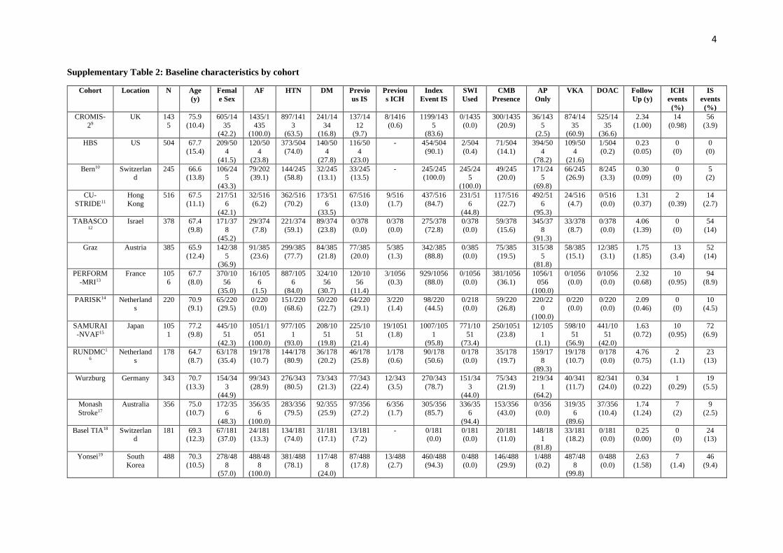
Supplementary Table 2: Baseline characteristics by cohort
Cohort Location N Age
(y)
Femal
e Sex
AF HTN DM Previo
us IS
Previou
s ICH
Index
Event IS
SWI
Used
CMB
Presence
AP
Only
VKA DOAC Follow
Up (y)
ICH
events
(%)
IS
events
(%)
CROMIS-
29
UK 143
5
75.9
(10.4)
605/14
35 (42.2)
1435/1
435 (100.0)
897/141
3 (63.5)
241/14
34 (16.8)
137/14
12 (9.7)
8/1416
(0.6)
1199/143
5 (83.6)
0/1435
(0.0)
300/1435
(20.9)
36/143
5 (2.5)
874/14
35 (60.9)
525/14
35 (36.6)
2.34
(1.00)
14
(0.98)
56
(3.9)
HBS US 504 67.7
(15.4)
209/50
4
(41.5)
120/50
4
(23.8)
373/504
(74.0)
140/50
4
(27.8)
116/50
4
(23.0)
- 454/504
(90.1)
2/504
(0.4)
71/504
(14.1)
394/50
4
(78.2)
109/50
4
(21.6)
1/504
(0.2)
0.23
(0.05)
0
(0)
0
(0)
Bern10 Switzerland
245 66.6 (13.8)
106/245
(43.3)
79/202 (39.1)
144/245 (58.8)
32/245 (13.1)
33/245 (13.5)
- 245/245 (100.0)
245/245
(100.0)
49/245 (20.0)
171/245
(69.8)
66/245 (26.9)
8/245 (3.3)
0.30 (0.09)
0 (0)
5 (2)
CU-
STRIDE11
Hong
Kong
516 67.5
(11.1)
217/51
6
(42.1)
32/516
(6.2)
362/516
(70.2)
173/51
6
(33.5)
67/516
(13.0)
9/516
(1.7)
437/516
(84.7)
231/51
6
(44.8)
117/516
(22.7)
492/51
6
(95.3)
24/516
(4.7)
0/516
(0.0)
1.31
(0.37)
2
(0.39)
14
(2.7)
TABASCO12
Israel 378 67.4
(9.8)
171/37
8
(45.2)
29/374
(7.8)
221/374
(59.1)
89/374
(23.8)
0/378
(0.0)
0/378
(0.0)
275/378
(72.8)
0/378
(0.0)
59/378
(15.6)
345/37
8
(91.3)
33/378
(8.7)
0/378
(0.0)
4.06
(1.39)
0
(0)
54
(14)
Graz Austria 385 65.9
(12.4)
142/38
5 (36.9)
91/385
(23.6)
299/385
(77.7)
84/385
(21.8)
77/385
(20.0)
5/385
(1.3)
342/385
(88.8)
0/385
(0.0)
75/385
(19.5)
315/38
5 (81.8)
58/385
(15.1)
12/385
(3.1)
1.75
(1.85)
13
(3.4)
52
(14)
PERFORM
-MRI13
France 105
6
67.7
(8.0)
370/10
56
(35.0)
16/105
6
(1.5)
887/105
6
(84.0)
324/10
56
(30.7)
120/10
56
(11.4)
3/1056
(0.3)
929/1056
(88.0)
0/1056
(0.0)
381/1056
(36.1)
1056/1
056
(100.0)
0/1056
(0.0)
0/1056
(0.0)
2.32
(0.68)
10
(0.95)
94
(8.9)
PARISK14 Netherlands
220 70.9 (9.1)
65/220 (29.5)
0/220 (0.0)
151/220 (68.6)
50/220 (22.7)
64/220 (29.1)
3/220 (1.4)
98/220 (44.5)
0/218 (0.0)
59/220 (26.8)
220/220
(100.0)
0/220 (0.0)
0/220 (0.0)
2.09 (0.46)
0 (0)
10 (4.5)
SAMURAI
-NVAF15
Japan 105
1
77.2
(9.8)
445/10
51
(42.3)
1051/1
051
(100.0)
977/105
1
(93.0)
208/10
51
(19.8)
225/10
51
(21.4)
19/1051
(1.8)
1007/105
1
(95.8)
771/10
51
(73.4)
250/1051
(23.8)
12/105
1
(1.1)
598/10
51
(56.9)
441/10
51
(42.0)
1.63
(0.72)
10
(0.95)
72
(6.9)
RUNDMC1
6
Netherland
s
178 64.7
(8.7)
63/178
(35.4)
19/178
(10.7)
144/178
(80.9)
36/178
(20.2)
46/178
(25.8)
1/178
(0.6)
90/178
(50.6)
0/178
(0.0)
35/178
(19.7)
159/17
8
(89.3)
19/178
(10.7)
0/178
(0.0)
4.76
(0.75)
2
(1.1)
23
(13)
Wurzburg Germany 343 70.7
(13.3)
154/34
3 (44.9)
99/343
(28.9)
276/343
(80.5)
73/343
(21.3)
77/343
(22.4)
12/343
(3.5)
270/343
(78.7)
151/34
3 (44.0)
75/343
(21.9)
219/34
1 (64.2)
40/341
(11.7)
82/341
(24.0)
0.34
(0.22)
1
(0.29)
19
(5.5)
Monash
Stroke17
Australia 356 75.0
(10.7)
172/35
6
(48.3)
356/35
6
(100.0)
283/356
(79.5)
92/355
(25.9)
97/356
(27.2)
6/356
(1.7)
305/356
(85.7)
336/35
6
(94.4)
153/356
(43.0)
0/356
(0.0)
319/35
6
(89.6)
37/356
(10.4)
1.74
(1.24)
7
(2)
9
(2.5)
Basel TIA18 Switzerland
181 69.3 (12.3)
67/181 (37.0)
24/181 (13.3)
134/181 (74.0)
31/181 (17.1)
13/181 (7.2)
- 0/181 (0.0)
0/181 (0.0)
20/181 (11.0)
148/181
(81.8)
33/181 (18.2)
0/181 (0.0)
0.25 (0.00)
0 (0)
24 (13)
Yonsei19 South
Korea
488 70.3
(10.5)
278/48
8 (57.0)
488/48
8 (100.0)
381/488
(78.1)
117/48
8 (24.0)
87/488
(17.8)
13/488
(2.7)
460/488
(94.3)
0/488
(0.0)
146/488
(29.9)
1/488
(0.2)
487/48
8 (99.8)
0/488
(0.0)
2.63
(1.58)
7
(1.4)
46
(9.4)

5
BIO-
STROKE/TIA20
Ireland 240 67.9
(13.3)
91/240
(37.9)
73/236
(30.9)
141/238
(59.2)
38/237
(16.0)
19/236
(8.1)
- 89/240
(37.1)
0/240
(0.0)
24/240
(10.0)
167/24
0 (69.6)
73/240
(30.4)
0/240
(0.0)
0.47
(0.35)
0
(0)
13
(5.4)
Kushiro
City21
Japan 631 71.5
(11.1)
257/63
1
(40.7)
86/631
(13.6)
407/631
(64.5)
182/63
1
(28.8)
115/63
1
(18.2)
17/631
(2.7)
631/631
(100.0)
0/631
(0.0)
268/631
(42.5)
568/63
1
(90.0)
63/631
(10.0)
0/631
(0.0)
0.15
(0.21)
20
(3.2)
99
(16)
IPAAC-Warfarin22
Hong Kong
81 71.3 (9.1)
40/81 (49.4)
81/81 (100.0)
56/81 (69.1)
27/81 (33.3)
25/81 (30.9)
1/81 (1.2)
65/81 (80.2)
71/81 (87.7)
24/81 (29.6)
0/81 (0.0)
81/81 (100.0)
0/81 (0.0)
2.10 (1.03)
3 (3.7)
5 (6.2)
CASPER23 Netherland
s
133 65.8
(10.6)
38/133
(28.6)
16/133
(12.0)
94/133
(70.7)
18/133
(13.5)
10/133
(7.5)
0/133
(0.0)
133/133
(100.0)
133/13
3
(100.0)
79/133
(59.4)
115/13
3
(86.5)
10/133
(7.5)
8/133
(6.0)
1.21
(0.17)
0
(0)
3
(2.3)
HERO24 Spain 935 77.6 (6.6)
487/935
(52.1)
856/933
(91.7)
693/933 (74.3)
212/932
(22.7)
246/933
(26.4)
8/933 (0.9)
803/925 (86.8)
0/935 (0.0)
247/935 (26.4)
1/934 (0.1)
623/934
(66.7)
310/934
(33.2)
1.92 (0.58)
18 (1.9)
32 (3.4)
HAGAKU
RE
Japan 350 73.1
(13.0)
141/35
0
(40.3)
102/35
0
(29.1)
263/347
(75.8)
116/35
0
(33.1)
50/350
(14.3)
10/349
(2.9)
317/350
(90.6)
28/350
(8.0)
127/350
(36.3)
197/35
0
(56.3)
93/350
(26.6)
60/350
(17.1)
2.15
(1.08)
9
(2.6)
23
(6.6)
Leuven25 Belgium 487 72.2
(9.4)
192/48
7
(39.4)
103/48
7
(21.1)
313/487
(64.3)
92/487
(18.9)
61/487
(12.5)
- 354/487
(72.7)
0/487
(0.0)
129/487
(26.5)
354/48
7
(72.7)
133/48
7
(27.3)
0/487
(0.0)
2.12
(0.72)
4
(0.82)
32
(6.6)
NOACISP Switzerlan
d
290 78.3
(9.1)
132/29
0 (45.5)
290/29
0 (100.0)
226/290
(77.9)
55/289
(19.0)
49/289
(17.0)
12/289
(4.2)
262/290
(90.3)
284/29
0 (97.9)
79/290
(27.2)
10/290
(3.4)
67/290
(23.1)
213/29
0 (73.4)
1.84
(0.74)
9
(3.1)
19
(6.6)
Min Lou26 China 106 64.4
(12.0)
34/106
(32.1)
16/106
(15.1)
80/106
(75.5)
- 18/106
(17.0)
7/106
(6.6)
106/106
(100.0)
106/10
6
(100.0)
36/106
(34.0)
92/106
(86.8)
7/106
(6.6)
7/106
(6.6)
0.39
(0.27)
0
(0)
2
(1.9)
MICRO27 Netherlands
397 65.3 (12.2)
165/397
(41.6)
30/396 (7.6)
218/397 (54.9)
54/397 (13.6)
35/397 (8.8)
0/397 (0.0)
35/397 (8.8)
0/397 (0.0)
72/397 (18.1)
357/397
(89.9)
40/397 (10.1)
0/397 (0.0)
3.25 (1.63)
11 (2.8)
21 (5.3)
Orken28 Turkey 452 71.9
(12.1)
233/45
2
(51.5)
353/45
2
(78.1)
356/452
(78.8)
150/45
2
(33.2)
123/45
2
(27.2)
0/452
(0.0)
432/452
(95.6)
250/45
2
(55.3)
132/452
(29.2)
0/452
(0.0)
321/45
2
(71.0)
131/45
2
(29.0)
2.59
(2.07)
3
(0.66)
8
(1.8)
CATCH29 Canada 392 67.6
(13.9)
154/39
2
(39.3)
26/392
(6.6)
218/392
(55.6)
54/392
(13.8)
0/392
(0.0)
0/392
(0.0)
236/392
(60.2)
0/392
(0.0)
62/392
(15.8)
325/39
2
(82.9)
67/392
(17.1)
0/392
(0.0)
0.26
(0.09)
1
(0.26)
13
(3.3)
MSS230 UK 209 66.4 (11.4)
82/209 (39.2)
21/209 (10.0)
157/209 (75.1)
- 29/209 (13.9)
0/209 (0.0)
209/209 (100.0)
199/209
(95.2)
34/209 (16.3)
188/209
(90.0)
21/209 (10.0)
0/209 (0.0)
1.08 (0.30)
0 (0)
31 (15)
Sainte-
Anne, Paris
France 302 78.6
(10.9)
154/30
2
(51.0)
302/30
2
(100.0)
215/302
(71.2)
56/302
(18.5)
39/302
(12.9)
6/302
(2.0)
302/302
(100.0)
0/279
(0.0)
80/302
(26.5)
0/302
(0.0)
122/30
2
(40.4)
180/30
2
(59.6)
1.53
(0.81)
5
(1.7)
20
(6.6)
STROKDE
M
France 178 64.0
(12.7)
68/178
(38.2)
12/178
(6.7)
100/178
(56.2)
23/178
(12.9)
20/178
(11.2)
1/178
(0.6)
178/178
(100.0)
0/178
(0.0)
23/178
(12.9)
130/17
8
(73.0)
40/178
(22.5)
8/178
(4.5)
3.32
(1.61)
0
(0)
16
(9)
NUS
(Chen)
Singapore 41 66.6
(10.2)
12/41
(29.3)
10/41
(24.4)
32/41
(78.0)
11/41
(26.8)
2/41
(4.9)
0/41
(0.0)
41/41
(100.0)
41/41
(100.0)
22/41
(53.7)
26/41
(63.4)
15/41
(36.6)
0/41
(0.0)
3.01
(1.32)
0
(0)
5
(12)
FUTURE Netherland
s
18 44.5
(5.3)
9/18
(50.0)
0/18
(0.0)
7/18
(38.9)
0/18
(0.0)
0/18
(0.0)
0/18
(0.0)
12/18
(66.7)
18/18
(100.0)
1/18
(5.6)
18/18
(100.0)
0/18
(0.0)
0/18
(0.0)
0.67
(0.72)
0
(0)
4
(22)

6
Heidelberg3
1
Germany 607 64.3
(14.0)
225/60
7 (37.1)
110/60
7 (18.1)
465/607
(76.6)
- 92/607
(15.2)
1/607
(0.2)
501/607
(82.5)
607/60
7 (100.0)
138/607
(22.7)
488/60
7 (80.4)
109/60
7 (18.0)
10/607
(1.6)
4.00
(1.27)
3
(0.49)
28
(4.6)
NNI Singapore 182 57.7
(11.5)
56/182
(30.8)
28/181
(15.5)
142/182
(78.0)
59/182
(32.4)
26/182
(14.3)
0/182
(0.0)
182/182
(100.0)
0/182
(0.0)
49/182
(26.9)
150/18
2
(82.4)
23/182
(12.6)
9/182
(4.9)
0.80
(0.63)
0
(0)
0
(0)
OXVASC32 UK 1067
68.3 (14.0)
508/1067
(47.6)
164/1066
(15.4)
581/1066
(54.5)
- - - 502/1067 (47.0)
0/1067 (0.0)
157/1067 (14.7)
949/1067
(88.9)
112/1067
(10.5)
6/1067 (0.6)
3.41 (1.53)
11 (1)
78 (7.3)
HKU32 Hong
Kong
966 68.9
(12.2)
388/96
6
(40.2)
124/96
6
(12.8)
628/966
(65.0)
272/96
6
(28.2)
93/966
(9.6)
12/966
(1.2)
966/966
(100.0)
966/96
6
(100.0)
433/966
(44.8)
862/96
6
(89.2)
63/966
(6.5)
41/966
(4.2)
2.90
(1.49)
19
(2)
89
(9.2)
Soo33 Hong
Kong
178 73.4
(9.6)
82/178
(46.1)
175/17
8
(98.3)
155/178
(87.1)
50/178
(28.1)
34/178
(19.1)
3/178
(1.7)
152/178
(85.4)
178/17
8
(100.0)
66/178
(37.1)
5/178
(2.8)
7/178
(3.9)
166/17
8
(93.3)
1.85
(1.44)
1
(0.56)
5
(2.8)
SIGNaL UK 206 72.4
(14.0)
85/206
(41.3)
65/206
(31.6)
146/206
(70.9)
49/206
(23.8)
55/206
(26.7)
8/206
(3.9)
185/206
(89.8)
140/20
6 (68.0)
92/206
(44.7)
163/20
6 (79.1)
9/206
(4.4)
34/206
(16.5)
0.60
(0.20)
1
(0.49)
24
(12)
Total 157
84
70.7
(12.2)
6697/1
5784
(42.4)
6882/1
5728
(43.8)
11222/1
5750
(71.3)
3208/1
3782
(23.3)
2300/1
4687
(15.7)
165/130
37
(1.3)
12804/15
774
(81.2)
4757/1
5759
(30.2)
4164/157
84
(26.4)
8733/1
5781
(55.3)
4759/1
5781
(30.2)
2289/1
5781
(14.5)
2.03
(1.53)
184
(1.2)
1048
(6.6)
Values shown are prevalence (%) or mean (SD). “ICH event (%)” and “IS event (%)” refer to the number and percentage of each cohort who experienced an event during
follow-up. Studies without references are unpublished. FUTURE: Follow-Up of Transient ischemic attack and stroke patients and Unelucidated Risk factor Evaluation study.
HAGAKURE: Hypertension, Amyloid, and aGe Associated Kaleidoscopic brain lesions on CT/MRI Undertaken with stroke REgistry. HBS: Heart Brain Interactions Study.
NNI: National Neuroscience Institute, Singapore. NOACISP: Novel Oral Anticoagulants in Stroke Patients, Basel; NCT02353585. SIGNaL: Stroke Investigation in North and
Central London. STROKDEM: Study of Factors Influencing Post-stroke Dementia.

7
Supplementary Table 3: Interaction terms
Each interaction was tested individually as an addition to a model comprising all candidate predictors for each
outcome. The association of each variable tested is shown at each level of the interacting variable, including the
interaction but not the main effect of the interacting variable. When testing interactions with antiplatelet vs
anticoagulant treatment, we omitted the three-level antithrombotic treatment to avoid collinearity. CMB recoded
describes CMB burden following recategorisation as a four-level variable to reduce sparseness.
A: Interactions with antithrombotic treatment
Variable Anticoagulant (HR, 95% CI) Antiplatelet (HR,
95% CI)
P-value for
interaction
ICH
CMB 0 1 1 0.36
CMB 1 2.11 (1.28 – 3.48) 1.72 (0.94 – 3.19)
CMB 2 - 4 2.01 (0.98 – 4.11) 2.33 (1.37 – 3.96)
CMB 5 - 10 1.25 (0.32 – 4.90) 4.66 (2.47 – 8.80)
CMB 11 - 19 5.67 (2.17 – 14.8) 4.53 (2.74 – 7.49)
CMB 20+ 3.15 (0.42 – 23.45) 15.01 (7.06 – 31.92)
Age (years) 1.04 (1.02 – 1.06) 1.03 (1.02 – 1.05) 0.66
Female sex 1.16 (0.81 – 1.66) 0.87 (0.54 – 1.40) 0.29
Presentation with ischaemic stroke 1.19 (0.49 – 2.89) 0.91 (0.34 – 2.48) 0.71
SWI MRI sequence used 0.73 (0.43 – 1.23) 0.42 (0.14 – 1.26) 0.28
Atrial fibrillation present 0.70 (0.29 – 1.73) 1.68 (0.96 – 2.91) 0.09
Hypertension present 0.85 (0.55 – 1.30) 1.17 (0.62 – 2.23) 0.41
Diabetes present 1.63 (0.97 – 2.73) 0.83 (0.45 – 1.50) 0.10
Ischaemic stroke before index event 1.30 (0.92 – 1.84) 1.41 (0.84 – 2.39) 0.81
Previous intracranial haemorrhage 1.98 (0.83 – 4.74) 7.39 (4.11 – 13.29) 0.011
East Asian population 1.17 (0.70 – 1.99) 3.21 (0.96 – 10.7) 0.09
Ischaemic stroke
CMB 0 1 1 054
CMB 1 0.92 (0.66 – 1.28) 1.16 (0.87 – 1.53)
CMB 2 - 4 1.16 (0.91 – 1.48) 1.32 (1.06 – 1.66)
CMB 5 - 10 0.95 (0.47 – 1.91) 1.98 (1.32 – 2.96)
CMB 11 - 19 1.43 (0.69 – 2.93) 1.44 (0.68 – 3.06)
CMB 20+ 1.85 (0.78 – 4.39) 1.94 (1.29 – 2.92)
Age (years) 1.02 (1.00 – 1.03) 1.02 (1.01 – 1.03) 0.69
Female sex 1.10 (0.88 – 1.38) 0.88 (0.71 – 1.09) 0.15
Presentation with ischaemic stroke 1.14 (0.71 – 1.82) 1.42 (0.93 – 2.15) 0.39
GRE MRI sequence used 0.94 (0.60 – 1.45) 0.50 (0.23 – 1.10) 0.04
Atrial fibrillation present 0.72 (0.43 – 1.21) 1.81 (1.30 – 2.50) 0.0040
Hypertension present 1.07 (0.79 – 1.45) 1.07 (0.74 – 1.55) 0.98
Diabetes present 1.37 (1.08 – 1.73) 1.25 (1.03 – 1.53) 0.53
Ischaemic stroke before index event 1.82 (1.22 – 2.71) 1.86 (1.48 – 2.34) 0.92
Previous intracranial haemorrhage 1.04 (0.49 – 2.22) 1.63 (0.79 – 3.35) 0.40
East Asian population 1.77 (1.12 – 2.81) 1.45 (0.49 – 4.28) 0.67
B: Interactions with MRI sequence type
Variable GRE SWI P-value for
interaction
ICH
CMB 0 1 1 0.50
CMB 1 2.28 (1.59 – 3.26) 1.09 (0.35 – 3.40)
CMB 2 - 4 2.50 (1.60 – 3.89) 1.32 (0.51 – 3.37)
CMB 5 - 10 2.91 (1.27 – 6.66) 3.40 (1.21 – 9.48)
CMB 11 - 19 5.09 (2.82 – 9.19) 4.20 (1.87 – 9.45)
CMB 20+ 9.14 (3.22 – 25.93) 8.35 (2.17 – 32.13)
Ischaemic stroke
CMB 0 1 1 0.0065
CMB 1 1.09 (0.80 – 1.49) 0.99 (0.69 – 1.41)
CMB 2 - 4 1.30 (1.12 – 1.50) 1.17 (0.75 – 1.81)
CMB 5 - 10 1.91 (1.24 – 2.93) 1.28 (0.77 – 2.14)
CMB 11 - 19 2.19 (1.14 – 4.21) 0.44 (0.21 – 0.95)
CMB 20+ 1.92 (1.29 – 2.84) 1.83 (0.95 – 3.50)
CMB recoded 0 1 1 0.18
CMB recoded 1 1.09 (0.80 – 1.49) 0.99 (0.69 – 1.41)
CMB recoded 2 – 4 1.30 (1.12 – 1.50) 1.17 (0.75 – 1.81)
CMB recoded 5+ 1.97 (1.38 – 2.82) 1.17 (0.75 – 1.83)
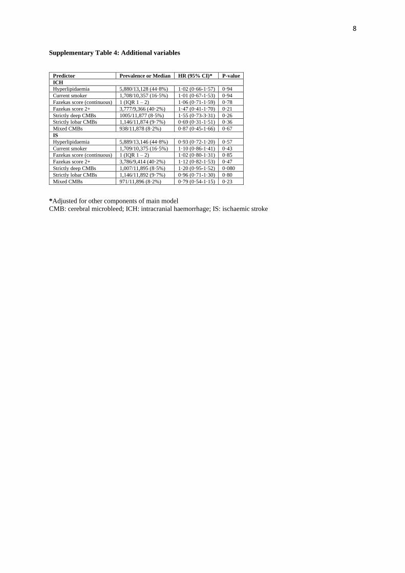
8
Supplementary Table 4: Additional variables
Predictor Prevalence or Median HR (95% CI)* P-value
ICH
Hyperlipidaemia 5,880/13,128 (44·8%) 1·02 (0·66-1·57) 0·94
Current smoker 1,708/10,357 (16·5%) 1·01 (0·67-1·53) 0·94
Fazekas score (continuous) 1 (IQR 1 – 2) 1·06 (0·71-1·59) 0·78
Fazekas score 2+ 3,777/9,366 (40·2%) 1·47 (0·41-1·70) 0·21
Strictly deep CMBs 1005/11,877 (8·5%) 1·55 (0·73-3·31) 0·26
Strictly lobar CMBs 1,146/11,874 (9·7%) 0·69 (0·31-1·51) 0·36
Mixed CMBs 938/11,878 (8·2%) 0·87 (0·45-1·66) 0·67
IS
Hyperlipidaemia 5,889/13,146 (44·8%) 0·93 (0·72-1·20) 0·57
Current smoker 1,709/10,375 (16·5%) 1·10 (0·86-1·41) 0·43
Fazekas score (continuous) 1 (IQR 1 – 2) 1·02 (0·80-1·31) 0·85
Fazekas score 2+ 3,786/9,414 (40·2%) 1·12 (0·82-1·53) 0·47
Strictly deep CMBs 1,007/11,895 (8·5%) 1·20 (0·95-1·52) 0·080
Strictly lobar CMBs 1,146/11,892 (9·7%) 0·96 (0·71-1·30) 0·80
Mixed CMBs 971/11,896 (8·2%) 0·79 (0·54-1·15) 0·23
*Adjusted for other components of main model
CMB: cerebral microbleed; ICH: intracranial haemorrhage; IS: ischaemic stroke

9
Supplementary Table 5: Characteristics of participants in highest-risk group for ICH and lower-risk group
for IS
Values show prevalence for categorical variables, and mean (SD) or median (IQR) for continuous variables.
Variable Group (n = 104) Remainder (n = 11,849)
Age 80·9 (8·11) 71·4 (11·7)
Female sex 57/104 (54·8%) 5,055/11,849 (42·7%)
East Asian population 4/104 (3·85%) 4,423/711,849 (37·3%)
Hypertension 81/104 (77·9%) 8,613/11,849 (72·8%)
Atrial fibrillation 102/104 (98·1%) 5,898/11,849 (49.8%)
Previous IS 1/104 (0·96%) 1,878/11,849 (15·6%)
Previous ICH 17/104 (16·4%) 131/11,849 (1·11%)
Diabetes mellitus 9/104 (8·7%) 2,825/11,849 (23·8%)
Hyperlipidaemia 42/104 (41·6%) 5,236/11,849 (44·7%)
CMB burden 0 13/104 (12·5%) 8,531/11,849 (72·0%)
1 60/104 (57·7%) 1,404/11,849 (11·9%)
2 – 4 28/104(26·9%) 1,207/11,849 (10·2%)
5 – 10 1/104 (1·0%) 382/11,849 (3·2%)
11 - 19 1/104 (1·0%) 178/11,849 (1·5%)
20 + 1/104 (1·0%) 147/11,849 (1·2%)
Antithrombotic treatment AP only 2/104 (1·9%) 8,670/11,849 (48.6%)
DOAC 19/104(18.3·3%) 2,215/11,849 (33·1%)
Warfarin/VKA 83/104 (79.8·8%) 4,612/11,849 (18·3%)
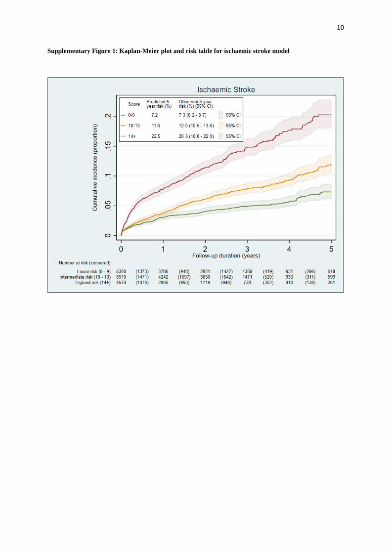
10
Supplementary Figure 1: Kaplan-Meier plot and risk table for ischaemic stroke model

11
Supplementary Figure 2: Model calibration – ischaemic stroke

12
Supplementary Figure 3: ICH model performance by MRI sequence type
Performance measure T2* GRE SWI
C-index (optimism-adjusted) 0·75 (0·70 – 0·79) 0·70 (0·62 – 0·79)
Calibration slope 0·94 (0·76 – 1·12) 0·94 (0·79 – 1·09)

13
Supplementary Figure 4: Comparative risks of symptomatic ICH and ischaemic stroke
Predicted five-year risks from ICH and ischaemic risk scores for all 11,953 participants with all variables available
without imputation. The red line indicates equality between predicted risks of ICH and IS; the orange line indicates
predicted IS risk twice that of ICH. For presentation, markers are translucent and jittered.

14
Supplementary Figure 5: Nomogram for symptomatic ICH risk
For each variable, select the appropriate category, then read the score for that variable from the scoring bar. After
summing the total score, select the corresponding ‘total score’ category and read the five-year ICH risk below.
0 21 43 5 6 7 8 9 10 11
Scoring
CMB count:0
0 1-4 5-10 11-19 20+
1-4 5-10 11-19 20+
GRESWI
Age:<70 70-79 80+
Population:Other East Asian
Previous ICH:
Previous IS:
No Yes
Antithrombotics:DOAC
No
APVKA
Yes
Total score:
Five-year ICH risk: 1.0% 2.3% 6.7%
8 +5 - 70 - 4
ICH

15
Supplementary Figure 6: Nomogram for ischaemic stroke risk
For each variable, select the appropriate category, then read the score for that variable from the scoring bar. After
summing the total score, select the corresponding ‘total score’ category and read the five-year IS risk below.
CMB count:0
0 1 5-19 20+
1 5-19 20+
GRESWI
Age:<60 60-69 70+
Population:Other East Asian
Previous IS:
Presentation:
No Yes
Antithrombotics:
TIA IS
Total score:
Five year IS risk: 7.2% 11.6% 22.5%
2-4
2-4
Diabetes:No Yes
OACAF
OACOther
APNo AF
APAF
0 21 43 5 6 7 8 9
Scoring
14 +10 - 130 - 9
Ischaemic stroke

16
Supplementary References
1 Vernooji MW, van der Lugt A, Ikram MA, et al. Prevalence and risk factors of cerebral microbleeds: the
Rotterdam Scan Study. Neurology 2008; 70(14):1208–14.
2 Mori N, Miki Y, Kikuta K, et al. Microbleeds in moyamoya disease: susceptibility-weighted imaging
versus T2*-weighted imaging at 3 Tesla. Invest Radiol 2008; 43(8):574–9.
3 Nandigam RNK, Viswanathan A, Delgado P, et al. MR imaging detection of cerebral microbleeds: effect
of susceptibility-weighted imaging, section thickness, and field strength. AJNR Am J Neuroradiol 2009; 30(20):
338.
4 Goos JDC, van der Flier WM, Knol DL, et al. Clinical relevance of improved microbleed detection by
susceptibility-weighted magnetic resonance imaging. Stroke 2011; 42(7): 1894–900.
5 Cheng AL, Batool S, McCreary CR, et al. Susceptibility-weighted imaging is more reliable than T2*-
weighted gradient-recalled echo MRI for detecting microbleeds. Stroke 2013; 44:2782–2786.
6 Guo LF, Wang G, Zhu XY, et al. Comparison of ESWAN, SWI-SPGR, and 2D T2*-weighted GRE
sequence for depicting cerebral microbleeds. Clin Neuroradiol 2013; 32(2):121–7.
7 Shams S, Martola J, Cavallin, et al. SWI or T2*: Which MRI sequence to use in the detection of cerebral
microbleeds? The Karolinska Imaging Dementia Study. Am J Neuroradiol 2015; 36(6): 1089–1095.
8 Shao L, Wang M, Ge X-H, et al. The use of susceptibility-weighted imaging to detect cerebral
microbleeds after lacunar infarction. Eur Rev Med Pharmacol Sci 2017; 21(13): 3105–3112.
9 Wilson D, Ambler G, Shakeshaft C, et al, and the CROMIS-2 collaborators. Cerebral microbleeds and
intracranial haemorrhage risk in patients anticoagulated for atrial fibrillation after acute ischaemic stroke or
transient ischaemic attack (CROMIS-2): a multicentre observational cohort study. Lancet Neurol 2018; 17: 539–
47
10 Gratz PP, El-Koussy M, Hsieh K, et al. Preexisting cerebral microbleeds on susceptibility-weighted
magnetic resonance imaging and post-thrombolysis bleeding risk in 392 patients. Stroke 2014; 45: 1684–88.
11 Yang J, Wong A, Wang Z, et al. Risk factors for incident dementia after stroke and transient ischemic
attack. Alzheimers Dement 2015; 11: 16–23.
12 Ben Assayag E, Korczyn AD, Giladi N, et al. Predictors for poststroke outcomes: the Tel Aviv Brain
Acute Stroke Cohort (TABASCO) study protocol. Int J Stroke 2012; 7: 341–47.
13 Chabriat H, Maeder P, Gass A, Michel P, Bracoud L, Hennerici M. Results of the PERFORM magnetic
resonance imaging study. J Neurol 2013; 260: 3071–76.
14 Truijman MT, Kooi ME, van Dijk AC, et al. Plaque At RISK (PARISK): prospective multicenter study
to improve diagnosis of high-risk carotid plaques. Int J Stroke 2014; 9: 747–54.
15 Yoshimura S, Koga M, Sato S, et al. Two-year outcomes of anticoagulation for acute ischemic stroke
with nonvalvular atrial fibrillation—SAMURAI-NVAF Study. Circ J 2018; 82: 1935–42.
16 van Leijsen EMC, van Uden IWM, Ghafoorian M, et al. Nonlinear temporal dynamics of cerebral small
vessel disease: The RUN DMC study. Neurology 2017; 89: 1569–77.
17 Karayiannis C, Soufan C, Chandra RV, et al. Prevalence of brain MRI markers of hemorrhagic risk in
patients with stroke and atrial fibrillation. Front Neurol 2016; 7: 151
18 Fluri F, Jax F, Amort M, et al. Significance of microbleeds in patients with transient ischaemic attack.
Eur J Neurol 2012; 19: 522–24.
19 Song TJ, Kim J, Song D, et al. Association of cerebral microbleeds with mortality in stroke patients
having atrial fibrillation. Neurology 2014; 83: 1308–15.
20 Callaly E, Ni Chroinin D, Hannon N, et al. Rates, predictors, and outcomes of early and late recurrence
after stroke: the north Dublin population stroke study. Stroke 2016; 47: 244–46.
21 Imaizumi T, Inamura S, Nomura T. The severities of white matter lesions possibly influence the
recurrences of several stroke types. J Stroke Cerebrovasc Dis 2014; 23: 1897–902.

17
22 Soo Y, Abrigo JM, Leung KT, et al, and the IPAAC study group. Risk of intracerebral haemorrhage in
Chinese patients with atrial fibrillation on warfarin with cerebral microbleeds: the IPAAC-Warfarin study. J
Neurol Neurosurg Psychiatry 2019; 90: 428–35.
23 Douven E, Schievink SHJ, Verhey FRJ, et al. The cognition and affect after stroke—a prospective
evaluation of risks (CASPER) study: rationale and design. BMC Neurol 2016; 16: 1–11.
24 Martí-Fàbregas J, Medrano-Martorell S, Merino E, et al. MRI predicts intracranial hemorrhage in
patients who receive long-term oral anticoagulation. Neurology 2019; 92(21):e2432-e2443
25 Thijs V, Lemmens R, Schoofs C, et al. Microbleeds and the risk of recurrent stroke. Stroke 2010; 41:
2005–09
26 Yan S, Jin X, Zhang X, Zhang S, Liebeskind DS, Lou M. Extensive cerebral microbleeds predict
parenchymal haemorrhage and poor outcome after intravenous thrombolysis. J Neurol Neurosurg Psychiatry
2015; 86: 1267–72.
27 Kwa VI, Algra A, Brundel M, Bouvy W, Kappelle LJ, and the MICRO Study Group. Microbleeds as a
predictor of intracerebral haemorrhage and ischaemic stroke after a TIA or minor ischaemic stroke: a cohort study.
BMJ Open 2013; 3: e002575.
28 Orken DN, Uysal E, Timer E, Kuloglu-Pazarcı N, Mumcu S, Forta H. New cerebral microbleeds in
ischemic stroke patients on warfarin treatment: two-year follow-up. Clin Neurol Neurosurg 2013; 115: 1682–85.
29 Zerna C, Modi J, Bilston L, Shoamanesh A, Coutts SB, Smith EE. Cerebral microbleeds and cortical
superficial siderosis in patients presenting with minor cerebrovascular events. Stroke 2016; 47: 2236–41.
30 Wardlaw J, Makin S, Valdés Hernández M, et al. Blood-brain barrier failure as a core mechanism in
cerebral small vessel disease and dementia: evidence from a cohort study. Alzheimers Dement 2017; 13: 634–43.
31 Horstmann S, Möhlenbruch M, Wegele C, et al. Prevalence of atrial fibrillation and association of
previous antithrombotic treatment in patients with cerebral microbleeds. Eur J Neurol 2015; 22: 1355–62.
32 Lau KK, Lovelock CE, Li L, et al. Antiplatelet treatment after transient ischemic attack and ischemic
stroke in patients with cerebral microbleeds in 2 large cohorts and an updated systematic review. Stroke 2018; 49:
1434–42.
33 Soo Y, Abrigo J, Leung KT, et al. Correlation of non-vitamin K antagonist oral anticoagulant exposure
and cerebral microbleeds in Chinese patients with atrial fibrillation. J Neurol Neurosurg Psychiatry 2018; 89:
680–86.