Embed Size (px)
Citation preview

الرحيم الرحمن الله بسم
Anatomy of ocular cranial nerves
Ocular cranial nerves
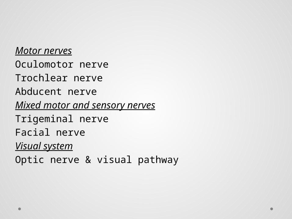
Motor nervesOculomotor nerveTrochlear nerveAbducent nerveMixed motor and sensory nervesTrigeminal nerveFacial nerveVisual systemOptic nerve & visual pathway
Oculomotor nerve(3rd)
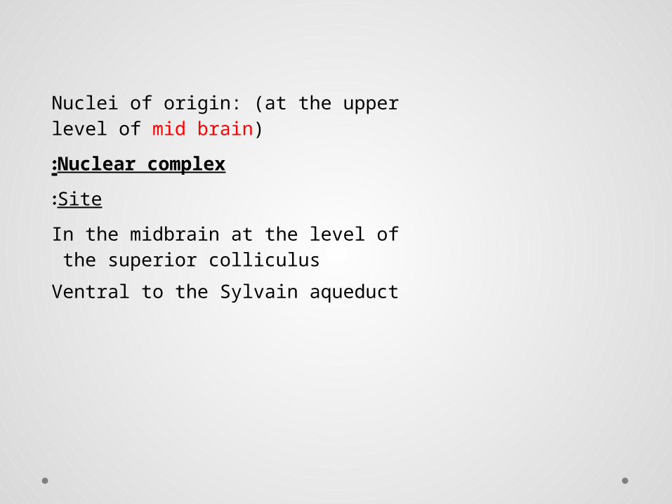
Nuclei of origin: (at the upper level of mid brain)
Nuclear complex:
Site:
In the midbrain at the level of the superior colliculus
Ventral to the Sylvain aqueduct

• Levator subnucleus
Unpaired
Innervates both levator muscles
• Superior rectus subnuclei
Paired
Innervates the contralateral superior rectus
• Medial rectus, inferior rectus & inferior oblique subnuclei
Paired
Innervate the ipsilateral muscles
Edinger-Westphal:
Paired
Give preganglionic parasympathetic -- ciliary ganglion.


Course and parts:
Fasciculus Third nerve nucleus (Efferent fibers) pass through the red nucleus----substantia nigra --- emerge at the medial aspect of the cerebral peduncle --- interpeduncular fossa.
Basilar partStarts as a series of rootlets which leave the midbrain on the medial aspect of the cerebral peduncle --- coalesce to form the main trunkPasses between posterior cerebral a. & superior
cerebellar a .Running lateral to and parallel with the posterior communicating a.

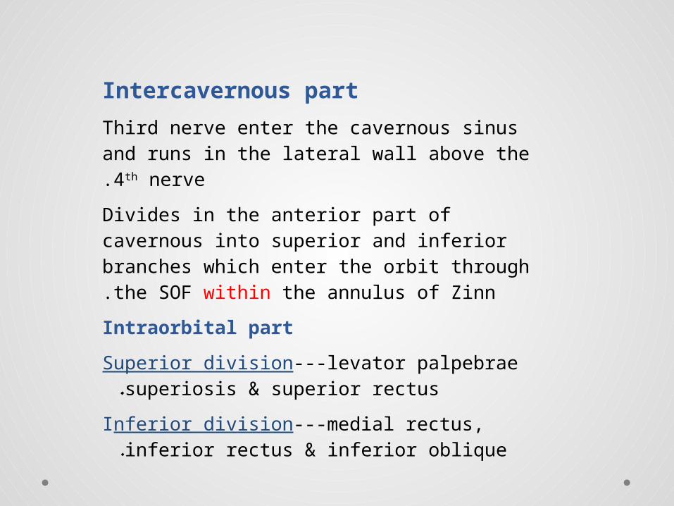
Intercavernous part
Third nerve enter the cavernous sinus and runs in the lateral wall above the 4th nerve.
Divides in the anterior part of cavernous into superior and inferior branches which enter the orbit through the SOF within the annulus of Zinn.
Intraorbital part
Superior division---levator palpebrae superiosis & superior rectus.
Inferior division---medial rectus, inferior rectus & inferior oblique.
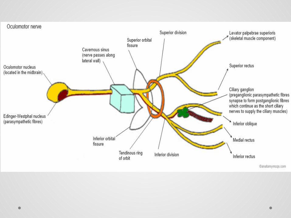
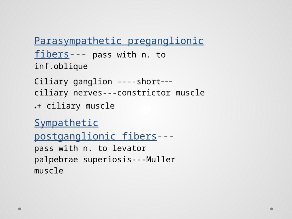
Parasympathetic preganglionic fibers--- pass with n. to inf.oblique
---Ciliary ganglion ----short ciliary nerves---constrictor muscle + ciliary
muscle.
Sympathetic postganglionic fibers---pass with n. to levator palpebrae superiosis---Muller muscle

Trochlear nerve(4th)
Nucleus
Site: In the midbrain at the level of the inferior colliculus.
Ventral to the Sylvain aqueduct
It is caudal to, and continuous with the 3rd nerve nuclear complex.

Course and parts
Fasciculus
Consists of axons which curve posteriorly & decussate around the cerebral aqueduct in the anterior medullary velum.
The trunk
It leave the brainstem on the dorsal surface just caudal to the inferior colliculus --- curve laterally around the brainstem--- runs forwards under the free edge of the tentorium--- pass between the posterior cerebral artery & the superior cerebellar artery .


Intracavernous part
The 4th nerve enters the cavernous sinus and runs in the lateral wall inferior to the 3rd nerve and above the 1st division of the 5th.
In the inferior part of the cavernous sinus it rises and passes through SOF above and lateral to the annulus of Zinn.
Intraorbital part
It passes anteriorly and medially crossing above the Oculomotor nerve, levator palpebral superiors muscle and the superior rectus muscle
It enters the superior oblique muscle on its orbital surface.


Abducent nerve

Nucleus
Site: at the lower level of the pons.
Ventral to the floor of the 4th ventricle.
The fasciculus of 7th nerve curve around
the Abducent nucleus and produce an
elevation in the floor of the 4th ventricle
(facial colliculus).


Course and parts
Fasciculus
Passes ventrally to leave the brainstem at the ponto medullary junction.
Basilar part
• Passes upward close to the base of the skull.
• Crossed by the anterior inferior cerebellar artery.
• Pierces the dura below the posterior clinoid.
• Crosses the apex of petrous bone.
• Enter the cavernous sinus.

Intracavernous part
Runs forwards inferolateral to I.C.A. at the lower level than 3rd & 4th nerves, as well as the ophthalmic n.
Intraorbital part
SOF within the annulus of Zinn (between the two heads of the LR muscle) --- lateral rectus muscle through its bulbar surface.

The trigeminal nerve

The trigeminal nerve has two roots, sensory and motor, and has a ganglion
:Origin
The two roots arise from the lateral border of the pons.
Course:
On emerging from brainstem --- push the dura at the apex of petrous bone ---- forming a dural pocket called Cavum Trigeminal.
Trigeminal ganglion
• Crescent-shaped mass of cells
• Lying in the trigeminal impression on the anterosuperior surface of petrous bone.
• Enclosed inside cavum trigeminal.
• It has 2 borders:
Posterior concave border ---- attached to sensory root.
Anterior convex border ---- attached to ophthalmic, maxillary & mandibular nerves.


The ophthalmic nerve
:Course
• It start at the trigeminal ganglion --- passes forward in the lateral wall of the cavernous sinus
• Just posterior to SOF, it receives the frontal, lacrimal, and nasociliary nerves
• The frontal and lacrimal nerves leave the orbit above the ligament of Zinn, while the nasociliary leaves through the annulus of the ligament.

Frontal nerve• The largest
• Runs over Levator palpebrae superiosis.
• Branches:
Supra trochlear n. --- sensory from the medial scalp, eyelid and conjunctiva.
Supra orbital n. --- sensory from the forehead, scalp & upper eyelid.

Nasociliary nerve• Medium sized
• Passes forward and medially (with the ophthalmic a.) crossing over optic n.
• Branches:
Sensory root to ciliary ganglion.
2 long ciliary nerves --- sensory to the globe + sympathetic to dilator ms.
Posterior ethmoidal n.
Anterior ethmoidal n.
Infratrochlear n. --- superior and inferior palpebral nerves.
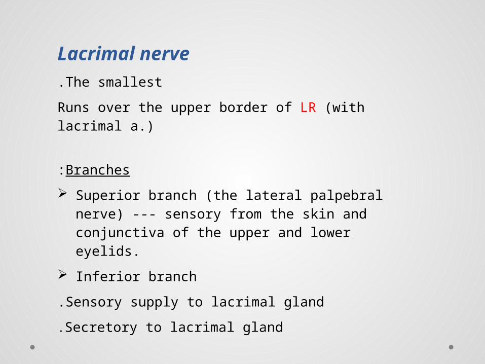
Lacrimal nerveThe smallest.
Runs over the upper border of LR (with lacrimal a.)
Branches:
Superior branch (the lateral palpebral nerve) --- sensory from the skin and conjunctiva of the upper and lower eyelids.
Inferior branch
Sensory supply to lacrimal gland.
Secretory to lacrimal gland.

The maxillary nerve
:Course
It start at the trigeminal ganglion --- pass forward in the lateral wall of the cavernous sinus --- foramen rotundum --- ptrego palatine fossa --- infratemporal fossa --- inferior orbital fissure --- continuous as infra orbital nerve.
It has 2 terminal branches:
Infraorbital nerve & Zygomatic nerve
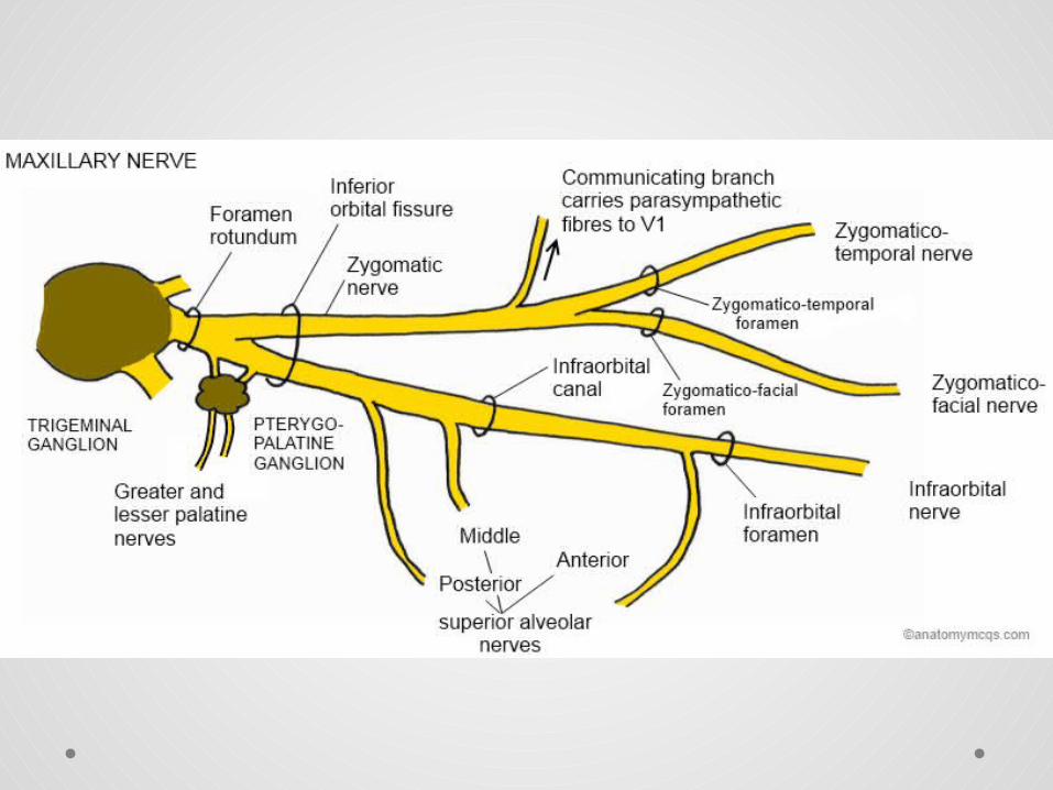

Infraorbital nerve:
It runs forwards from the inferior orbital fissure to --- infraorbital groove --- infraorbital canal --- infraorbital foramen
Branches:
• Middle superior alveolar n.
• Anterior superior alveolar n.
• Terminal ( Nasal , Labial , Palpebral )
The palpebral branch supply the skin and conjunctiva of lower lid.

Zygomatic nerve
It is divided into:
Zygomatico temporal and Zygomatico facial nerve
Zygomatico temporal nerve:
It runs in a groove on the lateral wall of orbit.
It gives a communicating branch to the lacrimal nerve which carries parasympathetic secretory fibers to the lacrimal gland.

Facial nerve
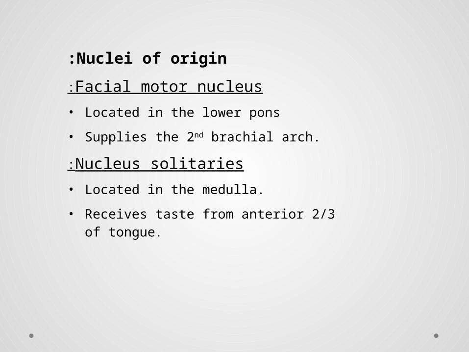
Nuclei of origin:
Facial motor nucleus:
• Located in the lower pons
• Supplies the 2nd brachial arch.
Nucleus solitaries:
• Located in the medulla.
• Receives taste from anterior 2/3 of tongue.

Parasympathetic nuclei:
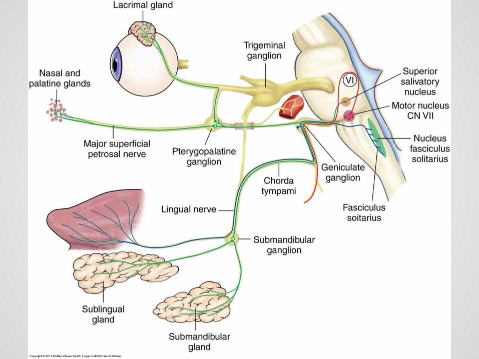
Lacrimatory nucleus --- parasympathetic preganglionic fibers to sphenopalatine ganglion --- lacrimal gland
It receives afferent fibers from hypothalamus (for emotional response) & from the sensory nuclei of the trigeminal nerve (for reflex lacrimation to irritation of the cornea or conjunctiva).

Course:
In the brain stem
It has 2 roots:
Motor root & nervus intermedius.
Both roots emerge from the brain stem at the inferior border of the pons, lateral to the Abducent nerve.
Intracranial & extracranial course
On emerge from the brain stem --- pass in the posterior cranial fossa --- internal auditory meatus --- facial canal.
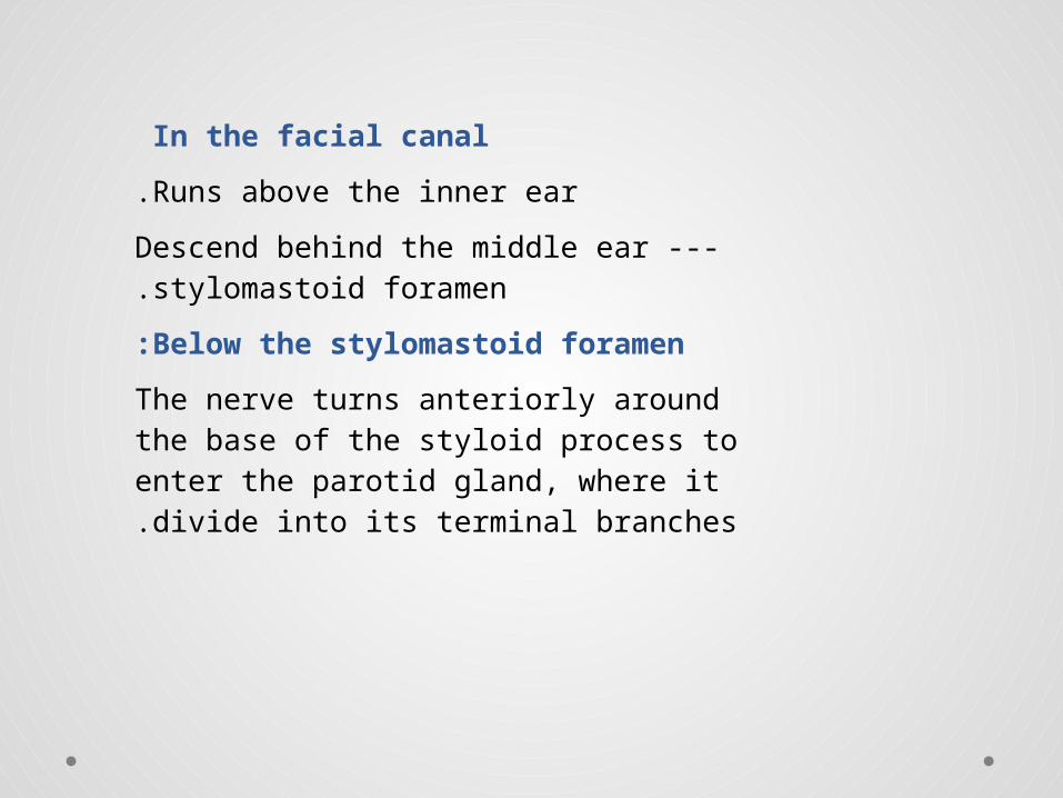
In the facial canal
Runs above the inner ear.
Descend behind the middle ear --- stylomastoid foramen.
Below the stylomastoid foramen:
The nerve turns anteriorly around the base of the styloid process to enter the parotid gland, where it divide into its terminal branches.

Branches:
In the facial canal:
Greater petrosal nerve :
• From geniculate ganglion
• Ptregopalatine fossa
• Sphenopalatine ganglion (relay) --- postganglionic parasympathetic fibers --- join maxillary n. --- Zygomatico temporal n. --- lacrimal n. --- lacrimal gland.
Nerve to Stapedius.
Chorda Tympani
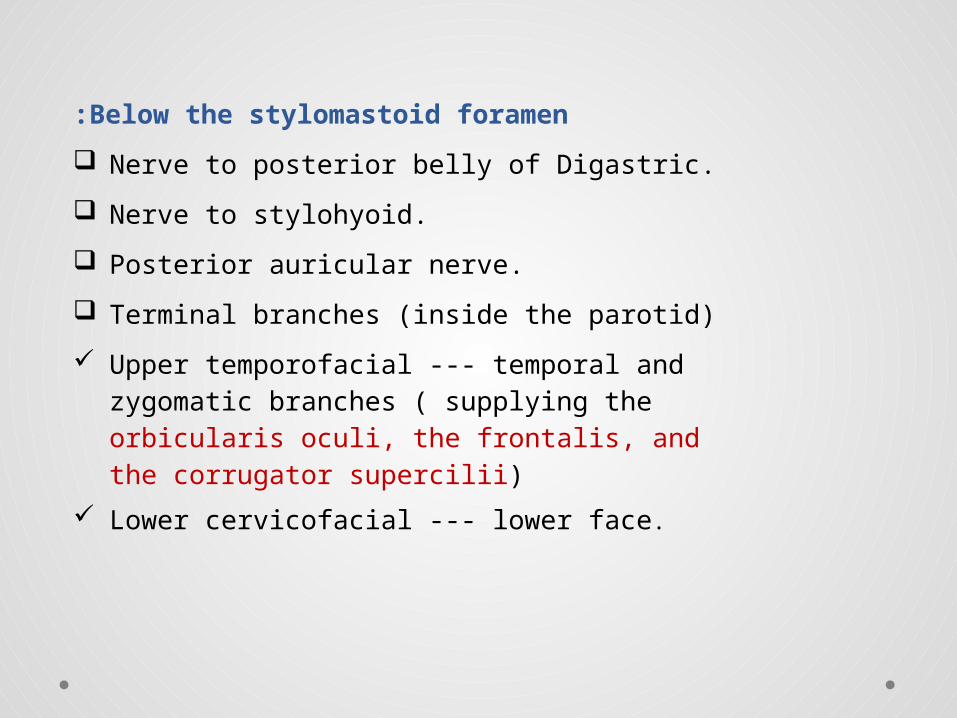
Below the stylomastoid foramen:
Nerve to posterior belly of Digastric.
Nerve to stylohyoid.
Posterior auricular nerve.
Terminal branches (inside the parotid)
Upper temporofacial --- temporal and zygomatic branches ( supplying the orbicularis oculi, the frontalis, and the corrugator supercilii)
Lower cervicofacial --- lower face.

Thank you