Embed Size (px)
Citation preview
Bioscience Reports (2018) 38 BSR20180470https://doi.org/10.1042/BSR20180470
Received: 27 March 2018Revised: 28 May 2018Accepted: 01 June 2018
Accepted Manuscript Online:05 June 2018Version of Record published:21 June 2018
Research Article
Nobiletin alleviates endometriosis viadown-regulating NF-κB activity in endometriosismouse modelXin Wei and Xu ShaoLiaocheng People’s Hospital, Dongchang West Road #67, Liaocheng 252000, China
Correspondence: Xu Shao ([email protected])
Nobiletin exhibits protective potential on inflammation and inhibits the activation of tran-scription factors nuclear factor-κB (NF-κB). However, its effects on the progression of en-dometriosis remain unsettled. The present study aimed to explore the in vivo alleviation ofnobiletin on endometriosis and its mechanism of action. The mouse model of endometrio-sis was established and administered with nobiletin. The ectopic lesion size was measuredand the hotplate test was performed to assess the amelioration of nobiletin on endometrio-sis. The expression of proliferation and angiogenesis relevant genes including proliferat-ing cell nuclear antigen (PCNA), vascular endothelial growth factor (VEGF), and E-cadherinwas measured by immunostaining and the mRNA expression of proinflammatory media-tors including interleukin (IL)-6, IL-1β, tumor necrosis factor (TNF)-α, matrix metallopro-teinases (MMP)-1, and MMP-3 was measured by RT-PCR. The change of NF-κB activity inendometriotic cells was evaluated by Western blotting and confirmed by luciferase assay.Administration of nobiletin significantly reduced lesions size and pain in endometriosis mice.Nobiletin significantly altered the expression of PCNA, VEGF, and E-cadherin in ectopic en-dometrium, as well as the levels of IL-6, IL-1β, TNF-α, MMP-1, and MMP-3. Nobiletin alsoshowed remarkably impairment on the activation of NF-κB in promoting endometriotic cells,likely targeting on the activity of IκB kinases (IKKs). The present study provides the first evi-dence that nobiletin exerts protection on endometriosis via inhibition the activation of NF-κB,specifically on the activity of IκB kinases.
IntroductionEndometriosis is defined as the presence of endometrial tissue outside the uterine cavity [1]. It is one of themost common gynecological disorders, affecting up to 10% of reproductive-aged women who suffer fromchronic pelvic pain, dysmenorrhea, dyspareunia, and subfertility [2,3]. New discoveries on the geneticand immune systems in the endometrium of women with endometriosis, and the secreted cytokines ofendometriotic lesions have given insights into the pathogenesis of this disorder, providing the foundationfor new treatments for disease-associated pain and infertility [4]. It is widely accepted that local inflamma-tion occurs in the peritoneal cavity of patients with endometriosis, which is characterized by the alterationof the immunologic components and inflammatory mediators in peritoneal fluid [5]. A number of studieshave shown that hormone such as gonadotropin-releasing hormone (GnRH) agonists and progestogensreduced endometriotic lesion development. Anti-inflammatory drugs including tumor necrosis factor(TNF)-α inhibitors, peroxisome proliferator-activated receptor-g (PPAR-g) agonists, and antibody againstmatrix metalloproteinases (anti-MMP) exert therapeutic effects on endometriosis [6–8]. Along with this,the presence of nuclear factor-κB (NF-κB) in human endometrium supports its role in the physiology and
c© 2018 The Author(s). This is an open access article published by Portland Press Limited on behalf of the Biochemical Society and distributed under the Creative Commons AttributionLicense 4.0 (CC BY).
1
Bioscience Reports (2018) 38 BSR20180470https://doi.org/10.1042/BSR20180470
pathophysiology of endometrial cells (ECs). Studies on the effects of NF-κB inhibitors in ECs and promoting en-dometriotic cells (EcCs) have shown that the inhibition of NF-κB could reduce endometriosis development andmaintenance [9]. Aberrant cytokine levels in the peritoneal fluid, which is regulated by NF-κB pathway, result ina proinflammatory local environment, promoting survival and growth of EcC in endometriosis patients [5]. NF-κBis a transcriptional factor that plays a crucial role in inflammation, immunity, cell adhesion, invasion, cellular prolif-eration, apoptosis, and angiogenesis [10]. Usage of NF-κB inhibitory agents in experimental endometriosis modelshas highlighted the role of the NF-κB pathway in endometriosis initiation and progression, making this pathway tobe an attractive target for the treatment and prevention of endometriosis.
Flavonoids have been reported to be functioned as anti-inflammatory agent and certain flavonoids affectstress/cytokine-induced NF-κB signal transduction. Among them, nobiletin is a polymethoxylated flavonoid foundin citrus fruit peel and has been widely used as herb medicine for centuries, playing important roles in tumor sup-pression, immune stimulation, anti-inflammation, antioxidation, as well as some cardiovascular disease mitigation[11,12]. For example, nobiletin inhibits the expression of the allergic cytokines, interleukin (IL)-4 and TNF-α as wellas the activation of their transcription factors NF-κB [13]. Given the inhibition of nobiletin on the NF-κB signalingwhich plays important roles in endometriosis pathogenesis, the objective of the present study was to investigate theeffects of nobiletin on the development of endometriotic lesions and how the process is regulated by NF-κB in an invivo experimental mice model of endometriosis.
Materials and methodsEthics statementAll animal procedures for these experiments were approved by the Institutional Animal and Use Committee atLiaocheng People’s Hospital. All mice were housed within environmentally controlled temperature and at a 12/12h (light/dark) schedule. All procedures were conducted in accordance with the approved animal protocol.
ReagentsNobiletin was isolated from Citrus depressa as described [14] and dissolved in saline containing 0.5% (v/v) Tween80 (Sigma-Aldrich, PA, U.S.A.). The following primary antibodies were used: Primary antibodies used in immuno-histochemistry against proliferating cell nuclear antigen (PCNA), vascular endothelial growth factor (VEGF), andE-cadherin were purchased from Santa Cruz Biotechnology (Santa Cruz, CA, U.S.A.). Rabbit monoclonal antibod-ies used in Western blotting against IκB kinase (IKK)α, p-IKKα, IκBα, p-IκBα, β-actin, and Lamin B were pur-chased from Abcam (Cambridge, MA, U.S.A.). Rabbit monoclonal antibody against p65 was purchased from Bey-otime (AN365, Jiangsu, China). The horseradish peroxidase-conjugated secondary antibodies were purchased fromThermofisher Scientific (Rockville, MD, U.S.A.).
Establishment of mouse model with endometriosisEndometriosis was experimentally induced as previously described [15]. Briefly, C57BL/6 female mice at 3–4 weekswere injected subcutaneously with pregnant mare serum gonadotropin (PMSG, 2 IU per mouse; Sigma-Aldrich).Approximately 42–44 h post PMSG injection, uteri tissues were harvested from these donor mice. Uterine stromaand epithelium (endometrium) were carefully separated from myometrium under dissecting microscope and werecut into 1 mm3 pieces. Further, endometrial pieces were suspended in saline, and 400 μl of suspension was injectedinto the peritoneal cavity of the recipient mice at 8–10 weeks. Recipient mice (2- to 4-month-old wild-type C57BL/6immunocompetent, reproductively intact females) were anesthetized with ketamine/xylazine (87.5 and 12.5 mg/kgbody weight), and Carprofen (5 mg/kg body weight) was given as analgesic upon surgery conclusion immediatelyand 48 h post-operatively. According to our preliminary study, after the establishment of endometriosis, the animalswere divided randomly into four groups (n=12): Sham control group, endometriosis (EM) group, EM plus nobiletinat 10 and 20 mg/kg groups. In the treatment group, mice were administered nobiletin intraperitoneally daily for 28consecutive days, and then killed through cervical dislocation and the total surface area of ectopic lesions in eachmouse was evaluated. Specifically, the abdominal cavity was immediately reopened, and all lesions were measuredwith two perpendicular diameters (the length D1 and the width D2). The surface area of each endometrial implanttissue was calculated by the formula: S = π × D1 × D2/4 (in mm2).
Hotplate proceduresThe sensitivity of the nociception was evaluated by the hotplate test [16] with a commercially available Hot PlateAnalgesia Meter (Model BME-480, Tianjin, China) consisting of a metal plate of 25 × 25 cm in size, which can be
2 c© 2018 The Author(s). This is an open access article published by Portland Press Limited on behalf of the Biochemical Society and distributed under the Creative Commons AttributionLicense 4.0 (CC BY).
Bioscience Reports (2018) 38 BSR20180470https://doi.org/10.1042/BSR20180470
heated to a constant temperature of 54.0 + 0.1◦C. As described in the manufacture’s instruction, the latency to respondto thermal stimulus is defined to be the time length (in second) elapsed from the moment when the mouse was placedin the cylinder to the time when it licked hind paws or jumped off the hot plate. The latency was calculated as themean of two readings recorded at intervals of 24 h. To assess the time-course progression in thermal latency as a resultof induced endometriosis, mice went through hotplate procedure at day 3, 7, 14, 21, and 28 respectively.
ImmunohistochemistryMice ectopic lesions were fixed with 10% neutral buffered formalin for 24 h at room temperature, followed by im-mersion in 70% ethanol overnight at 4◦C, then embedded in paraffin and sectioned at 4-μm thickness. For eachparaffin-embedded tissue block, slide was stained by hematoxylin and eosin (H&E) and immunostained for differentantibodies. For antigen retrieval, the slides were heated at 98◦C in the EDTA buffer (pH 8.0) or the citric acid solution(pH 6.0) for a total of 30 min and cooled naturally at room temperature. The slides were incubated with primary anti-bodies against PCNA, VEGF, and E-cadherin, diluted to 1:2,000, 1:50, and 1:100 respectively at 4◦C for 12 h. Further,after sufficient PBS wash, the slides were incubated with HRP-conjugated goat polyclonal antibody. The number andintensity of positive cells were counted by Image Pro-Plus 6.0 (Media Cybernetics, Inc., MD, U.S.A.). Images wereobtained with the microscope fitted with a digital camera (Olympus, SD, U.S.A.).
Quantitative real-time polymerase chain reaction (qRT-PCR)Ectopic endometrial samples were collected from the pelvic lesion region, and mRNA was extracted withRNeasy Mini Kit (Qiagen, Germany) according to manufacturer’s instruction. Concentration and quality oftotal RNA were assessed with a NanoDrop 2000 spectrophotometer (ThermoFisher Scientific, DE, U.S.A.).RNA was reverse transcribed using cDNA synthesis kits (Takara, Dalian, China), the expression levels of IL-6,IL-1β, TNF-α, MMP-1, and MMP-3 were determined using Taqman Real-Time PCR assays (ThermofisherScientific) on CFX96 TouchTM Real-Time PCR Detection System (Bio-Rad, CA, U.S.A.). The designed primerswere as follows: β-actin, F: 5′-CTGGGACGACATGGAGAAAA-3′, R: 5′-AAGGAAGGCTGGAAGAGTGC-3′;TNF-α, F: 5′-GCCACCACGCTCTTCTGTC-3′, R: 5′-TGCTCCTCCACTTGGTGGTT-3′; IL-1β,F: 5′-TTGACGGACCCCAAAAGATG-3′, R: 5′-AGCTGCCACAGCTTCTCCAC-3′; IL-6, F:5′-CCATCCAGTTGCCTTCTTGG-3′, R: 5′-TGCAAGTGCATCATCGTTGT-3′; MMP-1, F:5′-GGACTCTGAGCTCTTCTACC-3′; R: 5′-CACTAGAGACAAGAGTGGC-3′; MMP-3, F:5′-GGCAGAACCAAACAGGAGC-3′, R: 5′- GGCCCAGGAGTGCCTTCCCTCC-3′.
Cell culture and treatmentEndometrial and endometriotic stromal cells were isolated and characterized according to previous studies [17].Briefly, minced tissues were digested with type IV collagenase (Millipore, Shanghai, China), and stromal cells wereseparated from epithelial glands by passing through nylon meshes. The obtained stromal cells were then culturedin Dulbecco Modified Eagle Medium (Gibco, MD, U.S.A.) supplemented with 10% fetal bovine serum (FBS) andantibiotics at 37◦C.
Western blottingThe endometrial stromal cells were lysed and protein concentrations were determined by the Bradford method (Ther-mofisher Scientific, Gaithersburg, MD). Twenty-five micrograms of protein was loaded on 8% SDS-PAGE and trans-ferred onto a polyvinylidene difluoride membrane (Millipore, Bedford, MA). Nonspecific binding was blocked in5% nonfat milk at 4◦C overnight. Membrane was then incubated with rabbit monoclonal antibodies against IKKα,p-IKKα, IκBα, p-IκBα,β-actin, P65, and Lamin B at a 1:1000 dilution for 1 h at 37◦C. After washing with PBST bufferfor three times, membrane was further incubated with HRP-conjugated second antibody at a 1:25,000 dilutions for 1h at room temperature and detected by ECL substrate (Pierce, Rockville, MD).
Luciferase assayThe HEK293 cells (2 × 10⁵) were seeded on 24-well plates (Corning, Shanghai, China) and transfected with p65or IKKβ expression plasmids by standard calcium phosphate precipitation. After 34 h of transfection, cells wereincubated with nobiletin of 10 and 20 μg/ml for 2 h respectively. Then, cells were harvested for luciferase assays.Luciferase assays were performed with a dual-specific luciferase assay kit (Promega, Durham, NC).
c© 2018 The Author(s). This is an open access article published by Portland Press Limited on behalf of the Biochemical Society and distributed under the Creative Commons AttributionLicense 4.0 (CC BY).
3
Bioscience Reports (2018) 38 BSR20180470https://doi.org/10.1042/BSR20180470
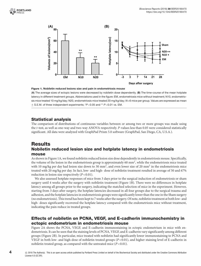
Figure 1. Nobiletin reduced lesions size and pain in endometriosis mouse
(A) The average sizes of ectopic lesions were decreased by nobiletin dose dependently. (B) The time-course of the mean hotplate
latency in different treatment groups. Abbreviations used in the figure: EM, endometriosis mice without treatment; N10, endometrio-
sis mice treated 10 mg/kg/day; N20, endometriosis mice treated 20 mg/kg/day; N=6 mice per group. Values are expressed as mean+− S.E.M. of three independent experiments; *P<0.05 and ** P<0.01 vs. EM.
Statistical analysisThe comparison of distributions of continuous variables between or among two or more groups was made usingthe t-test, as well as one-way and two-way ANOVA respectively. P-values less than 0.05 were considered statisticallysignificant. All data were analyzed with GraphPad Prism 5.0 software (GraphPad, San Diego, CA, U.S.A.).
ResultsNobiletin reduced lesion size and hotplate latency in endometriosismouseAs shown in Figure 1A, we found nobiletin reduced lesion size dose dependently in endometriosis mouse. Specifically,the volume of the lesion in the endometriosis group is approximately 60 mm2, while the endometriosis mice treatedwith 10 mg/kg per day had lesion size down to 30 mm2, and even lower size of 20 mm2 in the endometriosis micetreated with 20 mg/kg per day. In fact, low- and high- dose of nobiletin treatment resulted in average of 50 and 67%reduction in lesion size respectively (P<0.01).
We also assessed hotplate responses of mice from 3 days prior to the surgical induction of endometriosis or shamsurgery until 4 weeks after the surgery with nobiletin treatment (Figure 1B). There were no differences in hotplatelatency among all groups prior to the surgery, indicating the matched selection of mice in the experiment. However,starting from 3 days after surgery, the hotplate latencies decreased in all four groups due to the surgical trauma andadhesion, and the hotplate latencies in endometriosis groups were significantly lower than the one in the Sham surgery(no endometriosis). This trend has been kept to 7 weeks after the surgery. Of note, nobiletin treatment at both low- andhigh- doses significantly recovered the hotplate latency compared with the endometriosis mice without treatment,indicating the pain reduce in treated groups.
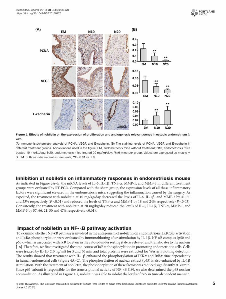
Effects of nobiletin on PCNA, VEGF, and E-cadherin immunochemistry inectopic endometrium in endometriosis mouseFigure 2A shows the PCNA, VEGF, and E-cadherin immunostaining in ectopic endometrium in mice with en-dometriosis. It can be seen that the staining levels of PCNA, VEGF, and E-cadherin vary significantly among differentgroups (Figure 2B). In particular, mice treated with nobiletin had significantly lower immunostaining to PCNA andVEGF in both low- and high-dose of nobiletin treated groups (P<0.01), and higher staining level of E-cadherin innobiletin treated group, as compared with the untreated mice (P<0.01).
4 c© 2018 The Author(s). This is an open access article published by Portland Press Limited on behalf of the Biochemical Society and distributed under the Creative Commons AttributionLicense 4.0 (CC BY).
Bioscience Reports (2018) 38 BSR20180470https://doi.org/10.1042/BSR20180470
Figure 2. Effects of nobiletin on the expression of proliferation and angiogenesis relevant genes in ectopic endometrium in
vivo
(A) Immunohistochemistry analysis of PCNA, VEGF, and E-cadherin. (B) The staining levels of PCNA, VEGF, and E-cadherin in
different treatment groups. Abbreviations used in the figure: EM, endometriosis mice without treatment; N10, endometriosis mice
treated 10 mg/kg/day; N20, endometriosis mice treated 20 mg/kg/day; N=6 mice per group. Values are expressed as means +−S.E.M. of three independent experiments; **P<0.01 vs. EM.
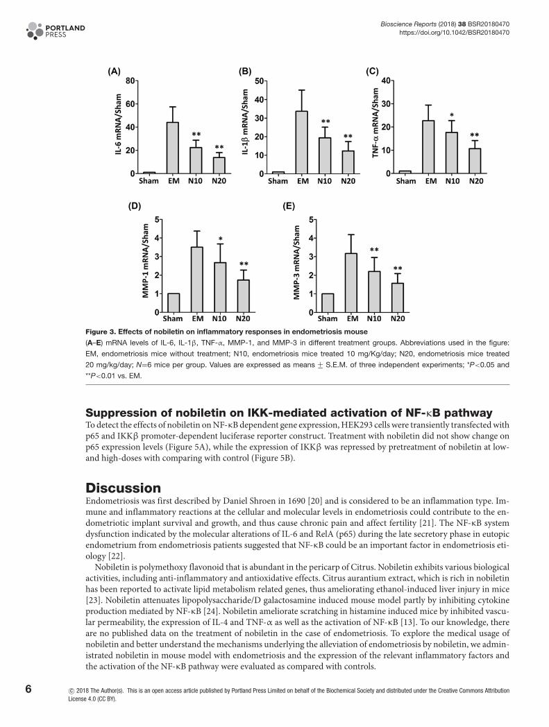
Inhibition of nobiletin on inflammatory responses in endometriosis mouseAs indicated in Figure 3A–E, the mRNA levels of IL-6, IL-1β, TNF-α, MMP-1, and MMP-3 in different treatmentgroups were evaluated by RT-PCR. Compared with the sham group, the expression levels of all these inflammatoryfactors were significant elevated in the endometriosis mice, suggesting the inflammation caused by the surgery. Asexpected, the treatment with nobiletin at 10 mg/kg/day decreased the levels of IL-6, IL-1β, and MMP-3 by 41, 30and 33% respectively (P<0.01) and reduced the levels of TNF-α and MMP-1 by 18 and 24% respectively (P<0.05).Consistently, the treatment with nobiletin at 20 mg/kg/day reduced the levels of IL-6, IL-1β, TNF-α, MMP-1, andMMP-3 by 57, 66, 21, 30 and 47% respectively<0.01).
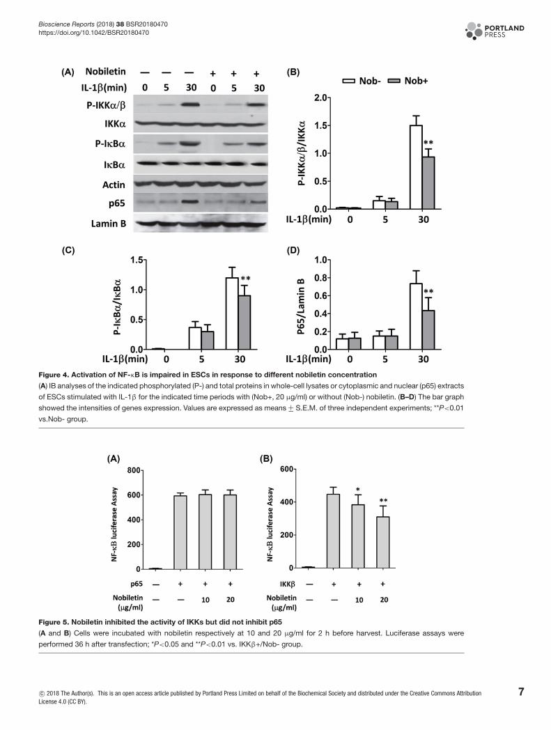
Impact of nobiletin on NF-κB pathway activationTo examine whether NF-κB pathway is involved in the antagonism of nobiletin on endometriosis, IKKα/β activationand IκBα phosphorylation were evaluated by immunoblotting after stimulation by IL-1β. NF-κB complex (p50 andp65), which is associated with IκB to retain in the cytosol under resting state, is released and translocates to the nucleus[18]. Therefore, we first investigated the time-course of IκBα phosphorylation in promoting endometriotic cells. Cellswere treated by IL-1β (10 ng/ml) for 5 and 30 min and total proteins were extracted for Western blotting detection.The results showed that treatment with IL-1β enhanced the phosphorylation of IKKα and IκBα time dependentlyin human endometrial cells (Figure 4A–C). The phosphorylation of nuclear extract (p65) is also enhanced by IL-1βstimulation. With the treatment of nobiletin, the phosphorylation of these factors was reduced significantly at 30 min.Since p65 subunit is responsible for the transcriptional activity of NF-κB [19], we also determined the p65 nuclearaccumulation. As illustrated in Figure 4D, nobiletin was able to inhibit the levels of p65 in time-dependent manner.
c© 2018 The Author(s). This is an open access article published by Portland Press Limited on behalf of the Biochemical Society and distributed under the Creative Commons AttributionLicense 4.0 (CC BY).
5
Bioscience Reports (2018) 38 BSR20180470https://doi.org/10.1042/BSR20180470
Figure 3. Effects of nobiletin on inflammatory responses in endometriosis mouse
(A–E) mRNA levels of IL-6, IL-1β, TNF-α, MMP-1, and MMP-3 in different treatment groups. Abbreviations used in the figure:
EM, endometriosis mice without treatment; N10, endometriosis mice treated 10 mg/Kg/day; N20, endometriosis mice treated
20 mg/kg/day; N=6 mice per group. Values are expressed as means +− S.E.M. of three independent experiments; *P<0.05 and
**P<0.01 vs. EM.
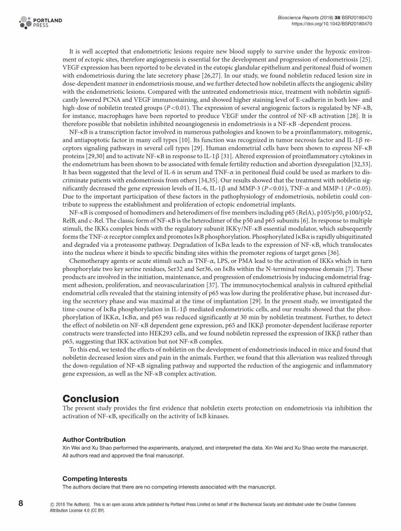
Suppression of nobiletin on IKK-mediated activation of NF-κB pathwayTo detect the effects of nobiletin on NF-κB dependent gene expression, HEK293 cells were transiently transfected withp65 and IKKβ promoter-dependent luciferase reporter construct. Treatment with nobiletin did not show change onp65 expression levels (Figure 5A), while the expression of IKKβ was repressed by pretreatment of nobiletin at low-and high-doses with comparing with control (Figure 5B).
DiscussionEndometriosis was first described by Daniel Shroen in 1690 [20] and is considered to be an inflammation type. Im-mune and inflammatory reactions at the cellular and molecular levels in endometriosis could contribute to the en-dometriotic implant survival and growth, and thus cause chronic pain and affect fertility [21]. The NF-κB systemdysfunction indicated by the molecular alterations of IL-6 and RelA (p65) during the late secretory phase in eutopicendometrium from endometriosis patients suggested that NF-κB could be an important factor in endometriosis eti-ology [22].
Nobiletin is polymethoxy flavonoid that is abundant in the pericarp of Citrus. Nobiletin exhibits various biologicalactivities, including anti-inflammatory and antioxidative effects. Citrus aurantium extract, which is rich in nobiletinhas been reported to activate lipid metabolism related genes, thus ameliorating ethanol-induced liver injury in mice[23]. Nobiletin attenuates lipopolysaccharide/D galactosamine induced mouse model partly by inhibiting cytokineproduction mediated by NF-κB [24]. Nobiletin ameliorate scratching in histamine induced mice by inhibited vascu-lar permeability, the expression of IL-4 and TNF-α as well as the activation of NF-κB [13]. To our knowledge, thereare no published data on the treatment of nobiletin in the case of endometriosis. To explore the medical usage ofnobiletin and better understand the mechanisms underlying the alleviation of endometriosis by nobiletin, we admin-istrated nobiletin in mouse model with endometriosis and the expression of the relevant inflammatory factors andthe activation of the NF-κB pathway were evaluated as compared with controls.
6 c© 2018 The Author(s). This is an open access article published by Portland Press Limited on behalf of the Biochemical Society and distributed under the Creative Commons AttributionLicense 4.0 (CC BY).
Bioscience Reports (2018) 38 BSR20180470https://doi.org/10.1042/BSR20180470
Figure 4. Activation of NF-κB is impaired in ESCs in response to different nobiletin concentration
(A) IB analyses of the indicated phosphorylated (P-) and total proteins in whole-cell lysates or cytoplasmic and nuclear (p65) extracts
of ESCs stimulated with IL-1β for the indicated time periods with (Nob+, 20 μg/ml) or without (Nob-) nobiletin. (B–D) The bar graph
showed the intensities of genes expression. Values are expressed as means +− S.E.M. of three independent experiments; **P<0.01
vs.Nob- group.
Figure 5. Nobiletin inhibited the activity of IKKs but did not inhibit p65
(A and B) Cells were incubated with nobiletin respectively at 10 and 20 μg/ml for 2 h before harvest. Luciferase assays were
performed 36 h after transfection; *P<0.05 and **P<0.01 vs. IKKβ+/Nob- group.
c© 2018 The Author(s). This is an open access article published by Portland Press Limited on behalf of the Biochemical Society and distributed under the Creative Commons AttributionLicense 4.0 (CC BY).
7
Bioscience Reports (2018) 38 BSR20180470https://doi.org/10.1042/BSR20180470
It is well accepted that endometriotic lesions require new blood supply to survive under the hypoxic environ-ment of ectopic sites, therefore angiogenesis is essential for the development and progression of endometriosis [25].VEGF expression has been reported to be elevated in the eutopic glandular epithelium and peritoneal fluid of womenwith endometriosis during the late secretory phase [26,27]. In our study, we found nobiletin reduced lesion size indose-dependent manner in endometriosis mouse, and we further detected how nobiletin affects the angiogenic abilitywith the endometriotic lesions. Compared with the untreated endometriosis mice, treatment with nobiletin signifi-cantly lowered PCNA and VEGF immunostaining, and showed higher staining level of E-cadherin in both low- andhigh-dose of nobiletin treated groups (P<0.01). The expression of several angiogenic factors is regulated by NF-κB,for instance, macrophages have been reported to produce VEGF under the control of NF-κB activation [28]. It istherefore possible that nobiletin inhibited neoangiogenesis in endometriosis is a NF-κB -dependent process.
NF-κB is a transcription factor involved in numerous pathologies and known to be a proinflammatory, mitogenic,and antiapoptotic factor in many cell types [10]. Its function was recognized in tumor necrosis factor and IL-1β re-ceptors signaling pathways in several cell types [29]. Human endometrial cells have been shown to express NF-κBproteins [29,30] and to activate NF-κB in response to IL-1β [31]. Altered expression of proinflammatory cytokines inthe endometrium has been shown to be associated with female fertility reduction and abortion dysregulation [32,33].It has been suggested that the level of IL-6 in serum and TNF-α in peritoneal fluid could be used as markers to dis-criminate patients with endometriosis from others [34,35]. Our results showed that the treatment with nobiletin sig-nificantly decreased the gene expression levels of IL-6, IL-1β and MMP-3 (P<0.01), TNF-α and MMP-1 (P<0.05).Due to the important participation of these factors in the pathophysiology of endometriosis, nobiletin could con-tribute to suppress the establishment and proliferation of ectopic endometrial implants.
NF-κB is composed of homodimers and heterodimers of five members including p65 (RelA), p105/p50, p100/p52,RelB, and c-Rel. The classic form of NF-κB is the heterodimer of the p50 and p65 subunits [6]. In response to multiplestimuli, the IKKs complex binds with the regulatory subunit IKKγ/NF-κB essential modulator, which subsequentlyforms the TNF-α receptor complex and promotes IκB phosphorylation. Phosphorylated IκBα is rapidly ubiquitinatedand degraded via a proteasome pathway. Degradation of IκBα leads to the expression of NF-κB, which translocatesinto the nucleus where it binds to specific binding sites within the promoter regions of target genes [36].
Chemotherapy agents or acute stimuli such as TNF-α, LPS, or PMA lead to the activation of IKKs which in turnphosphorylate two key serine residues, Ser32 and Ser36, on IκBs within the N-terminal response domain [7]. Theseproducts are involved in the initiation, maintenance, and progression of endometriosis by inducing endometrial frag-ment adhesion, proliferation, and neovascularization [37]. The immunocytochemical analysis in cultured epithelialendometrial cells revealed that the staining intensity of p65 was low during the proliferative phase, but increased dur-ing the secretory phase and was maximal at the time of implantation [29]. In the present study, we investigated thetime-course of IκBa phosphorylation in IL-1β mediated endometriotic cells, and our results showed that the phos-phorylation of IKKα, IκBα, and p65 was reduced significantly at 30 min by nobiletin treatment. Further, to detectthe effect of nobiletin on NF-κB dependent gene expression, p65 and IKKβ promoter-dependent luciferase reporterconstructs were transfected into HEK293 cells, and we found nobiletin repressed the expression of IKKβ rather thanp65, suggesting that IKK activation but not NF-κB complex.
To this end, we tested the effects of nobiletin on the development of endometriosis induced in mice and found thatnobiletin decreased lesion sizes and pain in the animals. Further, we found that this alleviation was realized throughthe down-regulation of NF-κB signaling pathway and supported the reduction of the angiogenic and inflammatorygene expression, as well as the NF-κB complex activation.
ConclusionThe present study provides the first evidence that nobiletin exerts protection on endometriosis via inhibition theactivation of NF-κB, specifically on the activity of IκB kinases.
Author ContributionXin Wei and Xu Shao performed the experiments, analyzed, and interpreted the data. Xin Wei and Xu Shao wrote the manuscript.All authors read and approved the final manuscript.
Competing InterestsThe authors declare that there are no competing interests associated with the manuscript.
8 c© 2018 The Author(s). This is an open access article published by Portland Press Limited on behalf of the Biochemical Society and distributed under the Creative CommonsAttribution License 4.0 (CC BY).
Bioscience Reports (2018) 38 BSR20180470https://doi.org/10.1042/BSR20180470
FundingThe authors confirm that there are no sources of funding to be acknowledged
AbbreviationsEC, endometrial cell; EcC, endometriotic cell; ECL, enhanced chemiluminescene; EM, endometriosis; GnRH,gonadotropin-releasing hormone; H&E, hematoxylin and eosin; HRP, horseradish peroxidase; IKK, IκB kinase; IL, interleukin;MMP, matrix metalloproteinases; NF-κB, nuclear factor-κB; PCNA, proliferating cell nuclear antigen; PMSG, pregnant mareserum gonadotropin; qRT-PCR, quantitative real-time polymerase chain reaction; TNF, tumor necrosis factor; VEGF, vascularendothelial growth factor.
References1 Defrere, S., Gonzalez-Ramos, R., Lousse, J.C., Colette, S., Donnez, O., Donnez, J. et al. (2011) Insights into iron and nuclear factor-kappa B
(NF-kappaB) involvement in chronic inflammatory processes in peritoneal endometriosis. Histol. Histopathol. 26, 1083–10922 Holoch, K.J. and Lessey, B.A. (2010) Endometriosis and infertility. Clin. Obstet. Gynecol. 53, 429–438,
https://doi.org/10.1097/GRF.0b013e3181db7d713 Nasu, K., Nishida, M., Ueda, T., Yuge, A., Takai, N. and Narahara, H. (2007) Application of the nuclear factor-kappaB inhibitor BAY 11-7085 for the
treatment of endometriosis: an in vitro study. Am. J. Physiol. Endocrinol. Metab. 293, E16–E23, https://doi.org/10.1152/ajpendo.00135.20064 Giudice, L.C. and Kao, L.C. (2004) Endometriosis. Lancet 364, 1789–1799, https://doi.org/10.1016/S0140-6736(04)17403-55 Lousse, J.C., Van Langendonckt, A., Gonzalez-Ramos, R., Defrere, S., Renkin, E. and Donnez, J. (2008) Increased activation of nuclear factor-kappa B
(NF-kappaB) in isolated peritoneal macrophages of patients with endometriosis. Fertil. Steril. 90, 217–220,https://doi.org/10.1016/j.fertnstert.2007.06.015
6 Bruner, K.L., Matrisian, L.M., Rodgers, W.H., Gorstein, F. and Osteen, K.G. (1997) Suppression of matrix metalloproteinases inhibits establishment ofectopic lesions by human endometrium in nude mice. J. Clin. Invest. 99, 2851–2857, https://doi.org/10.1172/JCI119478
7 Falconer, H., Mwenda, J.M., Chai, D.C., Wagner, C., Song, X.Y., Mihalyi, A. et al. (2006) Treatment with anti-TNF monoclonal antibody (c5N) reduces theextent of induced endometriosis in the baboon. Hum. Reprod. 21, 1856–1862, https://doi.org/10.1093/humrep/del044
8 Mihalyi, A., Simsa, P., Mutinda, K.C., Meuleman, C., Mwenda, J.M. and D’Hooghe, T.M. (2006) Emerging drugs in endometriosis. Expert Opin. Emerg.Drugs 11, 503–524, https://doi.org/10.1517/14728214.11.3.503
9 Gonzalez-Ramos, R., Van Langendonckt, A., Defrere, S., Lousse, J.C., Colette, S., Devoto, L. et al. (2010) Involvement of the nuclear factor-kappaBpathway in the pathogenesis of endometriosis. Fertil. Steril. 94, 1985–1994, https://doi.org/10.1016/j.fertnstert.2010.01.013
10 Viatour, P., Merville, M.P., Bours, V. and Chariot, A. (2005) Phosphorylation of NF-kappaB and IkappaB proteins: implications in cancer andinflammation. Trends Biochem. Sci. 30, 43–52, https://doi.org/10.1016/j.tibs.2004.11.009
11 Knekt, P., Jarvinen, R., Seppanen, R., Hellovaara, M., Teppo, L., Pukkala, E. et al. (1997) Dietary flavonoids and the risk of lung cancer and othermalignant neoplasms. Am. J. Epidemiol. 146, 223–230, https://doi.org/10.1093/oxfordjournals.aje.a009257
12 Li, S., Yu, H. and Ho, C.T. (2006) Nobiletin: efficient and large quantity isolation from orange peel extract. Biomed. Chromatogr. 20, 133–138,https://doi.org/10.1002/bmc.540
13 Jang, S.E., Ryu, K.R., Park, S.H., Chung, S., Teruya, Y., Han, M.J. et al. (2013) Nobiletin and tangeretin ameliorate scratching behavior in mice byinhibiting the action of histamine and the activation of NF-kappaB, AP-1 and p38. Int. Immunopharmacol. 17, 502–507,https://doi.org/10.1016/j.intimp.2013.07.012
14 Nagase, H., Yamakuni, T., Matsuzaki, K., Maruyama, Y., Kasahara, J., Hinohara, Y. et al. (2005) Mechanism of neurotrophic action of nobiletin in PC12Dcells. Biochemistry 44, 13683–13691, https://doi.org/10.1021/bi050643x
15 Nothnick, W.B., Graham, A., Holbert, J. and Weiss, M.J. (2014) miR-451 deficiency is associated with altered endometrial fibrinogen alpha chainexpression and reduced endometriotic implant establishment in an experimental mouse model. PLoS One 9, e100336,https://doi.org/10.1371/journal.pone.0100336
16 Bannon, A.W. and Malmberg, A.B. (2007) Models of nociception: hot-plate, tail-flick, and formalin tests in rodents. Curr. Protoc. 41, 8.9.1–8.9.1617 Hsiao, K.Y., Chang, N., Lin, S.C., Li, Y.H. and Wu, M.H. (2014) Inhibition of dual specificity phosphatase-2 by hypoxia promotes interleukin-8-mediated
angiogenesis in endometriosis. Hum. Reprod. 29, 2747–2755, https://doi.org/10.1093/humrep/deu25518 Perkins, N.D. (2007) Integrating cell-signalling pathways with NF-kappaB and IKK function. Nat. Rev. Mol. Cell Biol. 8, 49–62,
https://doi.org/10.1038/nrm208319 Tse, A.K., Wan, C.K., Shen, X.L., Yang, M. and Fong, W.F. (2005) Honokiol inhibits TNF-alpha-stimulated NF-kappaB activation and
NF-kappaB-regulated gene expression through suppression of IKK activation. Biochem. Pharmacol. 70, 1443–1457,https://doi.org/10.1016/j.bcp.2005.08.011
20 Knapp, V.J. (1999) How old is endometriosis? Late 17th- and 18th-century European descriptions of the disease. Fertil. Steril. 72, 10–1421 Kaponis, A., Iwabe, T., Taniguchi, F., Ito, M., Deura, I., Decavalas, G. et al. (2012) The role of NF-kappaB in endometriosis. Front. Biosci. 4, 1213–123422 Ponce, C., Torres, M., Galleguillos, C., Sovino, H., Boric, M.A., Fuentes, A. et al. (2009) Nuclear factor kappaB pathway and interleukin-6 are affected in
eutopic endometrium of women with endometriosis. Reproduction 137, 727–737, https://doi.org/10.1530/REP-08-040723 Choi, B.K., Kim, T.W., Lee, D.R., Jung, W.H., Lim, J.H., Jung, J.Y. et al. (2015) A polymethoxy flavonoids-rich Citrus aurantium extract ameliorates
ethanol-induced liver injury through modulation of AMPK and Nrf2-related signals in a binge drinking mouse model. Phytother. Res. 29, 1577–1584,https://doi.org/10.1002/ptr.5415
c© 2018 The Author(s). This is an open access article published by Portland Press Limited on behalf of the Biochemical Society and distributed under the Creative CommonsAttribution License 4.0 (CC BY).
9
Bioscience Reports (2018) 38 BSR20180470https://doi.org/10.1042/BSR20180470
24 He, Z., Li, X., Chen, H., He, K., Liu, Y., Gong, J. et al. (2016) Nobiletin attenuates lipopolysaccharide/Dgalactosamineinduced liver injury in mice byactivating the Nrf2 antioxidant pathway and subsequently inhibiting NFkappaBmediated cytokine production. Mol. Med. Rep. 14, 5595–5600,https://doi.org/10.3892/mmr.2016.5943
25 Rocha, A.L., Reis, F.M. and Taylor, R.N. (2013) Angiogenesis and endometriosis. Obstet. Gynecol. Int. 2013, 859619,https://doi.org/10.1155/2013/859619
26 Donnez, J., Smoes, P., Gillerot, S., Casanas-Roux, F. and Nisolle, M. (1998) Vascular endothelial growth factor (VEGF) in endometriosis. Hum. Reprod.13, 1686–1690, https://doi.org/10.1093/humrep/13.6.1686
27 Kupker, W., Schultze-Mosgau, A. and Diedrich, K. (1998) Paracrine changes in the peritoneal environment of women with endometriosis. Hum. Reprod.Update 4, 719–723, https://doi.org/10.1093/humupd/4.5.719
28 Kiriakidis, S., Andreakos, E., Monaco, C., Foxwell, B., Feldmann, M. and Paleolog, E. (2003) VEGF expression in human macrophages isNF-kappaB-dependent: studies using adenoviruses expressing the endogenous NF-kappaB inhibitor IkappaBalpha and a kinase-defective form of theIkappaB kinase 2. J. Cell Sci. 116, 665–674, https://doi.org/10.1242/jcs.00286
29 Laird, S.M., Tuckerman, E.M., Cork, B.A. and Li, T.C. (2000) Expression of nuclear factor kappa B in human endometrium; role in the control ofinterleukin 6 and leukaemia inhibitory factor production. Mol. Hum. Reprod. 6, 34–40, https://doi.org/10.1093/molehr/6.1.34
30 Page, M., Tuckerman, E.M., Li, T.C. and Laird, S.M. (2002) Expression of nuclear factor kappa B components in human endometrium. J. Reprod.Immunol. 54, 1–13, https://doi.org/10.1016/S0165-0378(01)00122-X
31 Cao, W.G., Morin, M., Metz, C., Maheux, R. and Akoum, A. (2005) Stimulation of macrophage migration inhibitory factor expression in endometrialstromal cells by interleukin 1, beta involving the nuclear transcription factor NFkappaB. Biol. Reprod. 73, 565–570,https://doi.org/10.1095/biolreprod.104.038331
32 von Wolff, M., Thaler, C.J., Strowitzki, T., Broome, J., Stolz, W. and Tabibzadeh, S. (2000) Regulated expression of cytokines in human endometriumthroughout the menstrual cycle: dysregulation in habitual abortion. Mol. Hum. Reprod. 6, 627–634, https://doi.org/10.1093/molehr/6.7.627
33 Kao, L.C., Germeyer, A., Tulac, S., Lobo, S., Yang, J.P., Taylor, R.N. et al. (2003) Expression profiling of endometrium from women with endometriosisreveals candidate genes for disease-based implantation failure and infertility. Endocrinology 144, 2870–2881, https://doi.org/10.1210/en.2003-0043
34 Bedaiwy, M.A., Falcone, T., Sharma, R.K., Goldberg, J.M., Attaran, M., Nelson, D.R. et al. (2002) Prediction of endometriosis with serum and peritonealfluid markers: a prospective controlled trial. Hum. Reprod. 17, 426–431, https://doi.org/10.1093/humrep/17.2.426
35 Cheong, Y.C., Shelton, J.B., Laird, S.M., Richmond, M., Kudesia, G., Li, T.C. et al. (2002) IL-1, IL-6 and TNF-alpha concentrations in the peritoneal fluidof women with pelvic adhesions. Hum. Reprod. 17, 69–75, https://doi.org/10.1093/humrep/17.1.69
36 Hoesel, B. and Schmid, J.A. (2013) The complexity of NF-kappaB signaling in inflammation and cancer. Mol. Cancer 12, 86,https://doi.org/10.1186/1476-4598-12-86
37 Lebovic, D.I., Mueller, M.D. and Taylor, R.N. (2001) Immunobiology of endometriosis. Fertil. Steril. 75, 1–10,https://doi.org/10.1016/S0015-0282(00)01630-7
10 c© 2018 The Author(s). This is an open access article published by Portland Press Limited on behalf of the Biochemical Society and distributed under the Creative Commons AttributionLicense 4.0 (CC BY).