Embed Size (px)
Citation preview
APPROACH TO PLEURAL EFFUSION
תל השומר ' נה מוחמד פנימית ד'מחאג

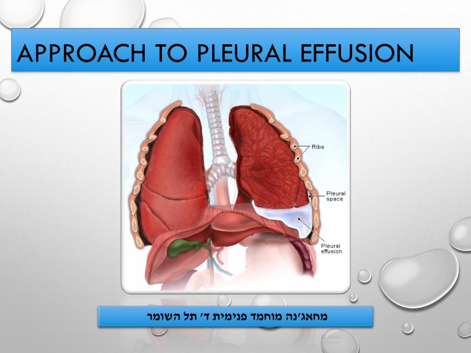
שמתלוננת על קוצר נשימה עם החמרה בימים 34אתם תורנים בחדר מיון מגיעה אישה צעירה בת
מדווחת שלאחרונה יש לה , כאבים בחזה בעיקר בצד ימין ,מלווה בחום וצמרמורת אמתית , האחרונים
.נוטלת גלולות, מכורה לאלכוהול , מעשנת : ברקע.הזעות לילה ושיעול פרודוקטיבי
:בקבלתה
עם דיספניית, 38.6חום , 140/80לחץ דם , קולות לב סדירים ללא אוושות הימודנאמיתיציבה
.בטן רכה למעט רגישות קלה בבטן עליונה עם הקרנה קלה לגב , באוויר חדר 93סטורציה
עם עמימות בניקוש , ירודפרימיטוס, כניסת אוויר מופחתת מימין , התפשטות בית חזה לא סימטרית
מצד ימין
גפיים תקינות ללא בצקות ללא סימני צלוליטיס .
אזיניל, קודיקאל: בקהילה קבלה , מדווחת שמזה חודש סובלת מכאבי חזה מתגברים בנשימה
. ללא שיפור משמעותי . ואיטופאן,טאריביד,

Differential diagnosis DOCs ?

• Definition and overview
• Pathophysiology
• Etiology
• Clinical manifestation
• Complications
• Lab tests and diagnosis
• Treatment and management
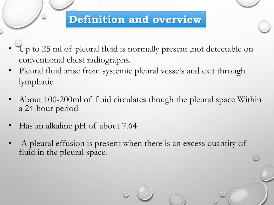
Definition and overview
• Up to 25 ml of pleural fluid is normally present ,not detectable on
conventional chest radiographs.
• Pleural fluid arise from systemic pleural vessels and exit through
lymphatic
• About 100-200ml of fluid circulates though the pleural space Within a 24-hour period
• Has an alkaline pH of about 7.64
• A pleural effusion is present when there is an excess quantity of fluid in the pleural space.

ETIOLOGY AND PATHOPHYSIOLOGY

Increased pulmonary capillary pressure (CHF)
Decreased intrapleural pressure (atelectasis)
Increased capillary permeability (Pneumonia)
Decreased plasma oncotic pressure (hypoalbuminemia)
Increased pleural membrane permeability (malignancy)
lymphatic obstruction (malignancy) , rupture
[chylothorax]
diaphragmatic defect , cirrhosis (hepatic hydrothorax)

PLEURAL EFFUSION TYPES
• THE 5 MAJOR TYPES OF PLEURAL EFFUSION
ARE:
1) TRANSUDATE
2) EXUDATE
3) EMPYEMA
4) HEMORRHAGIC PLEURAL EFFUSION OR
HEMOTHORAX
5) CHYLOUS OR CHYLIFORM EFFUSION.

Harrison’s Principles of Internal Medicine, 18th edition.
Fauci, Braunwald, Kasper, Hauser, Longo, Jameson, Loscalzo.

LEADING CAUSES OF PLEURAL EFFUSION IN USA
IN DECREASING ORDER OF INCIDENCE
1. CONGESTIVE HEART FAILURE
2. PNEUMONIA
3. CANCER
4. PULMONARY EMBOLISM
5. VIRAL DISEASE
6. CABG
7. CIRRHOSIS WITH ASCITES

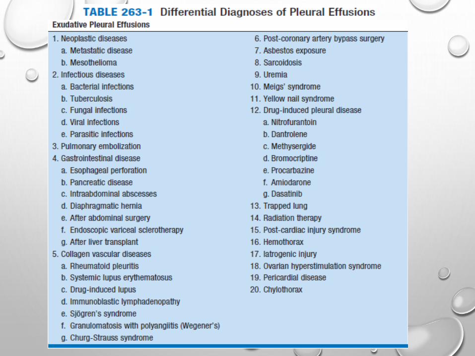
EXUDATIVE PLEURAL EFFUSIONS
Drug-induced pleural disease 1) Nitrofurantoin
2) Dantrolene
3) Methysergide
4) Bromocriptine
5) Procarbazine
6) Amiodarone

CLINICAL MANIFESTATIONPHYSICAL FINDINGSSYMPTOMS AND

• HISTORY:
• DYSPNEA , ORTHOPNEA
• PLEURITIC CHEST PAIN
• COUGH
• FEVER
• HEMOPTYSIS
• ARTHRALGIA , MYALGIA,
ARTHRITIS, OTHER AUTOIMMUNE
RELATED HISTORY
• WT. LOSS
• TRAUMA
• HISTORY OF CANCER
• SMOKING
• ORAL CONTRACEPTIVE
• CARDIAC SURGERY [E.G CABG]
• OCCUPATIONS
• PHYSICAL:
• DULLNESS TO PERCUSSION
• DECREASED BREATH SOUNDS
• ABSENT TACTILE FREMITUS
• OTHER FINDINGS: ASCITES, JVP,
PERIPHERAL EDEMA, FRICTION
RUB, UNILATERAL LEG
SWELLING

CLUES IN THE PHYSICAL TO THE COMMON ETIOLOGIES
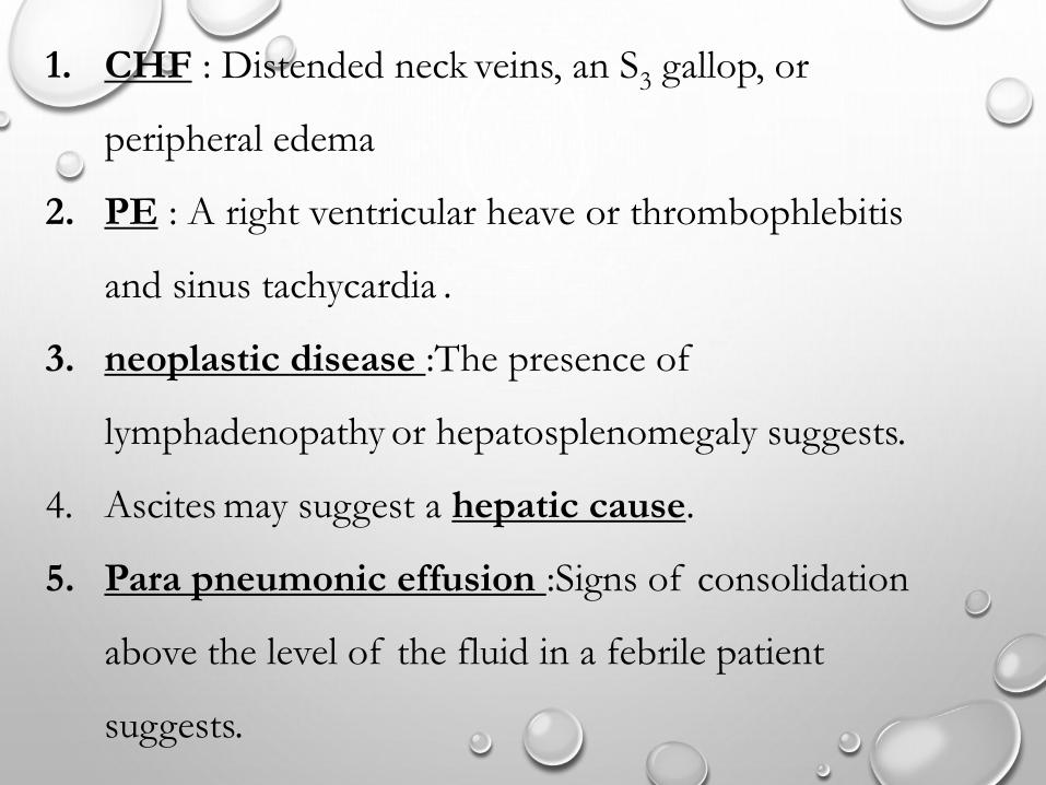
1. CHF : Distended neck veins, an S3 gallop, or
peripheral edema
2. PE : A right ventricular heave or thrombophlebitis
and sinus tachycardia .
3. neoplastic disease :The presence of
lymphadenopathy or hepatosplenomegaly suggests.
4. Ascites may suggest a hepatic cause.
5. Para pneumonic effusion :Signs of consolidation
above the level of the fluid in a febrile patient
suggests.
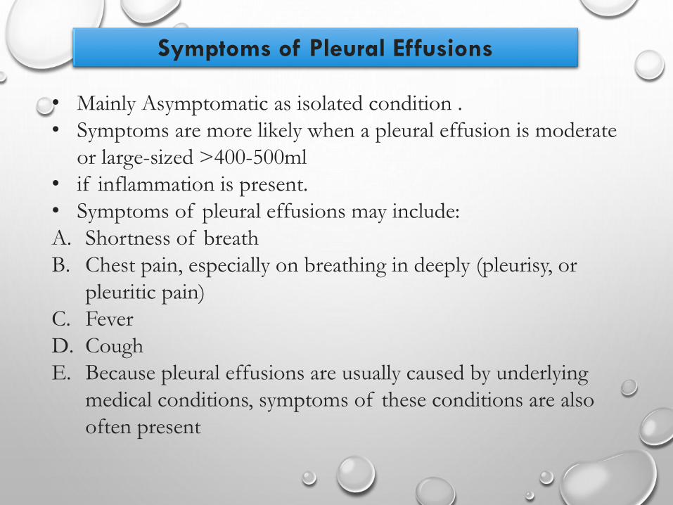
• Mainly Asymptomatic as isolated condition .
• Symptoms are more likely when a pleural effusion is moderate
or large-sized >400-500ml
• if inflammation is present.
• Symptoms of pleural effusions may include:
A. Shortness of breath
B. Chest pain, especially on breathing in deeply (pleurisy, or
pleuritic pain)
C. Fever
D. Cough
E. Because pleural effusions are usually caused by underlying
medical conditions, symptoms of these conditions are also
often present
Symptoms of Pleural Effusions

DIAGNOSIS
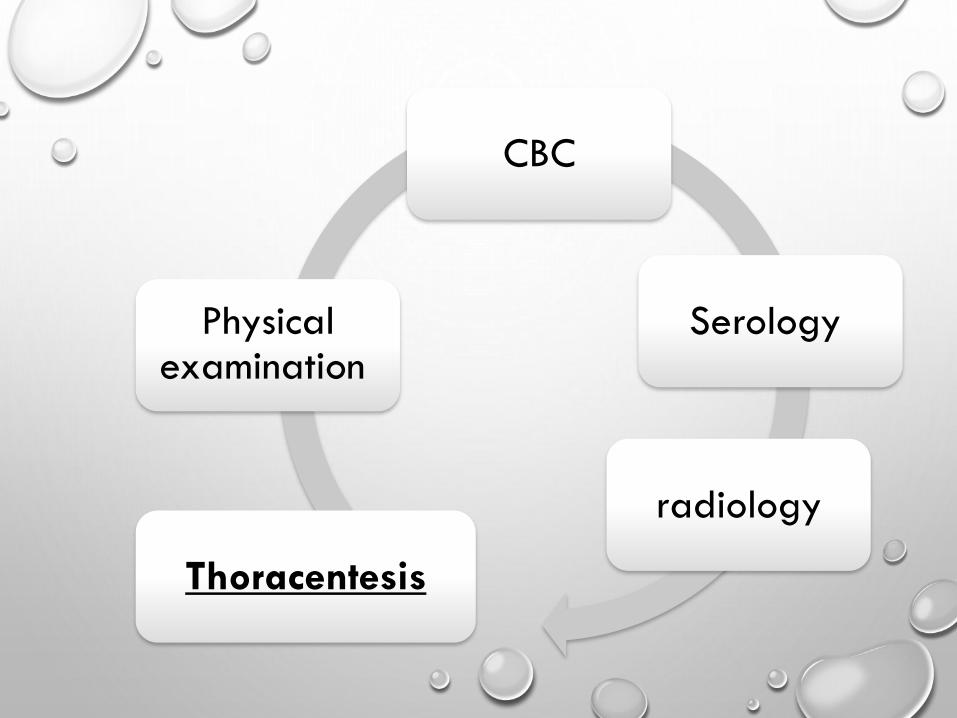
Physical examination
radiology
Serology
Thoracentesis
CBC

LIGHT’S CRITERIA
• SENSITIVITY 99%, SPECIFICITY 98%

Exceptions
These are processes that typically cause exudative effusions,
but may cause transudative effusions.
•Amyloidosis
•Chylothorax
•Constrictive pericarditis
•Hypothyroid pleural effusion
•Malignancy
•Pulmonary embolism
•Sarcoidosis
•Superior vena cava obstruction
•Trapped lung

SERUM
• Serology for autoimmune disorders : RF factor , Anti
CCP , ANA , ANTI dDNA ….
• Routine : RBC , hemoglobin , WBC , PMN …..
• Infectious : CRP , ESR , WBC , PMN , leukocytosis ,
acute phase proteins , LDH
• Albumin , total protein
• Renal function
• Liver enzymes , ALT , AST , GGT , ALP
• PT , PTT , INR* , PLT
RADIOLOGY
Effusions of more than 175 mL are usually apparent as
blunting of the costophrenic angle.
• Location : TB Vs CHF Vs cirrhosis
• Mediastinal shift
• Heart enlargement [ CHF]
• Amount
• Recent Vs previous
• Reccurent [ malignancy e.g. mesothelioma]
A. CXR [ PA , AP , lateral decubitus ]
B. Ultrasound
C. CT

LOCATION AMOUNT CORRELATION
75 mL barley detectable
175 mL obscure the lateral cost phrenic sulcus on an PA
500 mL obscure the diaphragmatic contour on an PA
1000 ml reaches the level of the 4th anterior rib,
On decubitus radiographs and CT scans, less than 10 mL can be
identified
PORCEL et al. AFP 2006; 73: 1212

QUANTITATION OF EFFUSION
Based on the decubitus films1. small effusions <1.5 cm
2. moderate =1.5 to 4.5 cm
3. large effusions >4.5 cm.
Effusions thicker than one 1cm are usually large enough
for sampling by thoracentesis, since at least 200 mL of
liquid are already present

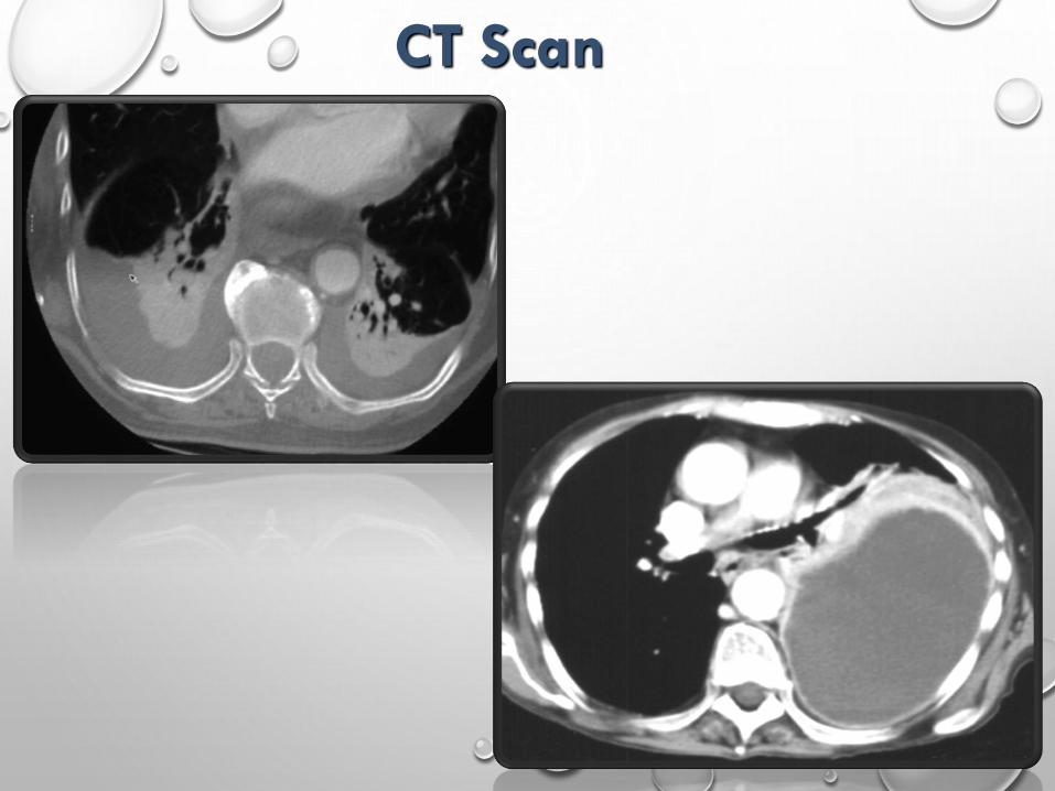
CT Scan

Thoracentesis for both diagnosis and treatment Pleural Fluid Analysis

INDICATIONS FOR THORACENTESIS
LIKELY INDICATED IN MOST PATIENTS!
> 1 CM LAYERING ON LATERAL DECUBITUS
CHF IS HIGHLY UNLIKELY [ E.GLARGE EFFUSION
RECURRENT PLEURAL EFFUSION , MALIGNANCY
PLEURAL EFFUSION AND FEVER: EMPYEMA
THERAPEUTIC THORACENTESIS: DYSPNEA, CHEST PAIN …
UNCLEAR ETIOLOGY OR OBVIOUS CAUSE WITH ATYPICAL PRESENTATION
CHF WITH ATYPICAL PRESENTATION [E.G. UNEQUAL BILATERAL EFFUSION]

CONTRAINDICATIONS
There are no absolute contraindications to thoracentesis
Benefit Vs risks
Caution if :
A. PTT , PT
B. Cr >6 mg/dL
C. decisions to reverse the coagulopathy or correct the
thrombocytopenia should be individualized
D. Anticoagulation or a bleeding diathesis

complications

• PAIN AT THE PUNCTURE SITE
• BLEEDING (HEMATOMA, HEMOTHORAX (1%) , OR
HEMOPERITONEUM)
• PNEUMOTHORAX ( 2-6%)
• SOFT TISSUE INFECTION
• SPLEEN OR LIVER PUNCTURE
• VASOVAGAL EVENTS
• SEEDING THE NEEDLE TRACT WITH TUMOR
• ADVERSE REACTIONS TO THE ANESTHETIC

Pleural fluid glucose, lactate, amylase, triglyceride, and/or tumor
markers
Microscopic examination –(WBCs) or (RBCs) or microorganisms.
WBC differential—determination of percentages of different types of
WBCs High PMN bacterial infection
High lymphocytes TB
Gram stain –Bacterial culture and susceptibility testing
Less commonly ordered tests for infectious diseases, such as tests
for viruses, mycobacteria (AFB smear and culture), and parasites.
Ph
RF factor
Cytology
Appearance : cloudy , milky , bloody . . . .
Pleural Fluid Analysis

PORCEL et al. AFP 2006; 73: 1212
Pleural Fluid Tests

PORCEL et al. AFP 2006; 73: 1212
Pleural Fluid Tests

PORCEL et al. AFP 2006; 73: 1212
Pleural Fluid Tests

PORCEL et al. AFP 2006; 73: 1212
Pleural Fluid Tests
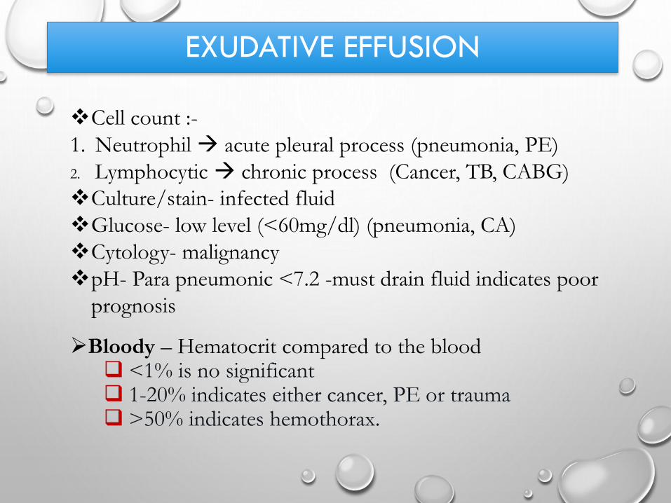
EXUDATIVE EFFUSION
Cell count :-
1. Neutrophil acute pleural process (pneumonia, PE)
2. Lymphocytic chronic process (Cancer, TB, CABG)
Culture/stain- infected fluid
Glucose- low level (<60mg/dl) (pneumonia, CA)
Cytology- malignancy
pH- Para pneumonic <7.2 -must drain fluid indicates poor
prognosis
Bloody – Hematocrit compared to the blood <1% is no significant 1-20% indicates either cancer, PE or trauma >50% indicates hemothorax.

EXUDATIVE EFFUSIONSOTHER TESTS
• SUSPECTED TB
• ADENOSINE DEAMINASE (> 50 IU/L)
• B2 - MICROGLOBULIN
• PCR (SENS 100%, SPEC 95%)
• PPD
• SUSPECTED RHEUMATOID
• PLEURAL RF
• LOW GLUCOSE
• SUSPECTED SLE
• SERUM COMPLEMENT
• PLEURAL ANA
• SUSPECTED PNEUMONIA
• PH
• SUSPECTED PANCREATITIS
• PLEURAL AMYLASE
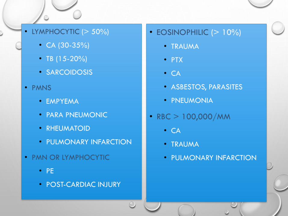
• LYMPHOCYTIC (> 50%)
• CA (30-35%)
• TB (15-20%)
• SARCOIDOSIS
• PMNS
• EMPYEMA
• PARA PNEUMONIC
• RHEUMATOID
• PULMONARY INFARCTION
• PMN OR LYMPHOCYTIC
• PE
• POST-CARDIAC INJURY
• EOSINOPHILIC (> 10%)
• TRAUMA
• PTX
• CA
• ASBESTOS, PARASITES
• PNEUMONIA
• RBC > 100,000/MM
• CA
• TRAUMA
• PULMONARY INFARCTION

MALIGNANT EFFUSIONS

Clinical features suggestive of malignancy:
1. Symptoms > one month
2. Absence of fever
3. Bloody tinged fluid
4. CT very suggestive for malignancy
5. Persistent pneumonia
6. Pts history : smoking , asbestosis , malignancy history
Pleural fluid:
A. Appearance : Mostly bloody
B. WBC differential : mainly lymphocytic
C. Glucose : mostly decreased <60 mg\dL , or normal
D. Elevated lactate >2/3 X serum lactate
E. PH < 7.2 typically
F. Cytology and tumor markers are positive**

Lung >breast > lymphoma/leukemia
metastatic adenocarcinoma positive
cytology 70%
Lymphoma 25-50%
Mesothelioma 10%
Squamous Cell Carcinoma 20%
Sarcoma within pleura 25%
Epidemiology

EMPYEMAPYOTHORAX OR PURULENT PLEURITIS

Typical symptoms include : cough, chest pain, shortness of
breath and fever , persistent pneumonia**
an accumulation of pus in the plural cavity along with :
a. Pleural PH < 7.2 with normal blood PH.
b. Pleural gluc< 60 mg\dl
c. Pleural lactate >2/3 serum lactate
d. Purulent , cloudy , yellow-brownish fluid
Treatment and management :
1. Thoracentesis
2. Chest tube
3. Antibiotics for 1-4 weeks or until improvement
4. Cipro , Flagyl , Penicillin's , clindamycin , vancomycin ,
gentamycin
Consider streptokinase ,
urokinase for fibrinolysis


CLINICAL SYMPTOMS
Shortness of breath, cough , chest pain-- common to pneumonia.
Febrile respiratory illness, accentuation, prolongation the symptoms in pneumonia-- alert the possibility of empyema.
Aerobic empyema-- acute febrile illness.
Anaerobic empyema-- more indolent, usually 10 days.

DRAINAGE

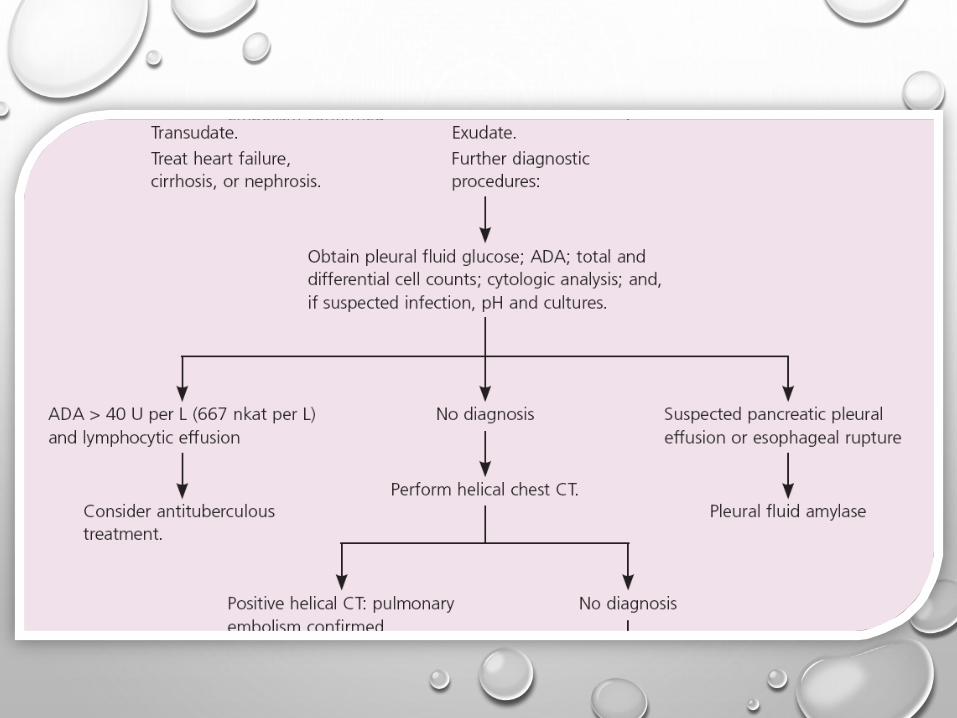
UNDIAGNOSED PLEURAL EFFUSIONS

• 15-20% of effusions
• Careful review of history, PE, meds, risk
factors
• Consider occult abdominal process
• Consider PE
Meigs' syndrome : triad of
ascites
pleural effusion
benign ovarian tumor .
It resolves after the resection of the tumor.

JOSÉ M. PORCEL, M.D., Arnau de Vilanova University Hospital, Lleida, SpainRICHARD W. LIGHT, M.D., Saint Thomas Hospital, Nashville, Tennessee
Am Fam Physician. 2006 Apr 1;73(7):1211-1220.


RESOURCES
1. REDUCING IATROGENIC RISK IN THORACENTESIS: ESTABLISHING BEST PRACTICE VIA EXPERIENTIAL
TRAINING IN A ZERO-RISK ENVIRONMENT.
DUNCAN DR, MORGENTHALER TI, RYU JH, DANIELS
CHEST. 2009;135(5): 1315
2. PNEUMOTHORAX FOLLOWING THORACENTESIS: A SYSTEMATIC REVIEW AND META-ANALYSIS.
GORDON CE, FELLER-KOPMAN D, BALK EM, SMETANA GW
ARCH INTERN MED. 2010;170(4):332
3. COMPLICATIONS ASSOCIATED WITH THORACENTESIS.
SENEFF MG, CORWIN RW, GOLD LH
CHEST 1986; 90:97-100
4. THORACENTESIS: COMPLICATONS, PATIENT EXPERIENCE AND DIAGNOSTIC VALUE.
COLLINS TR, SAHN SA. AM REVIEW RESPIRATORY DISEASE 1983; 127:A114
5. HARRISON’S PRINCIPLES OF INTERNAL MEDICINE, 18TH EDITION.
FAUCI, BRAUNWALD, KASPER, HAUSER, LONGO, JAMESON, LOSCALZO.
6. UPTODATE ONLINE. WWW.UPTODATE.COM.
7. PORCEL ET AL. AFP 2006; 73: 1212
8.


Thanks Docs