Embed Size (px)
Citation preview
Case ReportMetastatic Prostate Cancer of Hand
Akihito Nagano,1 Takatoshi Ohno,2 Koji Oshima,3 Daichi Ishimaru,1 Yutaka Nishimoto,4
Yoshiyuki Ohno,5 Akihiro Hirakawa,1 Tatsuhiko Miyazaki,6 and Haruhiko Akiyama1
1The Department of Orthopaedic Surgery, Gifu University School of Medicine, 1-1 Yanagido, Gifu 501-1193, Japan2The Department of Orthopaedic Surgery, Japanese Red Cross Gifu Hospital, 3-36 Iwakuracho, Gifu 502-0844, Japan3The Department of Orthopaedic Surgery, Ibi Kousei Hospital, 2547-4 Miwa, Ibigawa-cho, Gifu 501-0691, Japan4The Department of Nursing Course, Gifu University School of Medicine, 1-1 Yanagido, Gifu 501-1193, Japan5The Department of Orthopaedic Surgery, Gifu Municipal Hospital, 7-1 Kashima-cho, Gifu 500-8323, Japan6The Division of Pathology, Gifu University Hospital, 1-1 Yanagido, Gifu 501-1193, Japan
Correspondence should be addressed to Akihito Nagano; [email protected]
Received 3 August 2016; Accepted 5 October 2016
Academic Editor: Kaan Erler
Copyright © 2016 Akihito Nagano et al.This is an open access article distributed under theCreative CommonsAttribution License,which permits unrestricted use, distribution, and reproduction in any medium, provided the original work is properly cited.
Soft tissue metastases of prostate cancer to other sites are extremely rare, and, to our best knowledge, there have been no reports ofmetastasis to soft tissue of the hand. A 63-year-oldman was diagnosed with prostatic cancer. During treatment, bone and soft tissuemetastases to the right hand, appearing in the first web space, were observed.The tumor was resected, along with both the first andsecond metacarpal bones. The thumb was reconstructed by pollicization of the remaining index finger, enabling the patient to usethe pollicized thumb for activities of daily living. This is the first case report of prostate cancer metastasizing to the soft tissue inhand. After wide resection, pollicization was able to reconstruct a functional hand and thumb.
1. Introduction
Prostate cancer has been found to metastasize to the bones,regional lymph nodes, and lungs, with several previousreports describing metastases to other sites. Here, we presentthe first case of a prostate cancer that metastasized to the softtissue of the hand.
2. Case Presentation
During a routine check-up a 63-year-old man was found tohave a high prostate specific antigen (PSA) concentration(7.9 ng/mL). Transrectal fine needle aspiration (FNA) of theprostate provided a definitive diagnosis of poorly differenti-ated adenocarcinoma (cT3a, Gleason score 8 (4 + 4), 8/8 coresaffected). No metastases were detected, and treatment withboth the nonsteroidal antiandrogen bicalutamide (Casodex)and goserelin (Zoladex) reduced his PSA level to 0.2 ng/mLwithin three months. Five years later, however, despite hisPSA level remaining low, local extension to the bladder andmetastasis to the S1 vertebra were detected. Furthermore, hedeveloped a gradually enlarging painless mass in the first web
space of his right hand, adversely affecting his activities ofdaily living. Therefore he was referred to our department.
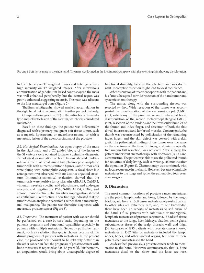
Physical examination showed a well-circumscribed, elas-tic soft mass located between the right thumb and index fin-ger. This mass, which was palpable but not mobile, measured5 × 4 × 3 cm in size. The skin over the mass was discolored,suggesting that the tumor had invaded the skin. Daily livingwas impaired due to restricted range of motion (ROM) of thethumb. Although no pain was associated with the mass, thepatient experienced sensory disturbance of the right thumb(Figure 1).
Laboratory tests showed elevation of alkaline phos-phatase but low PSA level (0.036 ng/mL).
2.1. Radiographic Findings. A roentgenogram of the righthand showed enlargement of first metacarpal interspace,indicating noncalcification of the soft tissue mass. The meta-carpal bones adjacent to the mass were normal without anybony destruction.
Magnetic resonance imaging (MRI) of the right handrevealed a well-defined, clearly circumscribedmass, with iso-
Hindawi Publishing CorporationCase Reports in OrthopedicsVolume 2016, Article ID 1472932, 5 pageshttp://dx.doi.org/10.1155/2016/1472932
2 Case Reports in Orthopedics
Figure 1: Soft tissue mass in the right hand.Themass was located in the first intercarpal space, with the overlying skin showing discoloration.
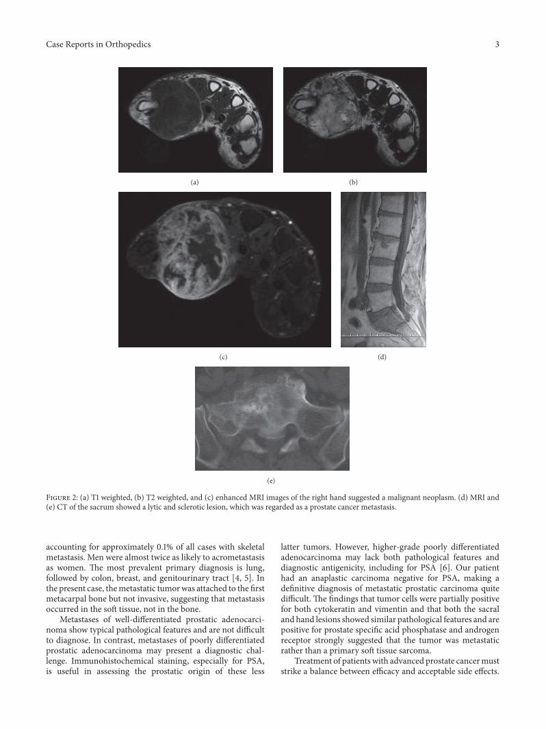
to low intensity on T1 weighted images and heterogeneouslyhigh intensity on T2 weighted images. After intravenousadministration of gadolinium-based contrast agent, the masswas well enhanced peripherally, but the central region waspoorly enhanced, suggesting necrosis.Themass was adjacentto the first metacarpal bone (Figure 2).
Thallium scintigraphy showed marked accumulation inthe right hand but no accumulation in other parts of the body.
Computed tomography (CT) of the entire body revealed alytic and sclerotic lesion of the sacrum, which was consideredmetastatic.
Based on these findings, the patient was differentiallydiagnosed with a primary malignant soft tissue tumor, suchas a myxoid liposarcoma or myxofibrosarcoma, or with ametastatic lesion of the adenocarcinoma of the prostate.
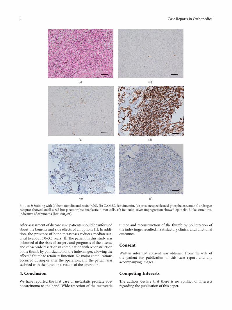
2.2. Histological Examination. An open biopsy of the massin the right hand and a CT-guided biopsy of the lesion ofthe S1 vertebra were obtained to make a definitive diagnosis.Pathological examination of both lesions showed multin-odular growth of small-sized but pleomorphic anaplastictumor cells with numerous mitotic figures. Some tumor cellswere plump with eosinophilic cytoplasm. A focal sheet-likearrangement was observed, with no distinct organoid struc-ture. Immunohistochemical evaluation showed that thetumor cells were positive for cytokeratin AE1/AE3, CAM5.2,vimentin, prostate specific acid phosphatase, and androgenreceptor and negative for PSA, S-100, CD34, CD68, andsmooth muscle actin. Reticulin silver impregnation showedan epithelioid-like structure.These findings indicated that thetumor was an anaplastic carcinoma rather than a mesenchy-mal malignancy. The patient was therefore diagnosed withmetastatic prostate cancer (Figure 3).
2.3. Treatment. The treatment of patient with cancer shouldbe performed on a case-by-case basis, depending on thepatient’s prognosis and functional capabilities especially inpatients with multiple metastasis. Generally, palliative treat-ment, such as radiation therapy, is chosen because of thedismal prognosis of patients with metastasis. In the presentcase, the prognosis was thought to be relatively better thanthe other cancer; in fact, the prognosis of prostate cancer withbone metastasis is reported as 3.0–3.5 years [1]. Furthermore,an amputation would bring about unacceptable degree of
functional disability, because the affected hand was domi-nant. Incomplete resection might lead to local recurrence.
After discussion of treatment optionswith the patient andhis family, he agreed to wide resection of the hand tumor andsystemic chemotherapy.
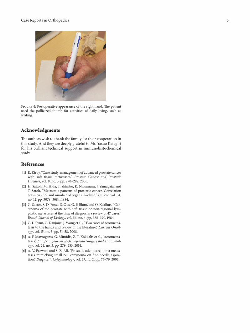
The tumor, along with the surrounding tissues, wasresected en bloc. Wide resection of the tumor was accom-panied by disarticulation of the carpometacarpal (CMC)joint, osteotomy of the proximal second metacarpal bone,disarticulation of the second metacarpophalangeal (MCP)joint, resection of the tendons and neurovascular bundles ofthe thumb and index finger, and resection of both the firstdorsal interosseous and lumbrical muscles. Concurrently, thethumb was reconstructed by pollicization of the remainingindex finger, and the skin defect was covered with a skingraft. The pathological findings of the tumor were the sameas the specimen at the time of biopsy, and microscopicallyfree margin (R0 resection) was achieved. After surgery, thepatient underwent chemotherapy with docetaxel (DTX) andestramustine.Thepatientwas able to use the pollicized thumbfor activities of daily living, such as writing, six months afterthe operation (Figure 4). Chemotherapy was continued, withno local recurrence in the hand.However, because ofmultiplemetastases to the lungs and spine, the patient died four yearsafter surgery.
3. Discussion
The most common locations of prostate cancer metastasesare the pelvic lymph nodes and bone, followed by the lungs,bladder, and liver [2]. Soft tissuemetastases of prostate cancerto other sites are extremely rare, and, to our knowledge,there have been no reports of metastasis to soft tissue ofthe hand. Of 47 patients with soft tissue or nonregionallymphaticmetastases of prostate carcinoma, 16 had soft tissuemetastasis to the lungs, liver, kidneys, bladder, penile glans,subcutaneous tissue of the scalp, thoracic wall, and thigh[3]. Autopsies of 1885 patients with prostate cancer showedmetastases in 1367. Sites of metastasis included the lymphnodes, bones, and other visceral organs, but none of thesepatients had metastases to the hand.
As described previously, a prostate cancer tends to meta-stasize to the bone. However, acrometastasis, that is, bonemetastasis distal to the elbow and the knee, are rare,
Case Reports in Orthopedics 3
(a) (b)
(c) (d)
(e)
Figure 2: (a) T1 weighted, (b) T2 weighted, and (c) enhanced MRI images of the right hand suggested a malignant neoplasm. (d) MRI and(e) CT of the sacrum showed a lytic and sclerotic lesion, which was regarded as a prostate cancer metastasis.
accounting for approximately 0.1% of all cases with skeletalmetastasis. Men were almost twice as likely to acrometastasisas women. The most prevalent primary diagnosis is lung,followed by colon, breast, and genitourinary tract [4, 5]. Inthe present case, themetastatic tumorwas attached to the firstmetacarpal bone but not invasive, suggesting that metastasisoccurred in the soft tissue, not in the bone.
Metastases of well-differentiated prostatic adenocarci-noma show typical pathological features and are not difficultto diagnose. In contrast, metastases of poorly differentiatedprostatic adenocarcinoma may present a diagnostic chal-lenge. Immunohistochemical staining, especially for PSA,is useful in assessing the prostatic origin of these less
latter tumors. However, higher-grade poorly differentiatedadenocarcinoma may lack both pathological features anddiagnostic antigenicity, including for PSA [6]. Our patienthad an anaplastic carcinoma negative for PSA, making adefinitive diagnosis of metastatic prostatic carcinoma quitedifficult. The findings that tumor cells were partially positivefor both cytokeratin and vimentin and that both the sacraland hand lesions showed similar pathological features and arepositive for prostate specific acid phosphatase and androgenreceptor strongly suggested that the tumor was metastaticrather than a primary soft tissue sarcoma.
Treatment of patients with advanced prostate cancermuststrike a balance between efficacy and acceptable side effects.
4 Case Reports in Orthopedics
(a) (b)
(c) (d)
(e) (f)
Figure 3: Staining with (a) hematoxylin and eosin (×20), (b) CAM5.2, (c) vimentin, (d) prostate specific acid phosphatase, and (e) androgenreceptor showed small-sized but pleomorphic anaplastic tumor cells. (f) Reticulin silver impregnation showed epithelioid-like structures,indicative of carcinoma (bar: 100 𝜇m).
After assessment of disease risk, patients should be informedabout the benefits and side effects of all options [1]. In addi-tion, the presence of bone metastases reduces median sur-vival to about 3.0–3.5 years [1]. The patient in this study wasinformed of the risks of surgery and prognosis of the diseaseand chose wide resection in combinationwith reconstructionof the thumb by pollicization of the index finger, allowing theaffected thumb to retain its function. Nomajor complicationsoccurred during or after the operation, and the patient wassatisfied with the functional results of the operation.
4. Conclusion
We have reported the first case of metastatic prostate ade-nocarcinoma to the hand. Wide resection of the metastatic
tumor and reconstruction of the thumb by pollicization ofthe index finger resulted in satisfactory clinical and functionaloutcomes.
Consent
Written informed consent was obtained from the wife ofthe patient for publication of this case report and anyaccompanying images.
Competing Interests
The authors declare that there is no conflict of interestsregarding the publication of this paper.
Case Reports in Orthopedics 5
Figure 4: Postoperative appearance of the right hand. The patientused the pollicized thumb for activities of daily living, such aswriting.
Acknowledgments
The authors wish to thank the family for their cooperation inthis study. And they are deeply grateful to Mr. Yasuo Katagirifor his brilliant technical support in immunohistochemicalstudy.
References
[1] R. Kirby, “Case study: management of advanced prostate cancerwith soft tissue metastases,” Prostate Cancer and ProstaticDiseases, vol. 8, no. 3, pp. 290–292, 2005.
[2] H. Saitoh, M. Hida, T. Shimbo, K. Nakamura, J. Yamagata, andT. Satoh, “Metastatic patterns of prostatic cancer. Correlationbetween sites and number of organs involved,” Cancer, vol. 54,no. 12, pp. 3078–3084, 1984.
[3] G. Saeter, S. D. Fossa, S. Ous, G. P. Blom, and O. Kaalhus, “Car-cinoma of the prostate with soft tissue or non-regional lym-phatic metastases at the time of diagnosis: a review of 47 cases,”British Journal of Urology, vol. 56, no. 4, pp. 385–390, 1984.
[4] C. J. Flynn, C. Danjoux, J. Wong et al., “Two cases of acrometas-tasis to the hands and review of the literature,” Current Oncol-ogy, vol. 15, no. 5, pp. 51–58, 2008.
[5] A. F. Mavrogenis, G. Mimidis, Z. T. Kokkalis et al., “Acrometas-tases,” European Journal of Orthopaedic Surgery and Traumatol-ogy, vol. 24, no. 3, pp. 279–283, 2014.
[6] A. V. Parwani and S. Z. Ali, “Prostatic adenocarcinoma metas-tases mimicking small cell carcinoma on fine-needle aspira-tion,” Diagnostic Cytopathology, vol. 27, no. 2, pp. 75–79, 2002.

Submit your manuscripts athttp://www.hindawi.com
Stem CellsInternational
Hindawi Publishing Corporationhttp://www.hindawi.com Volume 2014
Hindawi Publishing Corporationhttp://www.hindawi.com Volume 2014
MEDIATORSINFLAMMATION
of
Hindawi Publishing Corporationhttp://www.hindawi.com Volume 2014
Behavioural Neurology
EndocrinologyInternational Journal of
Hindawi Publishing Corporationhttp://www.hindawi.com Volume 2014
Hindawi Publishing Corporationhttp://www.hindawi.com Volume 2014
Disease Markers
Hindawi Publishing Corporationhttp://www.hindawi.com Volume 2014
BioMed Research International
OncologyJournal of
Hindawi Publishing Corporationhttp://www.hindawi.com Volume 2014
Hindawi Publishing Corporationhttp://www.hindawi.com Volume 2014
Oxidative Medicine and Cellular Longevity
Hindawi Publishing Corporationhttp://www.hindawi.com Volume 2014
PPAR Research
The Scientific World JournalHindawi Publishing Corporation http://www.hindawi.com Volume 2014
Immunology ResearchHindawi Publishing Corporationhttp://www.hindawi.com Volume 2014
Journal of
ObesityJournal of
Hindawi Publishing Corporationhttp://www.hindawi.com Volume 2014
Hindawi Publishing Corporationhttp://www.hindawi.com Volume 2014
Computational and Mathematical Methods in Medicine
OphthalmologyJournal of
Hindawi Publishing Corporationhttp://www.hindawi.com Volume 2014
Diabetes ResearchJournal of
Hindawi Publishing Corporationhttp://www.hindawi.com Volume 2014
Hindawi Publishing Corporationhttp://www.hindawi.com Volume 2014
Research and TreatmentAIDS
Hindawi Publishing Corporationhttp://www.hindawi.com Volume 2014
Gastroenterology Research and Practice
Hindawi Publishing Corporationhttp://www.hindawi.com Volume 2014
Parkinson’s Disease
Evidence-Based Complementary and Alternative Medicine
Volume 2014Hindawi Publishing Corporationhttp://www.hindawi.com