Embed Size (px)
Citation preview
Comparison of attenuation in ascites between antemortem and
postmortem computed tomography
Masanori Ishida, Wataru Gonoi, Hiroyuki Abe, Kotaro Fujimoto, Naomasa Okimoto, Tetsuo Ushiku, Osamu Abe
Department of Radiology, The University of Tokyo Hospital, Tokyo, Japan
email address; [email protected] (Masanori Ishida, MD,PhD)
ISFRI 2020 Virtual Poster Session

Conflict of interest; none
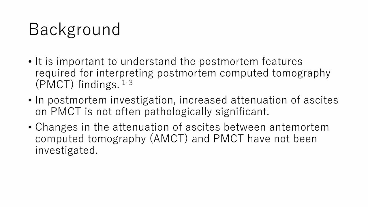
Background
• It is important to understand the postmortem features required for interpreting postmortem computed tomography (PMCT) findings. 1-3
• In postmortem investigation, increased attenuation of ascites on PMCT is not often pathologically significant.
• Changes in the attenuation of ascites between antemortem computed tomography (AMCT) and PMCT have not been investigated.
Objective
• To investigate the changes in ascites attenuation between antemortem (AMCT) and PMCT analyses of the same subjects.
Methods
• This study was approved by the Ethical Committee of the participating institution.
• Informed consent for the use of the cadavers in our study was obtained from the families of the deceased subjects.
• The potential subjects had been subjected to PMCT and conventional cadaver autopsy after death from non-traumatic causes at our academic tertiary care hospital between April 2009 and December 2018.
• Figure 1 summarizes the subject inclusion and exclusion criteria.
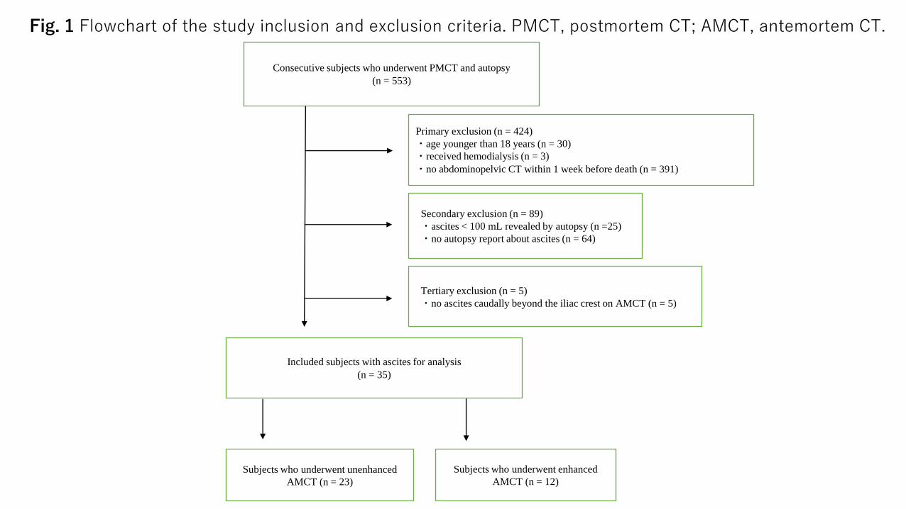
Consecutive subjects who underwent PMCT and autopsy
(n = 553)
Primary exclusion (n = 424)
・age younger than 18 years (n = 30)
・received hemodialysis (n = 3)
・no abdominopelvic CT within 1 week before death (n = 391)
Included subjects with ascites for analysis
(n = 35)
Secondary exclusion (n = 89)
・ascites < 100 mL revealed by autopsy (n =25)
・no autopsy report about ascites (n = 64)
Subjects who underwent unenhanced
AMCT (n = 23)
Subjects who underwent enhanced
AMCT (n = 12)
Tertiary exclusion (n = 5)
・no ascites caudally beyond the iliac crest on AMCT (n = 5)
Fig. 1 Flowchart of the study inclusion and exclusion criteria. PMCT, postmortem CT; AMCT, antemortem CT.
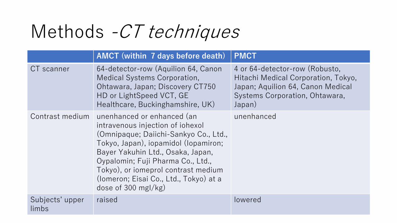
Methods -CT techniquesAMCT (within 7 days before death) PMCT
CT scanner 64-detector-row (Aquilion 64, Canon Medical Systems Corporation, Ohtawara, Japan; Discovery CT750 HD or LightSpeed VCT, GE Healthcare, Buckinghamshire, UK)
4 or 64-detector-row (Robusto, Hitachi Medical Corporation, Tokyo, Japan; Aquilion 64, Canon Medical Systems Corporation, Ohtawara, Japan)
Contrast medium unenhanced or enhanced (an intravenous injection of iohexol(Omnipaque; Daiichi-Sankyo Co., Ltd., Tokyo, Japan), iopamidol (Iopamiron; Bayer Yakuhin Ltd., Osaka, Japan, Oypalomin; Fuji Pharma Co., Ltd., Tokyo), or iomeprol contrast medium (Iomeron; Eisai Co., Ltd., Tokyo) at a dose of 300 mgI/kg)
unenhanced
Subjects’ upper limbs
raised lowered
Methods -Image analysis
• CT images were reviewed on a two-dimensional workstation (RadiAnt DICOM Viewer, Medixant, Poznan, Poland).
• The attenuation (in Hounsfield units, HU) of ascites was measured manually in 100-mm2 circular regions of interest (ROIs).
• ROIs were placed at similar sites on pairs of AMCT and PMCT images from each subject (Fig. 2). 4,5
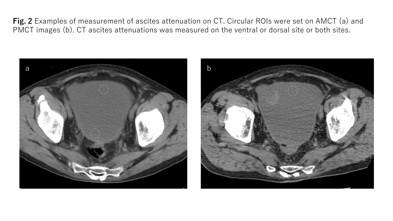
Fig. 2 Examples of measurement of ascites attenuation on CT. Circular ROIs were set on AMCT (a) and PMCT images (b). CT ascites attenuations was measured on the ventral or dorsal site or both sites.
ba
Methods -Autopsy technique
• Autopsies using conventional procedures were performed by board-certified pathologists immediately after PMCT.
• The pathologists were informed about the patients’ clinical histories and details regarding the circumstances of death and, but they were unaware of the PMCT findings.
• The pathologists noted the findings of ascites, including the volume and appearance (i.e., serous, hemorrhagic, proteinous, chylous, and/or fecal).
Methods -Clinical and autopsy data
• The following data were collected:
・Serum albumin concentration and estimated glomerular filtration rate (eGFR) within 1 day before AMCT
・History of hemodialysis and cardiopulmonary resuscitation
・Time intervals between AMCT and PMCT and between death and PMCT
・Ascites findings (the volume and appearance)
・Presence of carcinomatous peritonitis
Methods –Data and statistical analysis
• Mann–Whitney U test; to compare the demographic, clinical, imaging, and autopsy characteristics between subjects who underwent unenhanced and enhanced AMCT.
• Paired t-test; to evaluate differences in the attenuations of ascites between AMCT and PMCT.
• A multiple regression analysis; to evaluate potential factors contributing to changes in attenuation between AMCT and PMCT in subjects who exhibited significant changes in ascites attenuation.
• Spearman's rank correlation coefficient; to analyze a relationship between the time interval between enhanced AMCT and PMCT and elevated CT attenuation of ascites on PMCT.
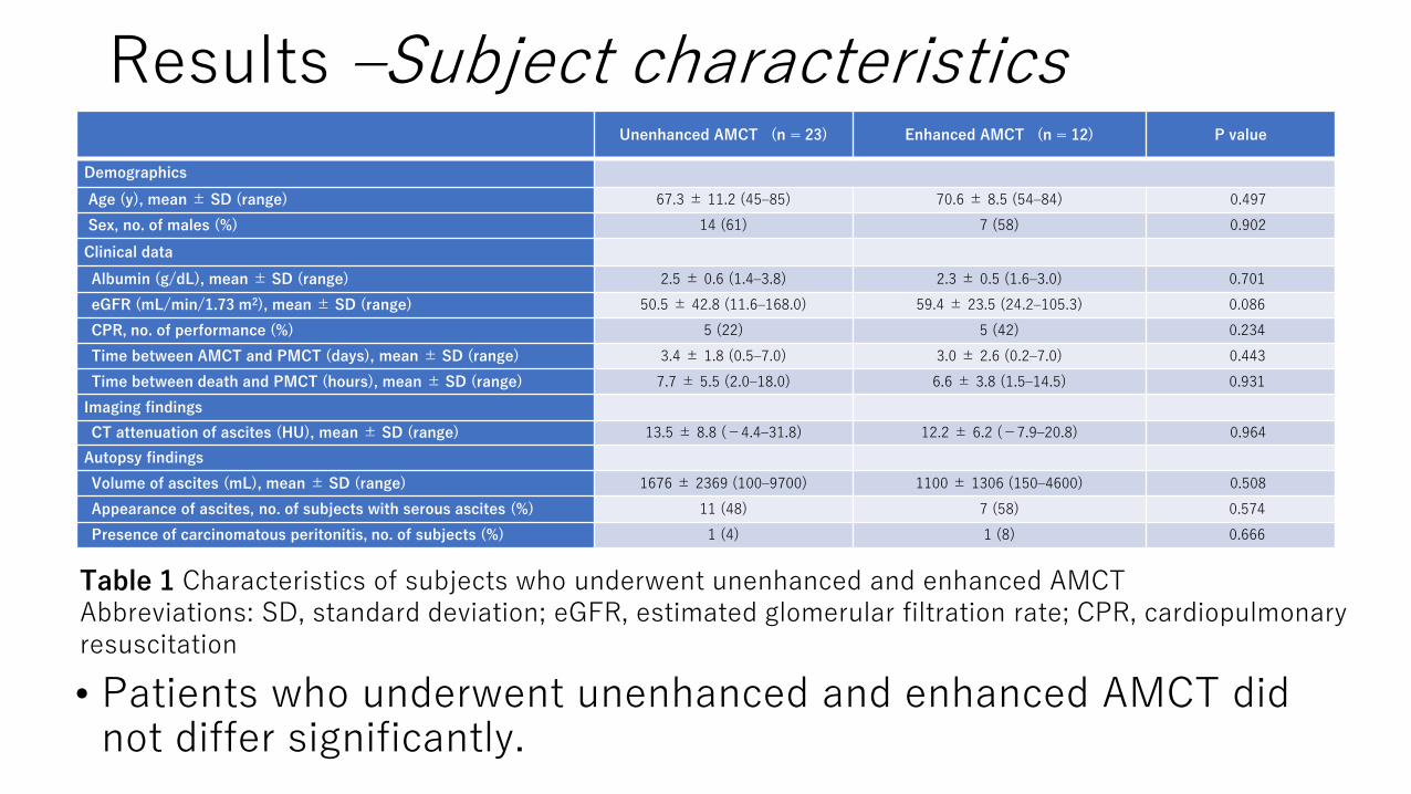
Results –Subject characteristicsUnenhanced AMCT (n = 23) Enhanced AMCT (n = 12) P value
Demographics
Age (y), mean ± SD (range) 67.3 ± 11.2 (45–85) 70.6 ± 8.5 (54–84) 0.497
Sex, no. of males (%) 14 (61) 7 (58) 0.902
Clinical data
Albumin (g/dL), mean ± SD (range) 2.5 ± 0.6 (1.4–3.8) 2.3 ± 0.5 (1.6–3.0) 0.701
eGFR (mL/min/1.73 m2), mean ± SD (range) 50.5 ± 42.8 (11.6–168.0) 59.4 ± 23.5 (24.2–105.3) 0.086
CPR, no. of performance (%) 5 (22) 5 (42) 0.234
Time between AMCT and PMCT (days), mean ± SD (range) 3.4 ± 1.8 (0.5–7.0) 3.0 ± 2.6 (0.2–7.0) 0.443
Time between death and PMCT (hours), mean ± SD (range) 7.7 ± 5.5 (2.0–18.0) 6.6 ± 3.8 (1.5–14.5) 0.931
Imaging findings
CT attenuation of ascites (HU), mean ± SD (range) 13.5 ± 8.8 (−4.4–31.8) 12.2 ± 6.2 (−7.9–20.8) 0.964
Autopsy findings
Volume of ascites (mL), mean ± SD (range) 1676 ± 2369 (100–9700) 1100 ± 1306 (150–4600) 0.508
Appearance of ascites, no. of subjects with serous ascites (%) 11 (48) 7 (58) 0.574
Presence of carcinomatous peritonitis, no. of subjects (%) 1 (4) 1 (8) 0.666
Table 1 Characteristics of subjects who underwent unenhanced and enhanced AMCTAbbreviations: SD, standard deviation; eGFR, estimated glomerular filtration rate; CPR, cardiopulmonary resuscitation
• Patients who underwent unenhanced and enhanced AMCT did not differ significantly.
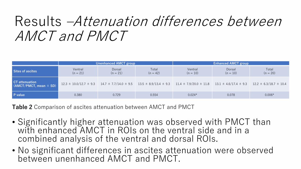
Results –Attenuation differences between AMCT and PMCT
Table 2 Comparison of ascites attenuation between AMCT and PMCT
Unenhanced AMCT group Enhanced AMCT group
Sites of ascitesVentral(n = 21)
Dorsal(n = 21)
Total(n = 42)
Ventral(n = 10)
Dorsal(n = 10)
Total(n = 20)
CT attenuation (AMCT/PMCT, mean ± SD)
12.3 ± 10.0/12.7 ± 9.3 14.7 ± 7.7/14.0 ± 9.5 13.5 ± 8.9/13.4 ± 9.3 11.4 ± 7.9/20.0 ± 11.8 13.1 ± 4.6/17.4 ± 9.3 12.2 ± 6.3/18.7 ± 10.4
P value 0.380 0.729 0.554 0.024* 0.078 0.006*
• Significantly higher attenuation was observed with PMCT than with enhanced AMCT in ROIs on the ventral side and in a combined analysis of the ventral and dorsal ROIs.
• No significant differences in ascites attenuation were observed between unenhanced AMCT and PMCT.
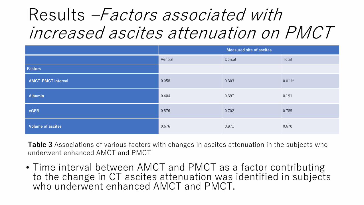
Results –Factors associated with increased ascites attenuation on PMCT
Table 3 Associations of various factors with changes in ascites attenuation in the subjects who underwent enhanced AMCT and PMCT
Measured site of ascites
Ventral Dorsal Total
Factors
AMCT-PMCT interval 0.058 0.303 0.011*
Albumin 0.404 0.397 0.191
eGFR 0.876 0.702 0.785
Volume of ascites 0.676 0.971 0.670
• Time interval between AMCT and PMCT as a factor contributing to the change in CT ascites attenuation was identified in subjects who underwent enhanced AMCT and PMCT.
Discussion
• An increase in the PMCT attenuation of ascites was observed in subjects who had undergone enhanced AMCT.
- Previous reports described the increased CT attenuation of ascites in a living body at a particular time interval after a previous enhanced CT scan.4,6 However, the mechanism of the delayed increase in CT attenuation of ascites was not determined.
- Increased vascular permeability may occur once circulation stops after death, leading to the postmortem leakage of plasma components from blood vessels.
- Water-soluble iodinated contrast medium comprises small molecules that diffuse readily through the pores of blood vessels into the extravascular and extracellular spaces.7-9
- Therefore, contrast medium may leak from the blood vessels into the ascites near and after death, which would support the observed increase in ascites attenuation on PMCT in the present study.
Discussion
• Significantly higher attenuation was observed with PMCT than with enhanced AMCT in ROIs on the ventral side and in a combined analysis of the ventral and dorsal ROIs.
- We consider that this result may be attributable to the dorsal sedimentation due to high specific gravity of contrast medium or the uneven distribution of leaked contrast medium in the ascites, which is repeatedly produced and absorbed.
• We identified the time interval between enhanced AMCT and PMCT as a factor associated inversely with the change in the CT attenuation of ascites.
- This observation suggests that considerable amounts of the injected contrast medium leaked from the blood vessels into the ascites immediately after injection and during the early postmortem state.
Discussion –Limitation
• AMCT and PMCT scans were conducted on different scanners. This is an important but inevitable limitation at institutions where different scanners are used for clinical patients and corpses.
• AMCT was performed with arms raised and PMCT was performed with arms at the sides.
• The number of subjects was small.
• There was a lack of enough consideration of the distribution of ascites.
Conclusions
• We confirmed an elevated level of ascites attenuation on PMCT relative to AMCT in subjects who underwent enhanced AMCT shortly before death.
References
1. Levy AD, Harcke HT, Mallak CT. Postmortem imaging: MDCT features of postmortem change and decomposition. Am J Forensic Med
Pathol. 2010;31:12–17.
2. Ishida M, Gonoi W, Okuma H, et al. Common postmortem computed tomography findings following atraumatic death: Differentiation
between normal postmortem changes and pathologic lesions. Korean J Radiol. 2015;16:798–809.
3. Wagensveld IM, Blokker BM, Wielopolski PA, et al. Total-body CT and MR features of postmortem change in in-hospital deaths. PLoS
One. 2017;12:e0185115.
4. Benedetti N, Aslam R, Wang ZJ, et al. Delayed enhancement of ascites after IV contrast material administration at CT: Time course and
clinical correlation. AJR Am J Roentgenol. 2009;193:732–737.
5. Seishima R, Okabayashi K, Hasegawa H, et al. Computed tomography attenuation values of ascites are helpful to predict perforation site.
World J Gastroenterol. 2015;21:1573–1579.
6. Cooper C, Silverman PM, Davros WJ, et al. Delayed contrast enhancement of ascitic fluid on CT: Frequency and significance. AJR Am J
Roentgenol. 1993;161:787–790.
7. Hammerman AM, Oberle PA, Susman N. Opacification of ascitic fluid on delayed contrast computed tomography scans. Clin Imaging.
1990;14:221–224.
8. Wise SW, DeMeo JH, Austin RF. Enhancing ascites: An aid to CT diagnosis. Abdom Imaging. 1996;21:67–68.
9. Helbich TH, Roberts TPL, Gossmann A, et al. Quantitative gadopentetate-enhanced MRI of breast tumors: Testing of different analytic
methods. Magn Reson Med. 2000;44:915–924.