Embed Size (px)
Citation preview
DMD #81737
1
Functional characterization of 21 allelic variants of
dihydropyrimidine dehydrogenase identified in 1,070 Japanese
individuals
Eiji HISHINUMA, Yoko NARITA, Sakae SAITO, Masamitsu MAEKAWA, Fumika
AKAI, Yuya NAKANISHI, Jun YASUDA, Masao NAGASAKI, Masayuki
YAMAMOTO, Hiroaki YAMAGUCHI, Nariyasu MANO, Noriyasu HIRASAWA,
Masahiro HIRATSUKA
Laboratory of Pharmacotherapy of Life-Style Related Diseases, Graduate School of
Pharmaceutical Sciences, Tohoku University, Sendai 980-8578, Japan (E.H., Y.N., F.A.,
Y.N., N.H., M.H.)
Department of Pharmaceutical Sciences, Tohoku University Hospital, Sendai 980-8574,
Japan (M.M., H.Y., N.M.)
Tohoku Medical Megabank Organization, Tohoku University, Sendai, 980-8575, Japan
(S.S., J.Y., M.N., M.Y., M.H.)
This article has not been copyedited and formatted. The final version may differ from this version.DMD Fast Forward. Published on May 16, 2018 as DOI: 10.1124/dmd.118.081737
at ASPE
T Journals on Septem
ber 1, 2018dm
d.aspetjournals.orgD
ownloaded from
DMD #81737
2
Running title: Functional characterization of DPYD allelic variants
Corresponding author:
Dr. Masahiro Hiratsuka, Ph.D.
Laboratory of Pharmacotherapy of Life-Style Related Diseases, Graduate School of
Pharmaceutical Sciences, Tohoku University, Sendai, Japan, 6-3, Aoba, Aramaki, Aoba-
ku, Sendai 980-8578, Japan
Tel & Fax: +81-22-717-7049
E-mail: [email protected]
Number of text pages: 41
Number of figures: 6
Number of tables: 3
Number of references: 28
Number of words in the Abstract: 240
Number of words in the Introduction: 465
Number of words in the Discussion: 932
This article has not been copyedited and formatted. The final version may differ from this version.DMD Fast Forward. Published on May 16, 2018 as DOI: 10.1124/dmd.118.081737
at ASPE
T Journals on Septem
ber 1, 2018dm
d.aspetjournals.orgD
ownloaded from
DMD #81737
3
Abbreviations: DPD, dihydropyrimidine dehydrogenase; 5-FU, 5-fluorouracil; FUH2,
dihydro-5-fluorouracil; β-NADPH, β-Nicotinamide adenine dinucleotide phosphate;
FAD, flavin adenine dinucleotide; FMN, flavin mono nucleotide; DTT, dithiothreitol;
GAPDH, glyceraldehyde 3-phosphate dehydrogenase
This article has not been copyedited and formatted. The final version may differ from this version.DMD Fast Forward. Published on May 16, 2018 as DOI: 10.1124/dmd.118.081737
at ASPE
T Journals on Septem
ber 1, 2018dm
d.aspetjournals.orgD
ownloaded from
DMD #81737
4
Abstract
Dihydropyrimidine dehydrogenase (DPD, EC 1.3.1.2), encoded by the DPYD
gene, is the rate-limiting enzyme in the degradation pathway of endogenous pyrimidine
and fluoropyrimidine drugs such as 5-fluorouracil (5-FU). DPD catalyzes the reduction
of uracil, thymine, and 5-FU. In Caucasians, DPYD mutations, including DPYD*2A,
DPYD*13, c.2846A>T, and c.1129-5923C>G/hapB3, are known to contribute to
interindividual variations in the toxicity of 5-FU. However, none of these DPYD
polymorphisms have been identified in the Asian population. Recently, 21 DPYD allelic
variants, including some novel single-nucleotide variants (SNVs), were identified in
1,070 healthy Japanese individuals by analyzing their whole-genome sequences (WGS),
but the functional alterations caused by these variants remain unknown. In this study, in
vitro analysis was performed on 22 DPD allelic variants by transiently expressing wild-
type DPD and 21 DPD variants in 293FT cells and characterizing their enzymatic
activities, using 5-FU as a substrate. DPD expression levels and dimeric forms were
determined using immunoblotting and blue native-PAGE, respectively. Additionally, the
values of three kinetic parameters, the Michaelis constant (Km), maximum velocity (Vmax),
and intrinsic clearance (CLint = Vmax/Km), were determined for the reduction of 5-FU. We
found that 11 variants exhibited significantly decreased intrinsic clearance in comparison
This article has not been copyedited and formatted. The final version may differ from this version.DMD Fast Forward. Published on May 16, 2018 as DOI: 10.1124/dmd.118.081737
at ASPE
T Journals on Septem
ber 1, 2018dm
d.aspetjournals.orgD
ownloaded from
DMD #81737
5
to wild-type DPD. Moreover, the band patterns observed in the immunoblots of blue
native gels indicated that DPD dimerization is required for enzymatic activity in DPD.
Thus, the detection of rare DPYD variants might facilitate severe adverse effect prediction
of 5-FU-based chemotherapy in the Japanese population.
This article has not been copyedited and formatted. The final version may differ from this version.DMD Fast Forward. Published on May 16, 2018 as DOI: 10.1124/dmd.118.081737
at ASPE
T Journals on Septem
ber 1, 2018dm
d.aspetjournals.orgD
ownloaded from
DMD #81737
6
Introduction
5-Fluorouracil (5-FU) is the anticancer drug most frequently prescribed for use
in the treatment of various solid tumors, such as those found in the gastrointestinal tract,
breast, head, and neck (Ide et al., 2013; Okuma et al., 2016). 5-FU has a narrow
therapeutic index, and approximately 10%–30% of patients treated with 5-FU-based
regimens develop early-onset severe or life-threatening toxicity (Rothenberg et al., 2001;
Twelves et al., 2005; Saltz et al., 2007). Therefore, a biomarker for the prediction of
adverse effects before 5-FU administration is necessary.
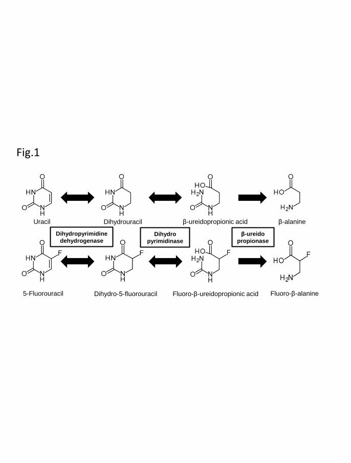
More than 80% of the administered dose of 5-FU is rapidly degraded by three
consecutive enzymes of the endogenous pyrimidine, uracil, and thymine catabolic
pathway (Fig. 1) (Daher et al., 1990; Kim et al., 2015; Kunicka et al., 2016). First,
dihydropyrimidine dehydrogenase (DPD, EC 1.3.1.2), the rate-limiting enzyme,
catalyzes the reduction of 5-FU to dihydro-5-fluorouracil (FUH2) mainly in the liver.
Following this step, dihydropyrimidinase (DHP, EC 3.5.2.2) catalyzes the hydrolytic ring
opening of FUH2. Lastly, the resulting fluoro-β-ureidopropionic acid (FUPA) is converted
into fluoro-β-alanine by β-ureidopropionase (β-UP, EC 3.5.1.6).
The DPD gene, DPYD, is located on chromosome 1p21, comprises 23 exons,
and features a 3,078-bp open reading frame (Lu et al., 1992). The gene encodes a
This article has not been copyedited and formatted. The final version may differ from this version.DMD Fast Forward. Published on May 16, 2018 as DOI: 10.1124/dmd.118.081737
at ASPE
T Journals on Septem
ber 1, 2018dm
d.aspetjournals.orgD
ownloaded from

DMD #81737
7
polypeptide containing 1,025 amino acid residues. A partial or complete DPD deficiency
leads to severe adverse effects in patients who receive 5-FU-based treatments (van
Kuilenburg, 2004; Al-Sanna'a et al., 2005). Currently, more than 450 DPYD
polymorphisms have been identified as the cause of 5-FU-related toxicity in cancer-
related treatments (Sistonen et al., 2012; Thomas et al., 2016; Vaudo et al., 2016; van
Kuilenburg et al., 2017). Some of these variants are known to alter mRNA splicing or the
protein sequence, resulting in reduced enzymatic activity. In the context of 5-FU, four
DPYD variants identified in Caucasians are known to have an impact on enzyme function
and 5-FU-related toxicity risk: c.1905+1G>A (IVS14+1G>A, DPYD*2A), which results
in exon 14 skipping; c.1679T>G (DPYD*13, p.I560S); c.1129-5923C>G/hapB3; and
c.2846A>T (p.D949V) (Froehlich et al., 2015; Meulendijks et al., 2015). However, none
of these DPYD polymorphisms have been identified in the Asian population (van
Kuilenburg, 2004; Maekawa et al., 2007).
Recently, the Tohoku Medical Megabank Organization (ToMMo) has reported
the whole-genome sequences (WGS) of 1,070 healthy Japanese individuals and has
constructed a Japanese population reference panel (1KJPN) (Nagasaki et al., 2015).
Twenty-one DPYD allelic variants, including some novel single-nucleotide variants
(SNVs), were identified in these individuals, but the resulting functional alterations
This article has not been copyedited and formatted. The final version may differ from this version.DMD Fast Forward. Published on May 16, 2018 as DOI: 10.1124/dmd.118.081737
at ASPE
T Journals on Septem
ber 1, 2018dm
d.aspetjournals.orgD
ownloaded from
DMD #81737
8
remain unknown. It is possible that rare DPYD variants could become novel genetic
markers that are used to predict the adverse effects of 5-FU in the Japanese population
before therapeutic administration.
In this study, we conducted in vitro assays on 21 DPD variants in 293FT cells
identified in 1,070 Japanese individuals under identical conditions, using 5-FU as a
substrate. We determined the values of kinetic parameters associated with DPD activities
involved in the reduction of 5-FU and investigated the mechanism underlying the
observed reduction in activity using blue native-PAGE and three-dimensional (3-D)
modeling. Our results help elucidate how alterations found in the amino acid sequence of
DPD affect its function and support the hypothesis that rare DPYD variants could
potentially serve as pharmacogenomic markers to predict severe 5-FU-related toxicity in
cancer patients receiving 5-FU-based treatments in the Japanese population.
This article has not been copyedited and formatted. The final version may differ from this version.DMD Fast Forward. Published on May 16, 2018 as DOI: 10.1124/dmd.118.081737
at ASPE
T Journals on Septem
ber 1, 2018dm
d.aspetjournals.orgD
ownloaded from
DMD #81737
9
Materials and Methods
Chemicals
The following reagents were purchased from the listed sources: sodium sulfide
pentahydrate (Na2S), Wako (Osaka, Japan); 5-fluorodihydropyrimidine-2,4-dione
(FUH2), Toronto Research Chemicals (North York, ON, Canada); β-nicotinamide adenine
dinucleotide phosphate (β-NADPH), Oriental Yeast (Tokyo, Japan); 5-fluorouracil (5-
FU), flavin adenine dinucleotide (FAD), flavin mono nucleotide (FMN), ammonium iron
(III) citrate, and dithiothreitol (DTT), Nacalai Tesque (Kyoto, Japan); Uracil-15N2, Santa
Cruz Biotechnology (Santa Cruz, CA, USA); polyclonal anti-human DPD antibody,
Millipore (Tokyo, Japan); polyclonal anti-GAPDH (glyceraldehyde 3-phosphate
dehydrogenase) antibody, Sigma-Aldrich (St. Louis, MO, USA); and horseradish
peroxidase (HRP)-conjugated goat anti-rabbit IgG, DakoCytomation (Glostrup,
Denmark) and Santa Cruz Biotechnology. All other chemicals and reagents were of the
highest quality commercially available.
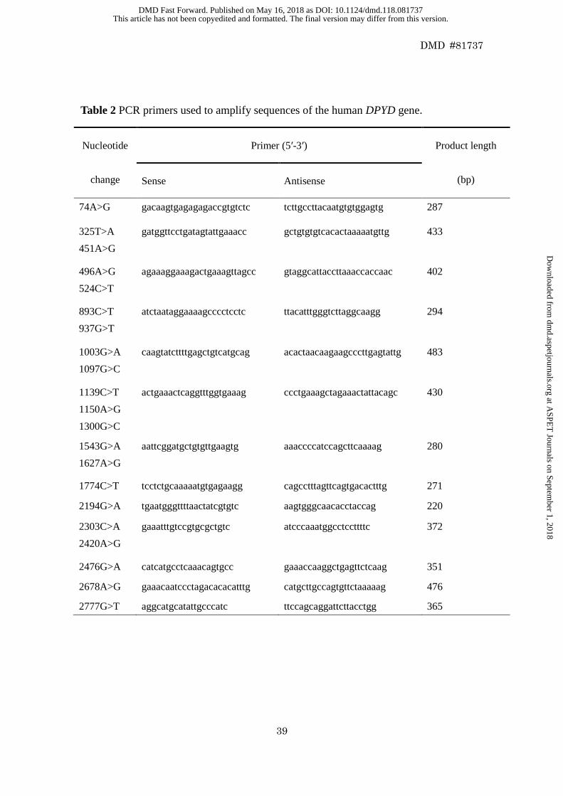
DPYD analysis by Sanger sequencing
To confirm the sequence alterations identified by WGS, we performed Sanger
sequencing analysis of DPYD by PCR amplification, using peripheral blood leukocyte
This article has not been copyedited and formatted. The final version may differ from this version.DMD Fast Forward. Published on May 16, 2018 as DOI: 10.1124/dmd.118.081737
at ASPE
T Journals on Septem
ber 1, 2018dm
d.aspetjournals.orgD
ownloaded from
DMD #81737
10
genomic DNA from Japanese subjects who participated in a community-based cohort
study conducted by ToMMo. The local Ethics Committee of Tohoku University School
of Medicine approved the study, and all cohort participants provided written informed
consent. Genomic DNA was prepared from whole blood using a Gentra Puregene Blood
Kit (Qiagen, Hilden, Germany) as previously described (Nagasaki et al., 2015). The
primer pairs used to amplify sequences containing the SNVs of the DPYD gene are listed
in Table 2. The reaction mixture contained approximately 10 ng genomic DNA, 0.5 µM
of each primer, and AmpliTaq Gold 360 Master Mix (Applied Biosystems, Foster City,
CA, USA), in a total reaction volume of 20 µL. The PCR conditions consisted of initial
denaturation at 95°C for 10 min, followed by 30 cycles of denaturation at 95°C for 30 s,
annealing at 60°C for 30 s, extension at 72°C for 30 s, and then extension at 72°C for 7
min. The resulting amplification products were purified using the ExoSAP-IT PCR
Product Cleanup Reagent (Affymetrix, Cleveland, OH, USA). Dye-terminator cycle
sequencing was performed using the BigDye Terminator v3.1 Cycle Sequencing Kit
(Applied Biosystems, Foster City, CA, USA). The amplicons were sequenced in both
directions with an Applied Biosystems 3500xL Genetic Analyzer (Applied Biosystems).
DPYD cDNA cloning and construction of expression vectors
This article has not been copyedited and formatted. The final version may differ from this version.DMD Fast Forward. Published on May 16, 2018 as DOI: 10.1124/dmd.118.081737
at ASPE
T Journals on Septem
ber 1, 2018dm
d.aspetjournals.orgD
ownloaded from
DMD #81737
11
The plasmid containing wild-type human DPYD cDNA was constructed using
TOPO cloning to insert a plasmid comprising the complete coding region of the human
DPYD into the pENTR/D-TOPO vector (Thermo Fisher Scientific, Waltham, MA, USA).
The plasmid containing wild-type human DPYD cDNA was used as a template to generate
21 DPYD variant constructs using a QuikChange Lightning Site-Directed Mutagenesis
Kit (Agilent Technologies), according to the manufacturer’s instructions. All the prepared
constructs were confirmed by direct sequencing. Wild-type and variant DPYD cDNA
fragments were subsequently subcloned into the mammalian expression vector
pcDNA3.4 (Thermo Fisher Scientific).
Expression of DPD variants in 293FT cells
293FT cells were cultured in Dulbecco’s modified Eagle’s medium (Nacalai
Tesque) containing 10% fetal bovine serum at 37°C under 5% CO2. Cells were plated at
a density of 2.0 × 10⁶ cells/100-mm dish; 24 h after plating, cells were transfected with
plasmids carrying DPYD cDNA (7 μg) and lacZ (1 μg) using polyethylenimine Max
reagent (Polysciences Inc., Warrington, PA, USA), according to the manufacturer’s
instructions. Sodium sulfide (10 μM), ammonium ferric citrate (10 μM), FAD (10 μM),
and FMN (10 μM) were added at 6 h post-transfection. Following incubation for 42 h at
This article has not been copyedited and formatted. The final version may differ from this version.DMD Fast Forward. Published on May 16, 2018 as DOI: 10.1124/dmd.118.081737
at ASPE
T Journals on Septem
ber 1, 2018dm
d.aspetjournals.orgD
ownloaded from

DMD #81737
12
37°C, cells were scraped off, centrifuged at 1,500 × g for 5 min, and resuspended in a
homogenization buffer containing 10 mM Tris-HCl (pH 7.4), 1 mM EDTA, and 10%
glycerol. After cell homogenization, the soluble fractions were prepared by differential
centrifugation at 9,000 × g for 20 min, followed by centrifugation of the resulting
supernatant at 105,000 × g for 60 min. The protein concentration was determined using a
BCA Protein Assay Kit (Thermo Fisher Scientific). The β-galactosidase activity was
measured using the β-Galactosidase Enzyme Assay System (Promega Co., Madison, WI,
USA) to allow adjustment for transfection efficiency.
Determination of DPD protein expression through immunoblotting after SDS-PAGE
Soluble fractions (10 μg) of 293FT cells were separated on 10% ePAGEL
(ATTO, Tokyo, Japan) with an SDS buffer containing 25 mM Tris, 192 mM glycine, and
0.1% SDS. Immunoblotting for each DPD variant was performed in triplicate, in
accordance with standard procedures. DPD was detected using a polyclonal rabbit anti-
human DPD antibody (1:2,000) and HRP-conjugated goat anti-rabbit IgG (1:5,000;
DakoCytomation). The loading control used was GAPDH, which was detected using a
polyclonal rabbit anti-GAPDH antibody (1:5,000) and HRP-conjugated goat anti-rabbit
IgG (1:10,000; Santa Cruz Biotechnology). Immunoblots were visualized using a
This article has not been copyedited and formatted. The final version may differ from this version.DMD Fast Forward. Published on May 16, 2018 as DOI: 10.1124/dmd.118.081737
at ASPE
T Journals on Septem
ber 1, 2018dm
d.aspetjournals.orgD
ownloaded from
DMD #81737
13
SuperSignal West Dura Extended Duration Substrate (Thermo Fisher Scientific).
Chemiluminescence was quantified using a ChemiDoc XRS+ with Image Lab Software
(Bio-Rad Laboratories).
Immunoblotting after blue native-PAGE
Blue native-PAGE was performed using a 5%–20% ePAGEL (ATTO), with
running buffer containing 25 mM Tris and 192 mM glycine. Soluble fractions (9 μg)
mixed with sample buffer (62.5 mM Tris, 10% glycerol, and 0.125% G-250, pH 7.4) were
loaded into each gel lane in triplicate, and electrophoresis was performed at 20 mA for
2.5 h at room temperature. After electrophoresis, the gels were incubated for 10 min with
an SDS buffer (20 mM Tris, 150 mM glycine, and 0.1% SDS, pH 7.4). Proteins were later
transferred onto PVDF membranes and immunoblotted as described above.
NativeMark™ Unstained Protein Standard (Thermo Fisher Scientific) was used as the
molecular weight marker, and proteins were detected by Coomassie Brilliant Blue
staining after electrophoresis.
5-FU reduction assays
DPD-mediated reduction of 5-FU was measured by quantifying FUH2 using
This article has not been copyedited and formatted. The final version may differ from this version.DMD Fast Forward. Published on May 16, 2018 as DOI: 10.1124/dmd.118.081737
at ASPE
T Journals on Septem
ber 1, 2018dm
d.aspetjournals.orgD
ownloaded from
DMD #81737
14
liquid chromatography–tandem mass spectrometry (LC-MS/MS). The incubation
mixture consisted of the sample soluble fraction (50 μg), 1 mM DTT, 200 μM β-NADPH,
2.5 mM magnesium chloride, and 50 mM potassium phosphate buffer (pH 7.4) in a total
volume of 150 μL. Following pre-incubation at 37°C for 3 min, reactions were initiated
by adding 5-FU (0.1, 0.2, 0.5, 1, 2, 5, 10, or 20 μM). After incubating the mixtures at
37°C for 30 min, the reactions were terminated by adding 150 μL of acetonitrile
containing 1 μM uracil-15N2 as an internal standard. 5-FU reduction measurements
obtained using 20 μM 5-FU and 50 μg of the soluble fraction containing wild-type and
variant DPD proteins showed that FUH2 formation was linear for incubations of up to 30
min. When the reaction containing 20 μM 5-FU was incubated for 30 min, the formation
of FUH2 was linear in the presence of up to 50 μg of soluble protein (data not shown).
After removing proteins by centrifuging reaction mixtures at 14,000 × g for 5
min, 150 μL of the supernatant was vacuum-dried at 40°C for 1 h and dissolved in 75 μL
of 0.1% (v/v) formic acid in water. Subsequently, 5 μL of the solution was injected into
an LC-MS/MS system, and FUH2 was measured using the system in the positive ion-
detection mode on the electrospray ionization interface (API 5000 triple-quadrupole mass
spectrometer; SCIEX, Framingham, MA, USA). HPLC separation was performed using
a Prominence HPLC system (SHIMADZU, Kyoto, Japan) and a CAPCELL PAK ADME
This article has not been copyedited and formatted. The final version may differ from this version.DMD Fast Forward. Published on May 16, 2018 as DOI: 10.1124/dmd.118.081737
at ASPE
T Journals on Septem
ber 1, 2018dm
d.aspetjournals.orgD
ownloaded from
DMD #81737
15
S3 column (2.1 × 150 mm, 3.0-μm particle size; OSAKA SODA, Osaka, Japan)
maintained at 8°C. FUH2 was eluted isocratically at a flow rate of 200 μL/min using a
mobile phase consisting of 0.1% (v/v) formic acid in water. The LC-MS/MS system was
controlled by the Analyst 1.5 software (Sciex), which was also used to analyze the data.
The standard curve for FUH2 was constructed in the 0.025–10 μM range using authentic
metabolite standards. The enzymatic activity was normalized to the corresponding DPD
expression level, adjusted for transfection efficiency.
Data analysis
The kinetic data were analyzed using the Enzyme Kinetics Module of SigmaPlot
12.5 (Systat Software, Inc., Chicago, IL, USA), a curve-fitting program based on
nonlinear regression analysis. The values of the Michaelis constant (Km), maximum
velocity (Vmax), and intrinsic clearance (CLint = Vmax/Km) were determined using the
software. Vmax was calculated using the average DPD expression-level values in triplicate.
All values are expressed herein as means ± SD of experiments performed in triplicate.
Statistical analyses of the protein expression levels and kinetic parameters were
performed by analysis of variance using Dunnett’s T3 test or the Kruskal–Wallis method
(IBM SPSS Statistics Ver. 22, International Business Machines, Armonk, NY, USA).
This article has not been copyedited and formatted. The final version may differ from this version.DMD Fast Forward. Published on May 16, 2018 as DOI: 10.1124/dmd.118.081737
at ASPE
T Journals on Septem
ber 1, 2018dm
d.aspetjournals.orgD
ownloaded from
DMD #81737
16
Differences or correlations with P < 0.05 were considered significant.
3-D simulation modeling analysis
A homology model of human DPD was generated by SWISS-MODEL
(https://swissmodel.expasy.org/), based on the crystal structure of pig DPD (RSCB
Protein Data Bank, accession code 1H7W). The CDOCKER protocol for Discovery
Studio 2.5 (Accelrys, San Diego, CA, USA) was used to create docked DPD-5-FU
structures. The Discovery Studio 4.5 visualizer was used for the 3-D imaging of DPD.
This article has not been copyedited and formatted. The final version may differ from this version.DMD Fast Forward. Published on May 16, 2018 as DOI: 10.1124/dmd.118.081737
at ASPE
T Journals on Septem
ber 1, 2018dm
d.aspetjournals.orgD
ownloaded from
DMD #81737
17
Results
To confirm the sequence alterations identified by WGS, we performed Sanger
sequencing analysis of the DPYD. For exon SNVs, the WGS results were the same as
those from the Sanger sequencing. Twenty-one DPYD allelic variants, including some
novel SNVs, were identified in the Japanese subjects.
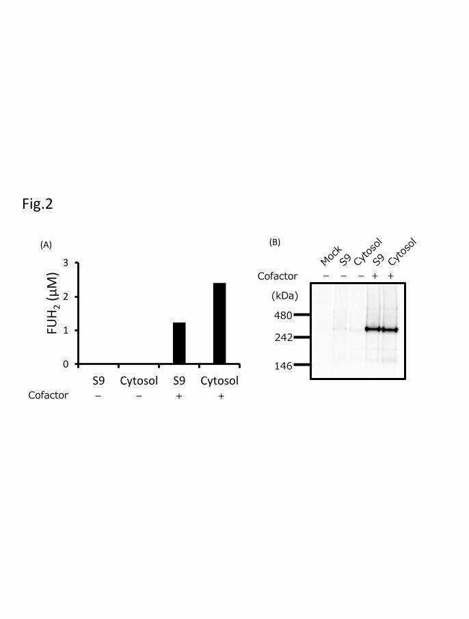
To analyze the effects of sodium sulfide, ammonium ferric citrate, FAD, and
FMN on DPD activity expressed in 293FT cells, we cultured 293FT cells with or without
these cofactors after transfection and measured DPD activity. The production of FUH2 by
DPD expressed with the four cofactors was observed using 50 µM 5-FU, whereas the
production of FUH2 of DPD expressed without cofactors could not be observed under the
same conditions (Fig. 2A). Furthermore, bands were detected in immunoblots after blue
native-PAGE only when DPD was expressed using the four cofactors (Fig. 2B).
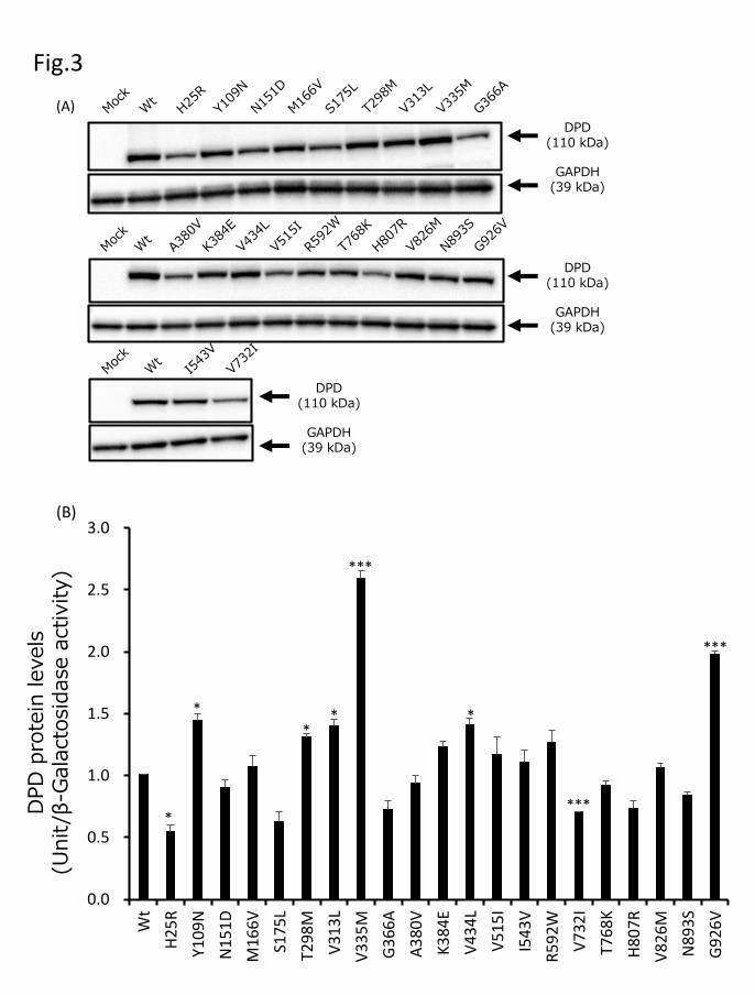
To functionally characterize DPYD variants, wild-type DPD and 21 DPD variant
proteins were transiently expressed in 293FT cells. The expression levels of these DPD
proteins were determined by performing quantitative immunoblotting after SDS-PAGE
with a polyclonal DPD antibody, which recognized all DPD variants expressed in 293FT
cells (Fig. 3A). GAPDH, used as a loading control, was stained at approximately the same
level in the soluble fractions of all transfected 293FT cells. Endogenous DPD protein was
This article has not been copyedited and formatted. The final version may differ from this version.DMD Fast Forward. Published on May 16, 2018 as DOI: 10.1124/dmd.118.081737
at ASPE
T Journals on Septem
ber 1, 2018dm
d.aspetjournals.orgD
ownloaded from
DMD #81737
18
not detected in 293FT cells transfected with an empty vector (mock). The average levels
of wild-type and variant DPD proteins, adjusted for transfection efficiency using the
previously measured β-galactosidase activity, are shown in Fig. 3B. We defined DPD
protein levels equal in intensity to 10 µg of wild-type DPD as 1 DPD unit. Relative to
wild-type, the DPD unit levels for Y109N, T298M, V313L, and V434L were slightly
elevated (P < 0.05), and those for V335M and G926V were considerably elevated (P <
0.005), while those for H25R were slightly lower (P < 0.05), and those for V732I were
considerably lower (P < 0.005). The levels of the remaining 13 variants did not differ
significantly from that of wild-type DPD.
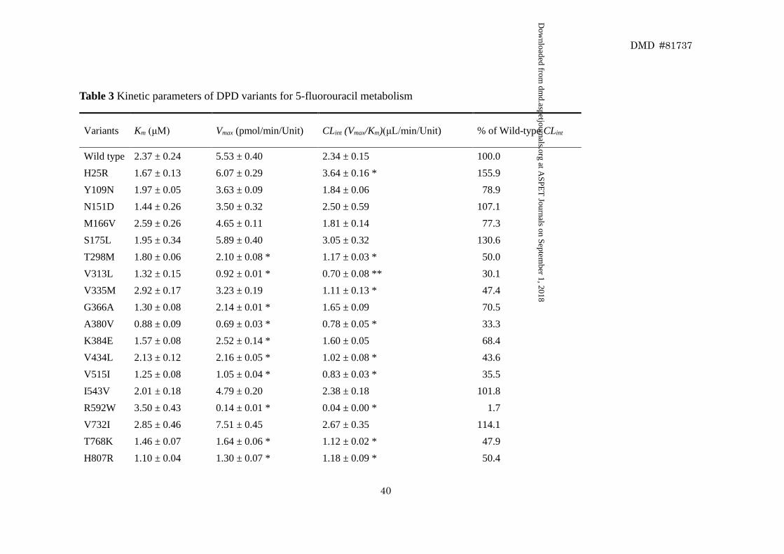
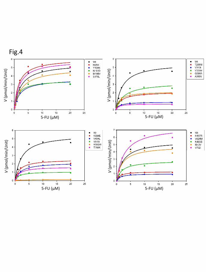
We determined the kinetic parameters for the reduction of 5-FU by wild-type
DPD and the 21 DPD variants (Table 3). The nonlinear regression curves for the
Michaelis–Menten kinetics are shown in Fig. 4. The Km, Vmax, and CLint values for the
reduction of 5-FU by wild-type DPD were 2.37 μM, 5.53 pmol∙min-1∙Unit-1, and 2.34
μL∙min-1∙Unit-1, respectively. For G926V, the kinetic parameters could not be determined
because no 5-FU reduction activity was detected. Relative to wild-type DPD, 12 variants
(T298M, V313L, G366A, A380V, K384E, V434L, V515I, R592W, T768K, H807R,
V826M, and N893S) exhibited significantly lower Vmax values (P < 0.05), and 10 variants
(T298M, V313L, V335M, A380V, V434L, V515I, R592W, T768K, H807R, and V826M)
This article has not been copyedited and formatted. The final version may differ from this version.DMD Fast Forward. Published on May 16, 2018 as DOI: 10.1124/dmd.118.081737
at ASPE
T Journals on Septem
ber 1, 2018dm
d.aspetjournals.orgD
ownloaded from
DMD #81737
19
exhibited significantly lower CLint values (P < 0.05).
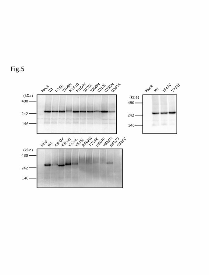
Immunoblotting after blue native-PAGE under non-SDS conditions revealed
approximately 242-kDa bands, estimated as dimeric forms of wild-type DPD (Fig. 5).
Identical bands of wild-type DPD were detected for H25R, Y109N, M166V, S175L,
T298M, V335M, K384E, V434L, I543V, and V732I. Compared with wild-type DPD, the
intensity of the dimeric forms of 10 DPD variants (N151D, V313L, G366A, A380V,
V515I, R592W, T768K, H807R, V826M, and N893S) was decreased. No dimeric band
was detected for the G926V DPD variant.
This article has not been copyedited and formatted. The final version may differ from this version.DMD Fast Forward. Published on May 16, 2018 as DOI: 10.1124/dmd.118.081737
at ASPE
T Journals on Septem
ber 1, 2018dm
d.aspetjournals.orgD
ownloaded from
DMD #81737
20
Discussion
DPD deficiency can result in severe toxicity during 5-FU-based cancer treatment
(Ezzeldin and Diasio, 2004; Mounier-Boutoille et al., 2010; Sahu et al., 2016). Twenty-
one DPYD allelic variants were identified in 1,070 Japanese individuals, but the
functional alterations caused by these variants remain unknown. In this study, we
characterized the enzymatic activity of wild-type DPD and 21 DPD variants using
recombinant proteins expressed in 293FT cells and determined the kinetic parameters of
these variant enzymes associated with 5-FU reduction activity. We also investigated the
mechanism underlying the reduced activity observed for these variants, using blue native-
PAGE and 3-D modeling-based structural analysis.
The effects of sodium sulfide, ammonium ferric citrate, FAD, and FMN on DPD
activity were analyzed using cultured 293FT cells with or without post-transfection
treatment with these cofactors. Enzymatic activity and an approximately 242-kDa band,
estimated as DPD dimeric forms, were observed only in DPD variants expressed with the
four cofactors. These results suggest that DPD enzymatic activity is dimerization-
dependent and consequently, that the four cofactors play a critical role in 5-FU reduction.
To characterize and assess the functional effects of the 21 DPYD mutations on
DPD activity in the Japanese population, wild-type and 21 variant DPD proteins were
This article has not been copyedited and formatted. The final version may differ from this version.DMD Fast Forward. Published on May 16, 2018 as DOI: 10.1124/dmd.118.081737
at ASPE
T Journals on Septem
ber 1, 2018dm
d.aspetjournals.orgD
ownloaded from
DMD #81737
21
transiently expressed in 293FT cells with the four cofactors. The kinetic parameters for
5-FU reduction activity were determined for wild-type and 20 DPD variants expressed in
293FT cells, with the exclusion of G926V. Ten DPD variants (T298M, V313L, V335M,
A380V, V434L, V515I, R592W, T768K, H807R, and V826M) exhibited significantly
reduced CLint values relative to wild-type DPD. Conversely, the metabolic activities of 5-
FU were almost eliminated in G926V. Offer et al. reported that out of six variants (N151D,
M166V, V515I, R592W, T768K, and N893S) expressed in HEK293T/c17 cells using 5-
FU as a substrate, only N151D, M166V, V515I, and T768K exhibited activity equal to
that of wild-type DPD (Offer et al., 2014). In contrast, R592W and N893S exhibited
reduced activity. The enzymatic activities of N151D, M166V, R592W, and N893S
measured using 293FT cells were similar to those previously reported, although the CLint
values obtained for V515I and T768K were significantly reduced in comparison to wild-
type DPD. This discrepancy might be due to differences in measurement conditions,
substrate concentrations, or expression systems. Collectively, these data indicate that
reduction in DPD enzymatic activity might induce accumulation of 5-FU in carriers of
inactive variant enzymes during 5-FU-based treatment administration. Variants exhibiting
altered activity could be responsible for interindividual differences observed in
pyrimidine metabolism and the appearance of severe adverse effects in cancer
This article has not been copyedited and formatted. The final version may differ from this version.DMD Fast Forward. Published on May 16, 2018 as DOI: 10.1124/dmd.118.081737
at ASPE
T Journals on Septem
ber 1, 2018dm
d.aspetjournals.orgD
ownloaded from
DMD #81737
22
chemotherapy.
The structure of human DPD exhibited a native enzyme composed of identical
dimer subunits containing one FMN, one FAD, and four FeS clusters (Lu et al., 1992;
Dobritzsch et al., 2001; Dobritzsch et al., 2002; Mattison et al., 2002; Schnackerz et al.,
2004). To determine the relationship between enzymatic activity and subunit dimerization,
we performed immunoblotting after blue native-PAGE, which revealed 242-kDa DPD
dimeric forms for all variants except the G926V variant. In contrast, no band was
observed for the inactive G926V variant. Our data suggest that DPD dimerization plays
a critical role in DPD-dependent 5-FU reduction.
Amino acid substitutions through non-synonymous DPYD mutations might
cause conformational changes in DPD active site or dimerization interaction site, and thus
DPD activity could be suppressed through dimerization inhibition. Thus, we used 3-D
modeling to analyze the effect of amino acid substitutions on DPD conformation. The
human DPD structure was generated by homology modeling, based on the crystal
structure for pig DPD. The DPD protein consists of five major domains (Figs. 6A and 6B)
(Dobritzsch et al., 2001; Dobritzsch et al., 2002; Mattison et al., 2002). Domain I (residues
27–172) contains two of the FeS clusters and has an exclusively α-helical secondary
structure. The FAD- and NADPH-binding domains II (residues 173–286, 442–524) and
This article has not been copyedited and formatted. The final version may differ from this version.DMD Fast Forward. Published on May 16, 2018 as DOI: 10.1124/dmd.118.081737
at ASPE
T Journals on Septem
ber 1, 2018dm
d.aspetjournals.orgD
ownloaded from
DMD #81737
23
III (residues 287–441) both contain a central parallel β-sheet surrounded by α-helices,
forming a Rossman-type nucleotide binding motif. Domain IV, which binds to FMN and
the substrate, belongs to the glycolate oxidase family of flavoproteins, with the typical
(β/α)8-barrel fold. Domain V (residues 1–26, 848–1025) contains two FeS clusters.
Within the dimer, the DPD dimer redox cofactors are organized in two electron transfer
chains connecting the binding sites for NADPH and the substrate (Lohkamp et al., 2010).
T298M, V313L, V335M, A380V, and V434L were located on the FAD- and NADPH-
binding sites (Fig. 6C). These amino acid substitutions might prevent binding of NADPH,
resulting in considerably decreased activity. In contrast, the R592W, T768K, H807R and
V826M substitutions affected the regions near the FMN and substrate-binding domains
(Fig. 6D), which caused an active site conformational change. Close-up views of the
V515I and G926V mutation sites are shown in Figs. 8E and 8F, respectively. V515
participates in a hydrophobic interaction with W501, H504, and K505 in the vicinity of
the FAD binding site and FeS clusters. The I515 mutation decreases activity by affecting
the binding ability or electron transfer of FAD. G926 is located on domain V, near the
active site. V926 substitution results in a hydrophobic interaction with L929 and inhibits
dimerization, which results in diminished enzymatic activity. Our data provide evidence
that the conformations of these domains and the active site might play a crucial role in
This article has not been copyedited and formatted. The final version may differ from this version.DMD Fast Forward. Published on May 16, 2018 as DOI: 10.1124/dmd.118.081737
at ASPE
T Journals on Septem
ber 1, 2018dm
d.aspetjournals.orgD
ownloaded from
DMD #81737
24
DPD activity.
In conclusion, wild-type and 21 variant DPD proteins were expressed in 293FT
cells, and their enzymatic activities were characterized in vitro. Eleven variants tested in
this study exhibited significantly decreased enzymatic activity because of dimerization
inhibition or conformational changes in each domain. These variants could contribute to
the significant interindividual variability observed in the pharmacokinetics and
pharmacodynamics of 5-FU and its oral prodrugs. Notably, rare DPYD variants could
potentially serve as novel pharmacogenomic markers associated with severe 5-FU
toxicity in Japanese and Asian patients. These findings provide insights that could
facilitate further genotype–phenotype correlation studies of interindividual differences in
drug efficacy and toxicity arising from disparities in DPD activity.
This article has not been copyedited and formatted. The final version may differ from this version.DMD Fast Forward. Published on May 16, 2018 as DOI: 10.1124/dmd.118.081737
at ASPE
T Journals on Septem
ber 1, 2018dm
d.aspetjournals.orgD
ownloaded from

DMD #81737
25
Acknowledgments: We thank the Biomedical Research Core of Tohoku University Graduate
School of Medicine for their technical support. We warmly thank Evelyn Marie Gutierrez Rico,
Tohoku University, for proofreading the manuscript.
This article has not been copyedited and formatted. The final version may differ from this version.DMD Fast Forward. Published on May 16, 2018 as DOI: 10.1124/dmd.118.081737
at ASPE
T Journals on Septem
ber 1, 2018dm
d.aspetjournals.orgD
ownloaded from
DMD #81737
26
Author Contributions
Participated in the research design: Hishinuma, Narita, Akai, Nakanishi and Hiratsuka
Conducted experiments: Hishinuma, Narita, Saito, Akai, Nakanishi, Yasuda, and
Yamamoto
Contributed new reagents or analytic tools: Saito, Maekawa, Yasuda, Nagasaki,
Yamamoto, Yamaguchi, Mano, Hirasawa, and Hiratsuka
Performed data analysis: Hishinuma, Narita, Saito, Maekawa, Akai, Nakanishi, Yasuda,
Nagasaki, Yamamoto, and Hiratsuka
Wrote or contributed to the writing of the manuscript: Hishinuma, Narita, Saito, Akai,
Nakanishi, Saito, Yasuda, Yamamoto, and Hiratsuka
This article has not been copyedited and formatted. The final version may differ from this version.DMD Fast Forward. Published on May 16, 2018 as DOI: 10.1124/dmd.118.081737
at ASPE
T Journals on Septem
ber 1, 2018dm
d.aspetjournals.orgD
ownloaded from
DMD #81737
27
References
Al-Sanna'a NA, Van Kuilenburg AB, Atrak TM, Abdul-Jabbar MA, and Van Gennip AH
(2005) Dihydropyrimidine dehydrogenase deficiency presenting at birth. Journal
of inherited metabolic disease 28:793-796.
Daher GC, Harris BE, and Diasio RB (1990) Metabolism of pyrimidine analogues and
their nucleosides. Pharmacology & therapeutics 48:189-222.
Dobritzsch D, Ricagno S, Schneider G, Schnackerz KD, and Lindqvist Y (2002) Crystal
structure of the productive ternary complex of dihydropyrimidine dehydrogenase
with NADPH and 5-iodouracil. Implications for mechanism of inhibition and
electron transfer. The Journal of biological chemistry 277:13155-13166.
Dobritzsch D, Schneider G, Schnackerz KD, and Lindqvist Y (2001) Crystal structure of
dihydropyrimidine dehydrogenase, a major determinant of the pharmacokinetics
of the anti-cancer drug 5-fluorouracil. The EMBO journal 20:650-660.
Ezzeldin H and Diasio R (2004) Dihydropyrimidine Dehydrogenase Deficiency, a
Pharmacogenetic Syndrome Associated with Potentially Life-Threatening
Toxicity Following 5-Fluorouracil Administration. Clinical Colorectal Cancer
4:181-189.
Froehlich TK, Amstutz U, Aebi S, Joerger M, and Largiader CR (2015) Clinical
This article has not been copyedited and formatted. The final version may differ from this version.DMD Fast Forward. Published on May 16, 2018 as DOI: 10.1124/dmd.118.081737
at ASPE
T Journals on Septem
ber 1, 2018dm
d.aspetjournals.orgD
ownloaded from
DMD #81737
28
importance of risk variants in the dihydropyrimidine dehydrogenase gene for the
prediction of early-onset fluoropyrimidine toxicity. International journal of
cancer 136:730-739.
Ide H, Kikuchi E, Hasegawa M, Hattori S, Yasumizu Y, Miyajima A, and Oya M (2013)
Therapeutic enhancement of S-1 with CPT-11 through down-regulation of
thymidylate synthase in bladder cancer. Cancer medicine 2:488-495.
Kim JY, Shin E, Kim JW, Lee HS, Lee DW, Kim SH, Lee JO, Kim YJ, Kim JH, Bang
SM, Ahn SH, Park DJ, Lee JS, Lee JS, Kim HH, and Lee KW (2015) Impact of
intratumoral expression levels of fluoropyrimidine-metabolizing enzymes on
treatment outcomes of adjuvant S-1 therapy in gastric cancer. PloS one
10:e0120324.
Kunicka T, Prochazka P, Krus I, Bendova P, Protivova M, Susova S, Hlavac V, Liska V,
Novak P, Schneiderova M, Pitule P, Bruha J, Vycital O, Vodicka P, and Soucek P
(2016) Molecular profile of 5-fluorouracil pathway genes in colorectal carcinoma.
BMC cancer 16:795.
Lohkamp B, Voevodskaya N, Lindqvist Y, and Dobritzsch D (2010) Insights into the
mechanism of dihydropyrimidine dehydrogenase from site-directed mutagenesis
targeting the active site loop and redox cofactor coordination. Biochimica et
This article has not been copyedited and formatted. The final version may differ from this version.DMD Fast Forward. Published on May 16, 2018 as DOI: 10.1124/dmd.118.081737
at ASPE
T Journals on Septem
ber 1, 2018dm
d.aspetjournals.orgD
ownloaded from
DMD #81737
29
biophysica acta 1804:2198-2206.
Lu ZH, Zhang R, and Diasio RB (1992) Purification and characterization of
dihydropyrimidine dehydrogenase from human liver. The Journal of biological
chemistry 267:17102-17109.
Maekawa K, Saeki M, Saito Y, Ozawa S, Kurose K, Kaniwa N, Kawamoto M, Kamatani
N, Kato K, Hamaguchi T, Yamada Y, Shirao K, Shimada Y, Muto M, Doi T, Ohtsu
A, Yoshida T, Matsumura Y, Saijo N, and Sawada J (2007) Genetic variations and
haplotype structures of the DPYD gene encoding dihydropyrimidine
dehydrogenase in Japanese and their ethnic differences. Journal of human
genetics 52:804-819.
Mattison LK, Johnson MR, and Diasio RB (2002) A comparative analysis of translated
dihydropyrimidine dehydrogenase cDNA; conservation of functional domains
and relevance to genetic polymorphisms. Pharmacogenetics 12:133-144.
Meulendijks D, Henricks LM, Sonke GS, Deenen MJ, Froehlich TK, Amstutz U,
Largiader CR, Jennings BA, Marinaki AM, Sanderson JD, Kleibl Z, Kleiblova P,
Schwab M, Zanger UM, Palles C, Tomlinson I, Gross E, van Kuilenburg AB, Punt
CJ, Koopman M, Beijnen JH, Cats A, and Schellens JH (2015) Clinical relevance
of DPYD variants c.1679T>G, c.1236G>A/HapB3, and c.1601G>A as predictors
This article has not been copyedited and formatted. The final version may differ from this version.DMD Fast Forward. Published on May 16, 2018 as DOI: 10.1124/dmd.118.081737
at ASPE
T Journals on Septem
ber 1, 2018dm
d.aspetjournals.orgD
ownloaded from
DMD #81737
30
of severe fluoropyrimidine-associated toxicity: a systematic review and meta-
analysis of individual patient data. The Lancet Oncology 16:1639-1650.
Mounier-Boutoille H, Boisdron-Celle M, Cauchin E, Galmiche JP, Morel A, Gamelin E,
and Matysiak-Budnik T (2010) Lethal outcome of 5-fluorouracil infusion in a
patient with a total DPD deficiency and a double DPYD and UTG1A1 gene
mutation. British journal of clinical pharmacology 70:280-283.
Nagasaki M, Yasuda J, Katsuoka F, Nariai N, Kojima K, Kawai Y, Yamaguchi-Kabata Y,
Yokozawa J, Danjoh I, Saito S, Sato Y, Mimori T, Tsuda K, Saito R, Pan X,
Nishikawa S, Ito S, Kuroki Y, Tanabe O, Fuse N, Kuriyama S, Kiyomoto H,
Hozawa A, Minegishi N, Douglas Engel J, Kinoshita K, Kure S, Yaegashi N, To
MJRPP, and Yamamoto M (2015) Rare variant discovery by deep whole-genome
sequencing of 1,070 Japanese individuals. Nature communications 6:8018.
Offer SM, Fossum CC, Wegner NJ, Stuflesser AJ, Butterfield GL, and Diasio RB (2014)
Comparative functional analysis of DPYD variants of potential clinical relevance
to dihydropyrimidine dehydrogenase activity. Cancer research 74:2545-2554.
Okuma Y, Hosomi Y, Miyamoto S, Shibuya M, Okamura T, and Hishima T (2016)
Correlation between S-1 treatment outcome and expression of biomarkers for
refractory thymic carcinoma. BMC cancer 16:156.
This article has not been copyedited and formatted. The final version may differ from this version.DMD Fast Forward. Published on May 16, 2018 as DOI: 10.1124/dmd.118.081737
at ASPE
T Journals on Septem
ber 1, 2018dm
d.aspetjournals.orgD
ownloaded from
DMD #81737
31
Rothenberg ML, Meropol NJ, Poplin EA, Cutsem EV, and Wadler S (2001) Mortality
Associated With Irinotecan Plus Bolus Fluorouracil/Leucovorin: Summary
Findings of an Independent Panel. Journal of Clinical Oncology 19:3801-3807.
Sahu A, Ramaswamy A, and Ostwal V (2016) Dihydro pyrimidine dehydrogenase
deficiency in patients treated with capecitabine based regimens: a tertiary care
centre experience. Journal of gastrointestinal oncology 7:380-386.
Saltz LB, Niedzwiecki D, Hollis D, Goldberg RM, Hantel A, Thomas JP, Fields AL, and
Mayer RJ (2007) Irinotecan fluorouracil plus leucovorin is not superior to
fluorouracil plus leucovorin alone as adjuvant treatment for stage III colon cancer:
results of CALGB 89803. Journal of clinical oncology : official journal of the
American Society of Clinical Oncology 25:3456-3461.
Schnackerz KD, Dobritzsch D, Lindqvist Y, and Cook PF (2004) Dihydropyrimidine
dehydrogenase: a flavoprotein with four iron-sulfur clusters. Biochimica et
biophysica acta 1701:61-74.
Sistonen J, Smith C, Fu Y-K, and Largiadèr CR (2012) A new DPYD genotyping assay
for improving the safety of 5-fluorouracil therapy. Clinica Chimica Acta 414:109-
111.
Thomas F, Hennebelle I, Delmas C, Lochon I, Dhelens C, Garnier Tixidre C, Bonadona
This article has not been copyedited and formatted. The final version may differ from this version.DMD Fast Forward. Published on May 16, 2018 as DOI: 10.1124/dmd.118.081737
at ASPE
T Journals on Septem
ber 1, 2018dm
d.aspetjournals.orgD
ownloaded from
DMD #81737
32
A, Penel N, Goncalves A, Delord JP, Toulas C, and Chatelut E (2016) Genotyping
of a family with a novel deleterious DPYD mutation supports the pretherapeutic
screening of DPD deficiency with dihydrouracil/uracil ratio. Clinical
pharmacology and therapeutics 99:235-242.
Twelves C, Wong A, Nowacki MP, Abt M, Burris HI, Carrato A, Cassidy J,
Cervantes A, Fagerberg J, Georgoulias V, Husseini F, Jodrell D,
Koralewski P, Kröning H, Maroun J, Marschner N, McKendrick J,
Pawlicki M, Rosso R, Schüller J, Seitz J-F, Stabuc B, Tujakowski J,
Van Hazel G, Zaluski J, and Scheithauer W (2005) Capecitabine as
Adjuvant Treatment for Stage III Colon Cancer. New England Journal of
Medicine 352:2696-2704.
van Kuilenburg AB (2004) Dihydropyrimidine dehydrogenase and the efficacy and
toxicity of 5-fluorouracil. European journal of cancer 40:939-950.
van Kuilenburg AB, Meijer J, Maurer D, Dobritzsch D, Meinsma R, Los M, Knegt LC,
Zoetekouw L, Jansen RL, Dezentje V, van Huis-Tanja LH, van Kampen RJ, Hertz
JM, and Hennekam RC (2017) Severe fluoropyrimidine toxicity due to novel and
rare DPYD missense mutations, deletion and genomic amplification affecting
DPD activity and mRNA splicing. Biochimica et biophysica acta 1863:721-730.
This article has not been copyedited and formatted. The final version may differ from this version.DMD Fast Forward. Published on May 16, 2018 as DOI: 10.1124/dmd.118.081737
at ASPE
T Journals on Septem
ber 1, 2018dm
d.aspetjournals.orgD
ownloaded from
DMD #81737
33
Vaudo CE, Gil B, Galuski K, Zarwan C, and Nugent FW (2016) Early-Onset 5-
Fluorouracil Toxicity in a Patient Negative for Dihydropyrimidine
Dehydrogenase Mutations: The Clinical Course of Reversal with Uridine
Triacetate. Pharmacotherapy 36:e178-e182.
This article has not been copyedited and formatted. The final version may differ from this version.DMD Fast Forward. Published on May 16, 2018 as DOI: 10.1124/dmd.118.081737
at ASPE
T Journals on Septem
ber 1, 2018dm
d.aspetjournals.orgD
ownloaded from
DMD #81737
34
Footnotes: This study was supported in part by the Foundation for Promotion of Cancer
Research in Japan [Grants 159 and 100], Tohoku Medical Megabank Project from the
Ministry of Education,Culture,Sports,Science and Technology (MEXT) and the Japan
Agency for Medical Research and Development (AMED) [Grant JP17km0105002].
This article has not been copyedited and formatted. The final version may differ from this version.DMD Fast Forward. Published on May 16, 2018 as DOI: 10.1124/dmd.118.081737
at ASPE
T Journals on Septem
ber 1, 2018dm
d.aspetjournals.orgD
ownloaded from
DMD #81737
35
Figure Legends
Fig. 1. Metabolic pathway of uracil and 5-fluorouracil. Uracil and 5-fluorouracil are
inactivated by dihydropyrimidine dehydrogenase, dihydropyrimidinase, and β-
ureidopropionase. β-Alanine and fluoro-β-alanine are the final metabolites in this
pathway.
Fig. 2. Effect of DPD cofactors (sodium sulfide, ammonium ferric citrate, FAD, and
FMN) on enzymatic activity. (A) DPD activity with or without the four cofactors was
determined at 50 μM 5-FU. (B) DPD proteins in 293FT cells with or without the four
cofactors were determined through blue native-PAGE and immunoblotting analysis.
Fig. 3. Expression levels of wild-type and variant DPD proteins. (A) DPD protein levels
were determined through SDS-PAGE and immunoblotting analysis. (B) DPD expression
levels were normalized relative to β-galactosidase activity in 293FT cells. Bars represent
means ± SD of three independent assays. *P < 0.05 and ***P < 0.005 compared to wild-
type DPD.
Fig. 4. Michaelis–Menten curves of DPD variants. The kinetic parameters Km, Vmax, and
This article has not been copyedited and formatted. The final version may differ from this version.DMD Fast Forward. Published on May 16, 2018 as DOI: 10.1124/dmd.118.081737
at ASPE
T Journals on Septem
ber 1, 2018dm
d.aspetjournals.orgD
ownloaded from
DMD #81737
36
intrinsic clearance of 5-fluorouracil reduction were determined.
Fig. 5. Blue native-PAGE and immunoblotting analysis showing immunoreactive DPD
variant proteins. Blue native PAGE was performed using tris-glycine buffer and 5%–20%
polyacrylamide gels; 9 μg of soluble fractions of DPD variant proteins were loaded into
each lane in triplicate. DPD variants were detected using polyclonal antibodies against
human DPD.
Fig. 6. DPD structural analysis. (A) Diagram showing the overall structure of human DPD,
FAD, NADPH, FMN, and FeS clusters. 5-Fluorouracil is colored yellow, FAD and FMN
are colored green, NADPH is colored blue, and the Fe-S clusters are shown as gray
spheres. (B) The DPD subunit. The distinct domains are colored differently. (C) Amino
acid substitutions for five variants (T298M, V313L, V335M, A380V, and V434L) within
the FAD binding site. (D) Amino acid substitutions of four variants (R592W, T768K,
H807R, and V826M) within the substrate-binding site. (E) Diagrams showing a fragment
of the crystal structures of wild-type DPD (left panel) and V515I (right panel). The V515
and I515 residues are shown in yellow. (F) The diagram of a fragment of the crystal
structure of DPD wild-type (left panel) and G926V (right panel). G926 and V926 are
This article has not been copyedited and formatted. The final version may differ from this version.DMD Fast Forward. Published on May 16, 2018 as DOI: 10.1124/dmd.118.081737
at ASPE
T Journals on Septem
ber 1, 2018dm
d.aspetjournals.orgD
ownloaded from
DMD #81737
37
shown in yellow.
This article has not been copyedited and formatted. The final version may differ from this version.DMD Fast Forward. Published on May 16, 2018 as DOI: 10.1124/dmd.118.081737
at ASPE
T Journals on Septem
ber 1, 2018dm
d.aspetjournals.orgD
ownloaded from

DMD #81737
38
Table 1 DPYD variants identified in 1,070 Japanese population
dbSNP rsID Location Nucleotide change Amino acid substitution Frequency (%)
– Exon 2 74A>G H25R 0.05
– Exon 5 325T>A Y109N 0.23
rs200562975 Exon 5 451A>G N151D 0.14
rs2297595 Exon 6 496A>G M166V 2.24
rs371792178 Exon 6 524C>T S175L 0.05
– Exon 9 893C>T T298M 0.05
– Exon 9 937G>T V313L 0.05
– Exon 10 1003G>A V335M 0.19
– Exon 10 1097G>C G366A 0.05
– Exon 11 1139C>T A380V 0.05
– Exon 11 1150A>G K384E 0.05
– Exon 11 1300G>C V434L 0.05
rs148994843 Exon 13 1543G>A V515I 0.05
rs1801159 Exon 13 1627A>G (DPYD*5) I543V 25.68
rs59086055 Exon 14 1774C>T R592W 0.05
rs1801160 Exon 18 2194G>A (DPYD*6) V732I 1.82
rs56005131 Exon 19 2303C>A T768K 2.20
– Exon 19 2420A>G H807R 0.05
– Exon 20 2476G>A V826M 0.14
rs188052243 Exon 21 2678A>G N893S 0.19
– Exon 22 2777G>T G926V 0.05
This article has not been copyedited and formatted. The final version may differ from this version.DMD Fast Forward. Published on May 16, 2018 as DOI: 10.1124/dmd.118.081737
at ASPE
T Journals on Septem
ber 1, 2018dm
d.aspetjournals.orgD
ownloaded from
DMD #81737
39
Table 2 PCR primers used to amplify sequences of the human DPYD gene.
Nucleotide
change
Primer (5′-3′) Product length
(bp) Sense Antisense
74A>G gacaagtgagagagaccgtgtctc tcttgccttacaatgtgtggagtg 287
325T>A
451A>G
gatggttcctgatagtattgaaacc gctgtgtgtcacactaaaaatgttg 433
496A>G
524C>T
agaaaggaaagactgaaagttagcc gtaggcattaccttaaaccaccaac 402
893C>T
937G>T
atctaataggaaaagcccctcctc ttacatttgggtcttaggcaagg 294
1003G>A
1097G>C
caagtatcttttgagctgtcatgcag acactaacaagaagcccttgagtattg 483
1139C>T
1150A>G
1300G>C
actgaaactcaggtttggtgaaag ccctgaaagctagaaactattacagc 430
1543G>A
1627A>G
aattcggatgctgtgttgaagtg aaaccccatccagcttcaaaag 280
1774C>T tcctctgcaaaaatgtgagaagg cagcctttagttcagtgacactttg 271
2194G>A tgaatgggttttaactatcgtgtc aagtgggcaacacctaccag 220
2303C>A
2420A>G
gaaatttgtccgtgcgctgtc atcccaaatggcctccttttc 372
2476G>A catcatgcctcaaacagtgcc gaaaccaaggctgagttctcaag 351
2678A>G gaaacaatccctagacacacatttg catgcttgccagtgttctaaaaag 476
2777G>T aggcatgcatattgcccatc ttccagcaggattcttacctgg 365
This article has not been copyedited and formatted. The final version may differ from this version.DMD Fast Forward. Published on May 16, 2018 as DOI: 10.1124/dmd.118.081737
at ASPE
T Journals on Septem
ber 1, 2018dm
d.aspetjournals.orgD
ownloaded from
DMD #81737
40
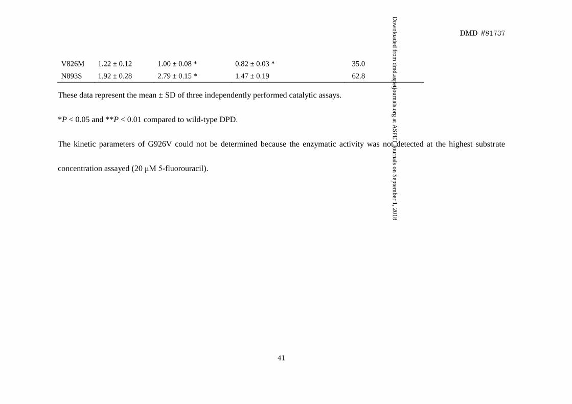
Table 3 Kinetic parameters of DPD variants for 5-fluorouracil metabolism
Variants Km (μM) Vmax (pmol/min/Unit) CLint (Vmax/Km)(μL/min/Unit) % of Wild-type CLint
Wild type 2.37 ± 0.24 5.53 ± 0.40 2.34 ± 0.15 100.0
H25R 1.67 ± 0.13 6.07 ± 0.29 3.64 ± 0.16 * 155.9
Y109N 1.97 ± 0.05 3.63 ± 0.09 1.84 ± 0.06 78.9
N151D 1.44 ± 0.26 3.50 ± 0.32 2.50 ± 0.59 107.1
M166V 2.59 ± 0.26 4.65 ± 0.11 1.81 ± 0.14 77.3
S175L 1.95 ± 0.34 5.89 ± 0.40 3.05 ± 0.32 130.6
T298M 1.80 ± 0.06 2.10 ± 0.08 * 1.17 ± 0.03 * 50.0
V313L 1.32 ± 0.15 0.92 ± 0.01 * 0.70 ± 0.08 ** 30.1
V335M 2.92 ± 0.17 3.23 ± 0.19 1.11 ± 0.13 * 47.4
G366A 1.30 ± 0.08 2.14 ± 0.01 * 1.65 ± 0.09 70.5
A380V 0.88 ± 0.09 0.69 ± 0.03 * 0.78 ± 0.05 * 33.3
K384E 1.57 ± 0.08 2.52 ± 0.14 * 1.60 ± 0.05 68.4
V434L 2.13 ± 0.12 2.16 ± 0.05 * 1.02 ± 0.08 * 43.6
V515I 1.25 ± 0.08 1.05 ± 0.04 * 0.83 ± 0.03 * 35.5
I543V 2.01 ± 0.18 4.79 ± 0.20 2.38 ± 0.18 101.8
R592W 3.50 ± 0.43 0.14 ± 0.01 * 0.04 ± 0.00 * 1.7
V732I 2.85 ± 0.46 7.51 ± 0.45 2.67 ± 0.35 114.1
T768K 1.46 ± 0.07 1.64 ± 0.06 * 1.12 ± 0.02 * 47.9
H807R 1.10 ± 0.04 1.30 ± 0.07 * 1.18 ± 0.09 * 50.4
This article has not been copyedited and formatted. The final version may differ from this version.DMD Fast Forward. Published on May 16, 2018 as DOI: 10.1124/dmd.118.081737
at ASPE
T Journals on Septem
ber 1, 2018dm
d.aspetjournals.orgD
ownloaded from
DMD #81737
41
V826M 1.22 ± 0.12 1.00 ± 0.08 * 0.82 ± 0.03 * 35.0
N893S 1.92 ± 0.28 2.79 ± 0.15 * 1.47 ± 0.19 62.8
These data represent the mean ± SD of three independently performed catalytic assays.
*P < 0.05 and **P < 0.01 compared to wild-type DPD.
The kinetic parameters of G926V could not be determined because the enzymatic activity was not detected at the highest substrate
concentration assayed (20 μM 5-fluorouracil).
This article has not been copyedited and formatted. The final version may differ from this version.DMD Fast Forward. Published on May 16, 2018 as DOI: 10.1124/dmd.118.081737
at ASPE
T Journals on Septem
ber 1, 2018dm
d.aspetjournals.orgD
ownloaded from
Fluoro-β-alanineDihydro-5-fluorouracil5-Fluorouracil Fluoro-β-ureidopropionic acid
Dihydro
pyrimidinase
Uracil Dihydrouracil β-ureidopropionic acid β-alanine
Dihydropyrimidine
dehydrogenaseβ-ureido
propionase
Fig.1
This article has not been copyedited and formatted. The final version may differ from this version.DMD Fast Forward. Published on May 16, 2018 as DOI: 10.1124/dmd.118.081737
at ASPE
T Journals on Septem
ber 1, 2018dm
d.aspetjournals.orgD
ownloaded from
0123
FUH
2(μM
)
146
(kDa)
242
480
Cofactor
- - - + +
- - + +
(A) (B)
0
1
2
3
S9 Cytosol S9 Cytosol
FUH
2(μM
)
146
(kDa)
242
480
Cofactor - - - + +
Cofactor - - + +
(A) (B)
Fig.2
This article has not been copyedited and formatted. The final version may differ from this version.DMD Fast Forward. Published on May 16, 2018 as DOI: 10.1124/dmd.118.081737
at ASPE
T Journals on Septem
ber 1, 2018dm
d.aspetjournals.orgD
ownloaded from
0.0
0.5
1.0
1.5
2.0
2.5
3.0
Wt
H2
5R
Y10
9N
N15
1D
M16
6V
S175
L
T29
8M
V31
3L
V33
5M
G3
66
A
A3
80V
K3
84E
V43
4L
V51
5I
I54
3V
R5
92W
V73
2I
T76
8K
H8
07R
V82
6M
N89
3S
G9
26
V
*
*
**
***
***
***
*
DPD
pro
tein
levels
(Unit/β
-Gala
cto
sidase
activity)
(B)
DPD(110 kDa)
GAPDH(39 kDa)
DPD(110 kDa)
GAPDH(39 kDa)
DPD(110 kDa)
GAPDH(39 kDa)
(A)
Fig.3
This article has not been copyedited and formatted. The final version may differ from this version.DMD Fast Forward. Published on May 16, 2018 as DOI: 10.1124/dmd.118.081737
at ASPE
T Journals on Septem
ber 1, 2018dm
d.aspetjournals.orgD
ownloaded from
5-FU (µM)
V(p
mo
l/m
in/U
nit
)
5-FU (µM)
V(p
mo
l/m
in/U
nit
)
5-FU (µM)
V(p
mo
l/m
in/U
nit
)
5-FU (µM)
V(p
mo
l/m
in/U
nit
)
Fig.4
This article has not been copyedited and formatted. The final version may differ from this version.DMD Fast Forward. Published on May 16, 2018 as DOI: 10.1124/dmd.118.081737
at ASPE
T Journals on Septem
ber 1, 2018dm
d.aspetjournals.orgD
ownloaded from
146
(kDa)
242
480
146
(kDa)
242
480
146
(kDa)
242
480
Fig.5
This article has not been copyedited and formatted. The final version may differ from this version.DMD Fast Forward. Published on May 16, 2018 as DOI: 10.1124/dmd.118.081737
at ASPE
T Journals on Septem
ber 1, 2018dm
d.aspetjournals.orgD
ownloaded from
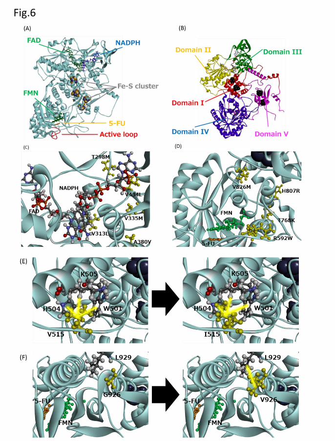
Fig.6
This article has not been copyedited and formatted. The final version may differ from this version.DMD Fast Forward. Published on May 16, 2018 as DOI: 10.1124/dmd.118.081737
at ASPE
T Journals on Septem
ber 1, 2018dm
d.aspetjournals.orgD
ownloaded from