Embed Size (px)
Citation preview
Intraoperative mass spectrometry mapping of anonco-metabolite to guide brain tumor surgerySandro Santagataa,b,c,1, Livia S. Eberlind,e,1, Isaiah Nortonf, David Calligarisf, Daniel R. Feldmana,f, Jennifer L. Idef,Xiaohui Liuf, Joshua S. Wileyd,e, Matthew L. Vestalf, Shakti H. Ramkissoona,b, Daniel A. Orringerf, Kristen K. Gilla,Ian F. Dunnf, Dora Dias-Santagatag, Keith L. Ligona,b,h, Ferenc A. Joleszi, Alexandra J. Golbyf, R. Graham Cooksd,e,2,and Nathalie Y. R. Agarc,f,i,2
Departments of aPathology, fNeurosurgery, and iRadiology, Brigham and Women’s Hospital and Harvard Medical School, Boston, MA 02115; bDepartment ofPathology, Boston Children’s Hospital and Harvard Medical School, Boston, MA 02115; cDepartment of Cancer Biology, Harvard Medical School and Dana–Farber Cancer Institute, Boston, MA 02115; dDepartment of Chemistry and eCenter for Analytical Instrumentation Development, Purdue University, WestLafayette, IN 47907; gDepartment of Pathology and Center for Cancer Research, Massachusetts General Hospital and Harvard Medical School, Boston,MA 02115; and hDepartment of Medical Oncology, Center for Molecular Oncologic Pathology, Dana–Farber Cancer Institute, Boston, MA 02215
Edited by Jerrold Meinwald, Cornell University, Ithaca, NY, and approved June 4, 2014 (received for review March 13, 2014)
For many intraoperative decisions surgeons depend on frozensection pathology, a technique developed over 150 y ago. Technicalinnovations that permit rapid molecular characterization of tissuesamples at the time of surgery are needed. Here, using de-sorption electrospray ionization (DESI) MS, we rapidly detect thetumor metabolite 2-hydroxyglutarate (2-HG) from tissue sectionsof surgically resected gliomas, under ambient conditions andwithout complex or time-consuming preparation. With DESI MS,we identify isocitrate dehydrogenase 1-mutant tumors with bothhigh sensitivity and specificity within minutes, immediately pro-viding critical diagnostic, prognostic, and predictive information.Imaging tissue sections with DESI MS shows that the 2-HG signaloverlaps with areas of tumor and that 2-HG levels correlate withtumor content, thereby indicating tumor margins. Mapping the 2-HG signal onto 3D MRI reconstructions of tumors allows theintegration of molecular and radiologic information for enhancedclinical decision making. We also validate the methodology and itsdeployment in the operating room: We have installed a massspectrometer in our Advanced Multimodality Image Guided Oper-ating (AMIGO) suite and demonstrate the molecular analysisof surgical tissue during brain surgery. This work indicatesthat metabolite-imaging MS could transform many aspects ofsurgical care.
mass spectrometry imaging | oncometabolite | intrasurgical diagnosis |brain cancer | IDH1
The microscopic review of tissue biopsies frequently remainsthe sole source of intraoperative diagnostic information, and
many important surgical decisions such as the extent of tumorresection are based on this information. This approach is time-consuming, requiring nearly 30 min between the moment a tissueis biopsied and the time the pathologist’s interpretation is com-municated back to the surgeon. Even after the report of the finalpathologic diagnosis is issued days later, a lot of diagnostic,prognostic, and predictive information is left undiscovered andunexamined within the tissue. Tools that provide more imme-diate feedback to the surgeon and the pathologist and that alsorapidly extract detailed molecular information could transformthe management of care for cancer patients.MS offers the possibility for the in-depth analysis of the pro-
teins and lipids that comprise tissues (1, 2). We have recentlyshown that desorption electrospray ionization (DESI) MS isa powerful methodology for characterizing lipids within tumorspecimens (3–6). The intensity profile of lipids ionized fromwithin tumors can be used for classifying tumors and for pro-viding valuable prognostic information such as tumor subtypeand grade. Because DESI MS is performed in ambient con-ditions with minimal pretreatment of the samples (7, 8), there isthe potential to provide diagnostic information rapidly within
the operating room (4, 6, 9). The ability to quickly acquire suchvaluable diagnostic information from lipids prompted us to deter-mine whether we could use DESI MS to detect additional mole-cules of diagnostic value within tumors, such as their metabolites.Recently, recurrent mutations have been described in the
genes encoding isocitrate dehydrogenases 1 and 2 (IDH1 andIDH2) in a number of tumor types including gliomas (10, 11),intrahepatic cholangiocarcinomas (12), acute myelogenous leu-kemias (13), and chondrosarcomas (14). These mutant en-zymes have the novel property of converting isocitrate to2-hydroxyglutarate (2-HG) (15). This oncometabolite haspleiotropic effects on DNA methylation patterns (16–18), on theactivity of prolyl hydroxylases (19), and on cellular differentia-tion and growth (20–22). Whereas 2-HG is present in vanishinglysmall amounts in normal tissues, concentrations are extremelyhigh in tumors with mutations in IDH1 and IDH2—severalmicromoles per gram of tumor have been reported (15). Severalgroups have reported that 2-HG can be detected by magnetic res-onance spectroscopy and imaging, hence providing a noninvasiveimaging approach for evaluating patients (23–27). Although such
Significance
The diagnosis of tumors during surgery still relies principally onan approach developed over 150 y ago: frozen section mi-croscopy. We show that a validated molecular marker—2-hydroxyglutarate generated from isocitrate dehydrogenase 1mutant gliomas—can be rapidly detected from tumors usinga form of ambient MS that does not require sample prepara-tion. We use the Advanced Multimodality Image GuidedOperating Suite at Brigham and Women’s Hospital to demon-strate that desorption electrospray ionization MS could beused to detect residual tumor that would have been left behindin the patient. The approach paves the way for the clinical test-ing of MS-based intraoperative monitoring of tumor metabolites,an advance that could revolutionize the care of surgicaloncology patients.
Author contributions: S.S., F.A.J., R.G.C., and N.Y.R.A. designed research; S.S., L.S.E., I.N.,D.R.F., J.L.I., X.L., J.S.W., S.H.R., D.A.O., K.K.G., I.F.D., D.D.-S., K.L.L., A.J.G., and N.Y.R.A.performed research; N.Y.R.A. contributed new reagents/analytic tools; L.S.E., I.N., D.C.,M.L.V., D.D.-S., R.G.C., and N.Y.R.A. analyzed data; and S.S., L.S.E., R.G.C., and N.Y.R.A.wrote the paper.
Conflict of interest statement: S.S. and N.Y.R.A. are scientific advisors to BayesianDx.
This article is a PNAS Direct Submission.
See Commentary on page 10906.1S.S. and L.S.E. contributed equally to this work.2To whom correspondence may be addressed. E-mail: [email protected] or [email protected].
This article contains supporting information online at www.pnas.org/lookup/suppl/doi:10.1073/pnas.1404724111/-/DCSupplemental.
www.pnas.org/cgi/doi/10.1073/pnas.1404724111 PNAS | July 29, 2014 | vol. 111 | no. 30 | 11121–11126
MED
ICALSC
IENCE
SCH
EMISTR
YSE
ECO
MMEN
TARY
Dow
nloa
ded
by g
uest
on
Janu
ary
12, 2
022
imaging approaches may provide information to plan surgeryand to follow the response to chemotherapeutics, applying themto guide decision making during an operation is currentlyimpractical.The ability to detect 2-HG intraoperatively would be partic-
ularly useful because infiltrating gliomas such as IDH1 and IDH2mutant gliomas are difficult to visualize with conventionalmeans, which contributes to the high prevalence of suboptimalsurgical resection. Multiple studies suggest that the more re-sidual tumor remains after surgery, the shorter the patient sur-vival for both low- and high-grade gliomas (28–32). Detectinginfiltrating glioma cells by microscopic review is challenging onwell-prepared H&E-stained permanent sections, and even moreso on H&E-stained frozen sections, which frequently harborprocessing artifacts. Thus, 2-HG detection could help to definesurgical margins, thereby allowing for more complete resectionand for longer survival (31, 32). Moreover, directing patientstoward appropriate clinical trials for targeted therapeutics (33)would be facilitated by more rapid molecular categorizationof tumors.Here, we show that 2-HG can be rapidly detected from glioma
samples using DESI MS under ambient conditions, withoutcomplex tissue preparation and during surgery, allowing rapidmolecular characterization and providing information that isunattainable by standard histopathology techniques. We alsopresent the first implementation, to our knowledge, of MS withinan operating room for the molecular characterization of tissue aspart of an image-guided therapy program. We cross-validate ourfindings using standard pathology techniques. Measuring specificmetabolites in tumor tissues with precise spatial distribution andunder ambient conditions provides a new paradigm for intra-operative surgical decision making, rapid diagnosis, and patientcare management.
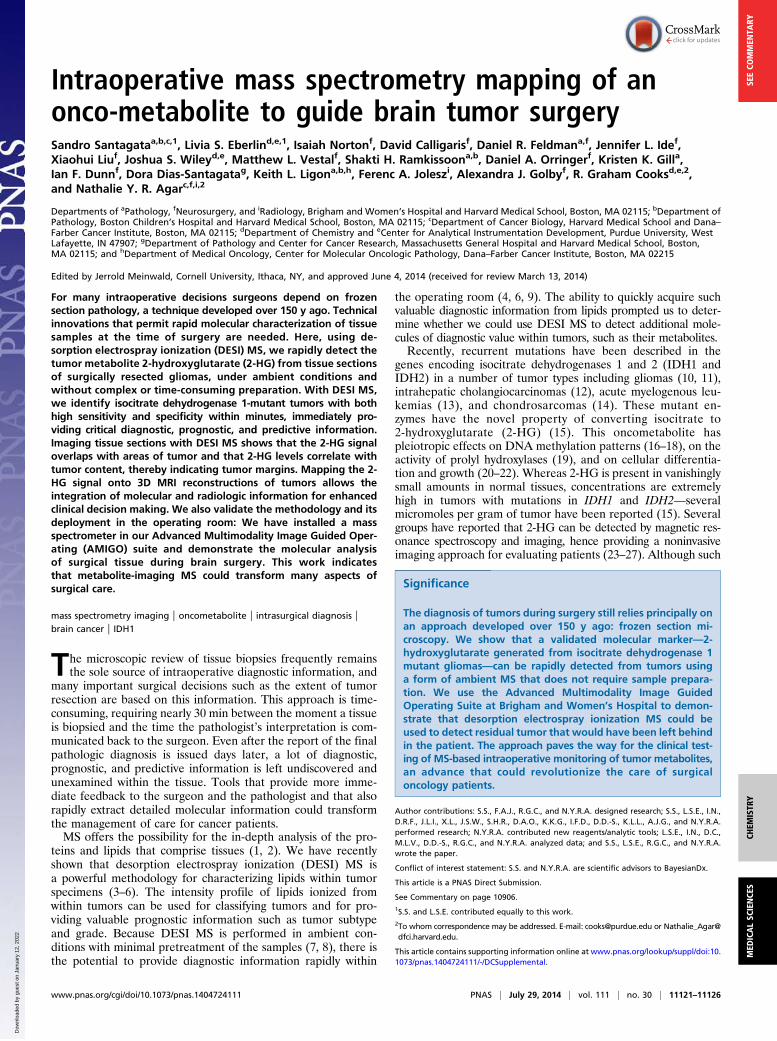
ResultsIdentification of 2-HG with DESI MS. To determine the conditionsfor detecting 2-HG from glioma frozen tissue sections by DESIMS, we first recorded the negative ion mode mass spectra fromtwo glioma samples: an oligodendroglioma with mutated IDH1(encoding the amino acid change R132H) and a glioblastomawith wild-type IDH1. The product of mutant IDH1, 2-HG, isa small organic acid containing two carboxylic acid functionalgroups in its structure. In the negative ion mode, the deproto-nated form of 2-HG should be detected at an m/z of 147.03(C5H7O5
−). Together with the rich diagnostic lipid informationcommonly observed from gliomas by DESI MS in the mass rangem/z 100–1,000, we detected a significant peak at m/z 147 in anIDH1 mutated sample (Fig. 1A), but not in an IDH1 wild-typesample (Fig. 1B).We used tandem MS analysis (MS2) with a linear ion trap
mass spectrometer to characterize the signal at m/z 147 (Fig. S1and Fig. 1 C–F). In an oligodendroglioma with the IDH1 R132Hmutation, the main fragment ion generated fromm/z 147 wasm/z129, which corresponds to loss of a water molecule from 2-HG(Fig. 1C). Further characterization of m/z 129 with an additionalstage of MS analysis (MS3) yielded two additional fragment ionsat m/z 101 and m/z 85, corresponding to neutral losses of CO andCO2, respectively (Fig. 1D). We obtained identical MS2 and MS3
fragmentation patterns when we subjected purified L-α-hydrox-yglutaric acid to tandem MS experiments (Fig. 1 E and F). Apurified standard of the wild-type IDH metabolite alpha-keto-glutarate was detected at m/z 145, and MS2 and MS3 fragmen-tation patterns yielded peaks at m/z 101 and at m/z 73 and m/z57, respectively (Fig. S2). We further characterized the 2-HGpeaks using a high-resolution and high-mass accuracy linear trapquadrupole (LTQ) Orbitrap mass spectrometer (Fig. S3). DESImass spectra from an IDH1 R132H mutant sample showeda prominent peak at m/z 147.0299 in the negative ion mode,
which matched the molecular formula of the deprotonated formof 2-HG (C5H7O5
−) with a mass accuracy of 0.3 ppm. In all,these results confirm the ability to reliably and rapidly detect2-HG from human glioma tissue sections with DESI MS.
Levels of 2-HG Correlate with Mutational Status and Tumor CellContent. We next monitored the levels of 2-HG using DESIMS in a panel of 35 human glioma specimens (Table S1) in-cluding primary and recurrent oligodendrogliomas, oligoas-trocytomas, and astrocytomas of different grades (3). We firstcharacterized the samples using a clinically validated antibodythat selectively recognizes the R132H mutant epitope and notthe wild-type epitope from IDH1 (34) (Table S1). Twenty-one ofthe 35 samples had the R132H mutation. We then measured2-HG levels in these samples directly from frozen tissue sectionsusing a linear ion trap LTQ DESI. In some samples we detecteda peak at m/z 147 and assigned it to 2-HG by MS2 analysis,thereby providing strong independent evidence that these sam-ples were mutated for one of the IDH genes. To account for thevariability in desorption and ionization efficiency throughout thetissue and between samples, we normalized the 2-HG signal tothe combined intensity of the 40 most abundant lipid species thatwere detected during each data acquisition (Table S1 and Sup-porting Information). In all of the 21 samples with the IDH1R132H mutation, we clearly detected 2-HG with a limit of de-tection estimated to be 3 μmol 2-HG/g of tissue (Fig. S4), whichis below the lowest concentration of 2-HG in tissue in IDH1mutant human gliomas as measured by HPLC-MS analysis (23).We also observed a correlation between the concentration of
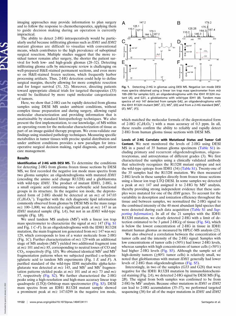
tumor cells and the intensity of the 2-HG signal: Samples withlow concentrations of tumor cells (<50%) had lower 2-HG levels,whereas samples with high concentrations of tumor cells (>50%)had higher 2-HG levels (Fig. S5). Although the sample set ofhigh-density tumors (≥80% tumor cells) is relatively small, wenoted that glioblastomas with mutant IDH1 generally had lowerlevels of 2-HG than oligodendrogliomas (Fig. S5).Interestingly, in two of the samples (G33 and G28) that were
negative for the IDH1 R132H mutation by immunohistochemi-cal staining (Fig. 2A), we detected 2-HG signal by DESI MS (Fig.2B). The signal from both samples was confirmed to be from2-HG by MS2 analysis. Because other mutations in IDH1 or IDH2can lead to 2-HG accumulation (35–37), we performed targetedsequencing (38) for all of the major mutations in IDH1 and IDH2
Fig. 1. Detecting 2-HG in gliomas using DESI MS. Negative ion mode DESImass spectra obtained using a linear ion trap mass spectrometer from m/z100–200 for samples G23, an oligodendroglioma with the IDH1 R132H mu-tant (A), and G31, a glioblastoma with wild-type IDH1 (B). Tandem massspectra of m/z 147 detected from sample G42, an oligodendroglioma withthe IDH1 R132H mutant [MS2, (C); MS3, (D)] and from a 2-HG standard [MS2,(E); MS3, (F)].
11122 | www.pnas.org/cgi/doi/10.1073/pnas.1404724111 Santagata et al.
Dow
nloa
ded
by g
uest
on
Janu
ary
12, 2
022
that have been described in gliomas (39). This analysis revealedthat both samples G33 and G28 harbored a less common butpreviously described IDH1 mutation that leads to substitution ofthe amino acid arginine with glycine at position 132 (R132G)(Fig. 2C). These results provide a clear example of how detecting2-HG with DESI MS allows rapid and accurate determination ofIDH1 status in human gliomas. Whereas the diagnostic antibodyonly recognizes one of the many IDH1 mutants (37), DESI MScaptures the presence of 2-HG independent of the underlyinggenetic mutation in IDH1. Notably, our results show that DESIMS can detect 2-HG with very high sensitivity and specificity: Wedetected 2-HG signal in all cases with mutant IDH1 (even whenthe tumor concentration was as low as 5%) and did not detect2-HG signal in any of the cases with wild-type IDH1.
DESI MS 2D Imaging of 2-HG in Glioma Sections Delineates TumorMargins. To further validate DESI MS as a tool for monitoring2-HG levels, we turned to 2D DESI MS imaging to study thespatial distribution of molecules across a tissue section (40).DESI MS imaging has recently been shown not to destroy asample as it is being analyzed when a histologically compatiblesolvent system is used (40). This relative preservation allows thesame tissue section to be stained with H&E following DESI MSdata acquisition, and the spatial molecular information derivedfrom DESI MS can then be overlaid onto the optical image ofthe tissue (40). As such, this approach provides a powerful way tocorrelate 2-HG levels with histopathology and, importantly, tovalidate the DESI MS observations.As a control, we acquired 2D DESI MS data from frozen
sections of human glioblastoma orthotopic xenograft models thathad been implanted into the brains of immunocompromisedmice (Fig. S6). A signal for 2-HG was not detected from xeno-grafts of a glioblastoma cell line (BT329) that has wild-typeIDH1 (Fig. S6A). Strikingly, however, a strong signal for 2-HGwas found throughout the tissue section of the mouse brain that
was diffusely infiltrated by a glioblastoma xenograft (BT116) thathas the IDH1 R132H mutation (Fig. S6B), as was similarly ob-served in an IDH1 R132H mutated oligodendroglioma xenograftmodel by liquid extraction surface analysis nano electrosprayionization-MS imaging (41).We next turned to tissue sections of human glioma specimens
that had been surgically resected. Using 2D DESI MS with bothan LTQ ion trap (Thermo Fisher Scientific) and an amaZonSpeed ion trap (Bruker Daltonics), we observed accumulation of2-HG within a densely cellular glioblastoma with mutated IDH1(Fig. S7). 2-HG was absent in an area of hemorrhage abuttingthe tumor (Fig. S7). In tissue specimens from two additionalglioma resections, we identified areas that contained regions oftumor as well as regions of brain with only scattered infiltratingglioma cells (i.e., the margin of the tumor). DESI MS revealedstrong 2-HG signals in the cellular portions of these samples butweaker signals in the portions of brain with scattered infiltratingtumor cells. By validating our DESI MS results directly withtissue histopathology, we show that monitoring 2-HG levels withDESI MS can help to readily discriminate tissue with dense tu-mor from tissue with only scattered tumor cells. Such discrimi-natory capacity can help define tumor margins.
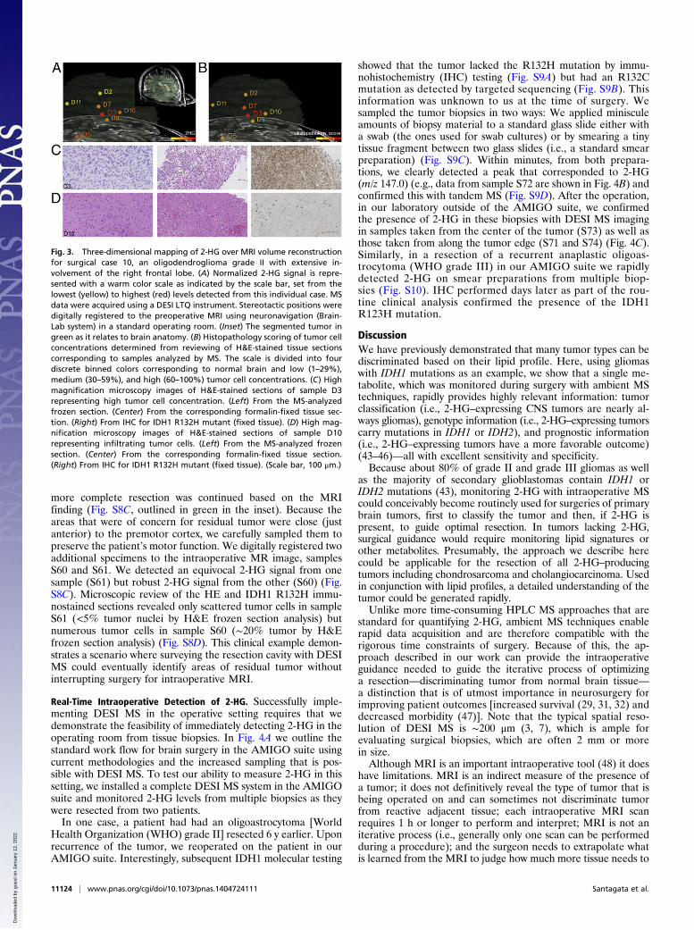
Three-Dimensional Mapping of 2-HG onto MRI Tumor Reconstructions.MRI information is critical for planning neurosurgical proce-dures. During the surgery, neuronavigation systems allow theneurosurgeon to register the position of surgical instrumentswith preoperative plans (i.e., confirming where the tools arerelative to the imaging findings). Surgeons can therefore digitallymark the site of a biopsy relative to the tumor in the MRI. Weresected two IDH1 mutated gliomas in this manner, using 3Dmapping, marking the positions of multiple biopsies in each case.In both cases, we measured the 2-HG content of each stereo-tactic specimen and normalized to its lipid signals (see Materialsand Methods and Supporting Information for details). We thencorrelated this information with the tumor cell content of eachstereotactic specimen, as determined by review of both H&E andimmunostains for IDH1 R132H.In the resection of an oligodendroglioma (Fig. 3), we identi-
fied strong 2-HG signals in the sample (D3) taken from thecenter of the tumor mass (Fig. 3A). This sample was composedof dense tumor (Fig. 3 B and C). Biopsies from the margins ofthe radiographic mass (e.g., D10, Fig. 3 B and D) contained lowconcentrations of infiltrating glioma cells (Fig. 3D). In suchsamples we detected low levels of 2-HG (Fig. 3A). Consistentwith our prior findings on a large panel of glioma specimens (Fig.S4 and Table S1), these stereotactic samples demonstrate thatthe normalized level of 2-HG correlates with the tumor cellconcentration and can help define samples that are at the in-filtrating border of the tumor.We performed a second surgical resection (Fig. S8) in the
Advanced Multimodality Image Guided Operating (AMIGO)suite (42) at Brigham and Women’s Hospital that is a part of theNational Center for Image-Guided Therapy. In this advancedsurgical and interventional environment, MRI can be performedduring the operation to see whether additional tumor remains insitu. This residual tumor can then be resected before the pro-cedure is completed.An oligoastrocytoma was resected in this second case. We
digitally registered the location of multiple biopsy pieces to thepreoperative MRI and measured 2-HG levels in each of them(Fig. S8A). The highest levels of 2-HG were detected in speci-mens that were taken from the center of the tumor mass, whichproved to be dense cellular tumor (Fig. S8B). We took anintraoperative MRI of the patient’s brain once it seemed that theentire tumor had been removed (i.e., following an apparent grosstotal resection). The T2-weighted intraoperative image revealeda region that was of concern for residual tumor, and surgery for
Fig. 2. Detecting 2-HG in glioblastoma with IDH1 R132G mutation. (A)Immunohistochemistry using an IDH1 R132H point mutation-specific anti-body on formalin-fixed and paraffin-embedded sections from glioma sam-ples (G23, G33, and G28). (Scale bar, 100 μm.) (B) Negative ion mode DESImass spectra obtained using a linear ion trap mass spectrometer for samplesG33 and G28 that are negative for IDH1 R132Hmutant immunohistochemistry.(C) Targeted mutational profiling using SNaPshot analysis on nucleic acidsextracted from gliobastoma archival specimens (G33 and G28) run in parallelwith a normal genomic DNA control, as indicated. The arrows point to the IDH1R132G (c.394C> G) mutant allele identified in both tumor samples. The assayedloci were as follows: (1) KRAS 35, (2) EGFR 2236_50del R, (3) PTEN 517, (4) TP53733, (5) IDH1 394, (6) PIK3CA 3139, (7) NOTCH1 4724, and (8) NOTCH1 4802.
Santagata et al. PNAS | July 29, 2014 | vol. 111 | no. 30 | 11123
MED
ICALSC
IENCE
SCH
EMISTR
YSE
ECO
MMEN
TARY
Dow
nloa
ded
by g
uest
on
Janu
ary
12, 2
022
more complete resection was continued based on the MRIfinding (Fig. S8C, outlined in green in the inset). Because theareas that were of concern for residual tumor were close (justanterior) to the premotor cortex, we carefully sampled them topreserve the patient’s motor function. We digitally registered twoadditional specimens to the intraoperative MR image, samplesS60 and S61. We detected an equivocal 2-HG signal from onesample (S61) but robust 2-HG signal from the other (S60) (Fig.S8C). Microscopic review of the HE and IDH1 R132H immu-nostained sections revealed only scattered tumor cells in sampleS61 (<5% tumor nuclei by H&E frozen section analysis) butnumerous tumor cells in sample S60 (∼20% tumor by H&Efrozen section analysis) (Fig. S8D). This clinical example demon-strates a scenario where surveying the resection cavity with DESIMS could eventually identify areas of residual tumor withoutinterrupting surgery for intraoperative MRI.
Real-Time Intraoperative Detection of 2-HG. Successfully imple-menting DESI MS in the operative setting requires that wedemonstrate the feasibility of immediately detecting 2-HG in theoperating room from tissue biopsies. In Fig. 4A we outline thestandard work flow for brain surgery in the AMIGO suite usingcurrent methodologies and the increased sampling that is pos-sible with DESI MS. To test our ability to measure 2-HG in thissetting, we installed a complete DESI MS system in the AMIGOsuite and monitored 2-HG levels from multiple biopsies as theywere resected from two patients.In one case, a patient had had an oligoastrocytoma [World
Health Organization (WHO) grade II] resected 6 y earlier. Uponrecurrence of the tumor, we reoperated on the patient in ourAMIGO suite. Interestingly, subsequent IDH1 molecular testing
showed that the tumor lacked the R132H mutation by immu-nohistochemistry (IHC) testing (Fig. S9A) but had an R132Cmutation as detected by targeted sequencing (Fig. S9B). Thisinformation was unknown to us at the time of surgery. Wesampled the tumor biopsies in two ways: We applied minisculeamounts of biopsy material to a standard glass slide either witha swab (the ones used for swab cultures) or by smearing a tinytissue fragment between two glass slides (i.e., a standard smearpreparation) (Fig. S9C). Within minutes, from both prepara-tions, we clearly detected a peak that corresponded to 2-HG(m/z 147.0) (e.g., data from sample S72 are shown in Fig. 4B) andconfirmed this with tandem MS (Fig. S9D). After the operation,in our laboratory outside of the AMIGO suite, we confirmedthe presence of 2-HG in these biopsies with DESI MS imagingin samples taken from the center of the tumor (S73) as well asthose taken from along the tumor edge (S71 and S74) (Fig. 4C).Similarly, in a resection of a recurrent anaplastic oligoas-trocytoma (WHO grade III) in our AMIGO suite we rapidlydetected 2-HG on smear preparations from multiple biop-sies (Fig. S10). IHC performed days later as part of the rou-tine clinical analysis confirmed the presence of the IDH1R123H mutation.
DiscussionWe have previously demonstrated that many tumor types can bediscriminated based on their lipid profile. Here, using gliomaswith IDH1 mutations as an example, we show that a single me-tabolite, which was monitored during surgery with ambient MStechniques, rapidly provides highly relevant information: tumorclassification (i.e., 2-HG–expressing CNS tumors are nearly al-ways gliomas), genotype information (i.e., 2-HG–expressing tumorscarry mutations in IDH1 or IDH2), and prognostic information(i.e., 2-HG–expressing tumors have a more favorable outcome)(43–46)—all with excellent sensitivity and specificity.Because about 80% of grade II and grade III gliomas as well
as the majority of secondary glioblastomas contain IDH1 orIDH2 mutations (43), monitoring 2-HG with intraoperative MScould conceivably become routinely used for surgeries of primarybrain tumors, first to classify the tumor and then, if 2-HG ispresent, to guide optimal resection. In tumors lacking 2-HG,surgical guidance would require monitoring lipid signatures orother metabolites. Presumably, the approach we describe herecould be applicable for the resection of all 2-HG–producingtumors including chondrosarcoma and cholangiocarcinoma. Usedin conjunction with lipid profiles, a detailed understanding of thetumor could be generated rapidly.Unlike more time-consuming HPLC MS approaches that are
standard for quantifying 2-HG, ambient MS techniques enablerapid data acquisition and are therefore compatible with therigorous time constraints of surgery. Because of this, the ap-proach described in our work can provide the intraoperativeguidance needed to guide the iterative process of optimizinga resection—discriminating tumor from normal brain tissue—a distinction that is of utmost importance in neurosurgery forimproving patient outcomes [increased survival (29, 31, 32) anddecreased morbidity (47)]. Note that the typical spatial reso-lution of DESI MS is ∼200 μm (3, 7), which is ample forevaluating surgical biopsies, which are often 2 mm or morein size.Although MRI is an important intraoperative tool (48) it does
have limitations. MRI is an indirect measure of the presence ofa tumor; it does not definitively reveal the type of tumor that isbeing operated on and can sometimes not discriminate tumorfrom reactive adjacent tissue; each intraoperative MRI scanrequires 1 h or longer to perform and interpret; MRI is not aniterative process (i.e., generally only one scan can be performedduring a procedure); and the surgeon needs to extrapolate whatis learned from the MRI to judge how much more tissue needs to
Fig. 3. Three-dimensional mapping of 2-HG over MRI volume reconstructionfor surgical case 10, an oligodendroglioma grade II with extensive in-volvement of the right frontal lobe. (A) Normalized 2-HG signal is repre-sented with a warm color scale as indicated by the scale bar, set from thelowest (yellow) to highest (red) levels detected from this individual case. MSdata were acquired using a DESI LTQ instrument. Stereotactic positions weredigitally registered to the preoperative MRI using neuronavigation (Brain-Lab system) in a standard operating room. (Inset) The segmented tumor ingreen as it relates to brain anatomy. (B) Histopathology scoring of tumor cellconcentrations determined from reviewing of H&E-stained tissue sectionscorresponding to samples analyzed by MS. The scale is divided into fourdiscrete binned colors corresponding to normal brain and low (1–29%),medium (30–59%), and high (60–100%) tumor cell concentrations. (C) Highmagnification microscopy images of H&E-stained sections of sample D3representing high tumor cell concentration. (Left) From the MS-analyzedfrozen section. (Center) From the corresponding formalin-fixed tissue sec-tion. (Right) From IHC for IDH1 R132H mutant (fixed tissue). (D) High mag-nification microscopy images of H&E-stained sections of sample D10representing infiltrating tumor cells. (Left) From the MS-analyzed frozensection. (Center) From the corresponding formalin-fixed tissue section.(Right) From IHC for IDH1 R132H mutant (fixed tissue). (Scale bar, 100 μm.)
11124 | www.pnas.org/cgi/doi/10.1073/pnas.1404724111 Santagata et al.
Dow
nloa
ded
by g
uest
on
Janu
ary
12, 2
022

be removed (without being able to ask specifically and directlywhether the exact tissue area in question in the surgical field istruly tumor tissue). Moreover, deformation of brain structuresoccurring following craniotomy (i.e., “brain shift”) renders pre-operative images inaccurate, as seen for the mapping of samplebiopsy sites for S92 and S95 in Fig. S9B. Importantly, performingan MRI is a major interruption to the surgical procedure. More-over, each operating room that contains an MRI machine costsover $10 million, so intraoperative MRIs are found in only themost advanced operating rooms and access to these importanttechnologies is somewhat restricted for many surgeons and patientsalike. DESI-MS platforms could be implemented in essentiallyany operating room facility at a very small fraction of the costs. Itis clear how characterizing 2-HG with DESI MS could play animportant role in neurosurgery.Other metabolites such as succinate and fumarate, which ac-
cumulate in specific tumor types (49), may similarly prove to bevaluable metabolite markers for guiding surgery with MS ap-proaches. As metabolomic discovery efforts intensify, the cadre
of useful metabolite markers will expand significantly. This willundoubtedly increase the breadth of applications and the di-agnostic utility of MS-based approaches that could use DESItechnologies or other ambient ionization methods (2, 50–52).Fluidly assessing molecular information, in a rapid timeframe,should allow more accurate determination of tumor marginsinformed by molecular cues (i.e., “molecular margins”), en-hancing the likelihood of achieving optimal tumor resection. Thelow tissue requirements for our methods also raise the possibility ofdetection in fine-needle aspirations, core-needle biopsies, or bone-marrow biopsies of a wide range of tumor types in both surgicaland nonsurgical settings, and some preliminary data supportingthis claim are available (53, 54).To date, surgery remains the first and most important treat-
ment modality for patients suffering from brain tumors. Becauseof the potential that we describe here, metabolite-imaging MS isa new tool with broad and powerful clinical and research appli-cations that could transform the surgical care of patients withbrain and other solid tumors.
Fig. 4. (A) Time course and work flow of patient care associated with a typical 5-h neurosurgery in the AMIGO, MRI-equipped, operative suite at Brighamand Women’s Hospital. See Supporting Information for additional description. (B) Negative ion mode DESI mass spectra obtained using an amaZon Speed iontrap from m/z 130–165 (Bruker Daltonics) from a swab (Left), a smear (Center), and a section (Right) for sample S72. (C) Normalized 2-HG signal is representedwith a warm color scale as indicated by the scale bar, set from the lowest (yellow) to highest (orange) levels detected from this individual case. Stereotacticpositions were digitally registered to the preoperative MRI using neuronavigation (BrainLab system) in a standard operating room. The 3D tumor volume isshown (Upper). Classification results of samples S74, S72, S73, and S71 are further visualized on axial sections (Lower).
Santagata et al. PNAS | July 29, 2014 | vol. 111 | no. 30 | 11125
MED
ICALSC
IENCE
SCH
EMISTR
YSE
ECO
MMEN
TARY
Dow
nloa
ded
by g
uest
on
Janu
ary
12, 2
022
Materials and MethodsTissue Samples. The tissue samples used in this study were obtained from theBrigham and Women’s Hospital/Dana–Farber Cancer Institute NeurooncologyProgram Biorepository collection as previously described (3) or from stereo-tactic surgical cases as described in Fig. 4 and Fig. S10. For additional detailssee Supporting Information. Genetic analysis was performed as described inSupporting Information.
Tissues were sectioned and immunostained as previously described (3). Fordetails see Supporting Information.
Identification of 2-HG by DESI MS. To determine whether 2-HG could bedetected directly from glioma tissue sections by DESI MS imaging, we analyzedhuman glioma samples by DESI MS in the negative ion mode using either anLTQ ion trap (Thermo Fisher Scientific) or an amaZon speed ion trap(Bruker Daltonics). For additional details see Supporting Information.
ACKNOWLEDGMENTS. We thank Marian Slaney, Sebastian Valentin, andTerri Woo for assistance with histology and immunohistochemistry andRevaz Machaidze for assistance with the project. We thank Dr. RebeccaFolkerth and the Brigham and Women’s Hospital Neuropathology Di-vision and Dr. Bill Richards and the Brigham and Women’s Tissue Bankfor facilitating access to archived tissue. This work was in part funded bythe James McDonnell Foundation (N.Y.R.A. and R.G.C.). N.Y.R.A. is sup-ported by the US Army Medical Research/Center for Integration of Med-icine and Innovative Technology Grant 2010A052245, the Daniel E.Ponton Fund for the Neurosciences, and the National Institutes ofHealth (NIH) Director’s New Innovator Award (Grant 1DP2OD007383‐01). Support to K.L.L. was provided by NIH Grant R01 RO1CA170592,the Sontag Foundation, and the Ivy Foundation. S.S. is supported byNIH Grant K08NS064168, the V Foundation, and the Jared Branfman Sun-flowers for Life Fund. The National Center for Image Guided Therapy GrantP41RR019703 provided support (to A.J.G. and N.Y.R.A.). R.G.C. is supportedby NIH Grant 1R21EB009459‐01.
1. Cornett DS, Reyzer ML, Chaurand P, Caprioli RM (2007) MALDI imaging mass spec-trometry: Molecular snapshots of biochemical systems. Nat Methods 4(10):828–833.
2. Harris GA, Galhena AS, Fernández FM (2011) Ambient sampling/ionization massspectrometry: Applications and current trends. Anal Chem 83(12):4508–4538.
3. Eberlin LS, et al. (2012) Classifying human brain tumors by lipid imaging with massspectrometry. Cancer Res 72(3):645–654.
4. Eberlin LS, et al. (2013) Ambient mass spectrometry for the intraoperative moleculardiagnosis of human brain tumors. Proc Natl Acad Sci USA 110(5):1611–1616.
5. Eberlin LS, et al. (2014) Molecular assessment of surgical-resection margins of gastriccancer by mass-spectrometric imaging. Proc Natl Acad Sci USA 111(7):2436–2441.
6. Calligaris D, et al. (2013) Mass spectrometry imaging as a tool for surgical decision-making. J Mass Spectrom 48(11):1178–1187.
7. Wiseman JM, Ifa DR, Venter A, Cooks RG (2008) Ambient molecular imaging by de-sorption electrospray ionization mass spectrometry. Nat Protoc 3(3):517–524.
8. Takáts Z, Wiseman JM, Gologan B, Cooks RG (2004) Mass spectrometry samplingunder ambient conditions with desorption electrospray ionization. Science 306(5695):471–473.
9. Agar NY, et al. (2011) Development of stereotactic mass spectrometry for brain tumorsurgery. Neurosurgery 68(2):280–289, discussion 290.
10. Parsons DW, et al. (2008) An integrated genomic analysis of human glioblastomamultiforme. Science 321(5897):1807–1812.
11. Losman JA, Kaelin WG, Jr (2013) What a difference a hydroxyl makes: Mutant IDH,(R)-2-hydroxyglutarate, and cancer. Genes Dev 27(8):836–852.
12. Borger DR, et al. (2012) Frequent mutation of isocitrate dehydrogenase (IDH)1 andIDH2 in cholangiocarcinoma identified through broad-based tumor genotyping.Oncologist 17(1):72–79.
13. Mardis ER, et al. (2009) Recurring mutations found by sequencing an acute myeloidleukemia genome. N Engl J Med 361(11):1058–1066.
14. Amary MF, et al. (2011) IDH1 and IDH2 mutations are frequent events in centralchondrosarcoma and central and periosteal chondromas but not in other mesen-chymal tumours. J Pathol 224(3):334–343.
15. Dang L, et al. (2009) Cancer-associated IDH1 mutations produce 2-hydroxyglutarate.Nature 462(7274):739–744.
16. Lu C, et al. (2012) IDH mutation impairs histone demethylation and results in a blockto cell differentiation. Nature 483(7390):474–478.
17. Turcan S, et al. (2012) IDH1 mutation is sufficient to establish the glioma hyper-methylator phenotype. Nature 483(7390):479–483.
18. Xu W, et al. (2011) Oncometabolite 2-hydroxyglutarate is a competitive inhibitor ofα-ketoglutarate-dependent dioxygenases. Cancer Cell 19(1):17–30.
19. Koivunen P, et al. (2012) Transformation by the (R)-enantiomer of 2-hydroxyglutaratelinked to EGLN activation. Nature 483(7390):484–488.
20. Wang F, et al. (2013) Targeted inhibition of mutant IDH2 in leukemia cells inducescellular differentiation. Science 340(6132):622–626.
21. Rohle D, et al. (2013) An inhibitor of mutant IDH1 delays growth and promotes dif-ferentiation of glioma cells. Science 340(6132):626–630.
22. Losman JA, et al. (2013) (R)-2-hydroxyglutarate is sufficient to promote leukemo-genesis and its effects are reversible. Science 339(6127):1621–1625.
23. Andronesi OC, et al. (2012) Detection of 2-hydroxyglutarate in IDH-mutated gliomapatients by in vivo spectral-editing and 2D correlation magnetic resonance spec-troscopy. Sci Transl Med 4(116):ra4.
24. Choi C, et al. (2012) 2-hydroxyglutarate detection by magnetic resonance spectros-copy in IDH-mutated patients with gliomas. Nat Med 18(4):624–629.
25. Elkhaled A, et al. (2012) Magnetic resonance of 2-hydroxyglutarate in IDH1-mutatedlow-grade gliomas. Sci Transl Med 4(116):ra5.
26. Pope WB, et al. (2012) Non-invasive detection of 2-hydroxyglutarate and other me-tabolites in IDH1 mutant glioma patients using magnetic resonance spectroscopy.J Neurooncol 107(1):197–205.
27. Andronesi OC, et al. (2013) Detection of oncogenic IDH1 mutations using magneticresonance spectroscopy of 2-hydroxyglutarate. J Clin Invest 123(9):3659–3663.
28. Sanai N, Berger MS (2008) Glioma extent of resection and its impact on patientoutcome. Neurosurgery 62(4):753–764, discussion 264–266.
29. Sanai N, Polley MY, McDermott MW, Parsa AT, Berger MS (2011) An extent of re-section threshold for newly diagnosed glioblastomas. J Neurosurg 115(1):3–8.
30. Smith JS, et al. (2008) Role of extent of resection in the long-term outcome of low-grade hemispheric gliomas. J Clin Oncol 26(8):1338–1345.
31. Beiko J, et al. (2014) IDH1 mutant malignant astrocytomas are more amenable tosurgical resection and have a survival benefit associated with maximal surgical re-section. Neuro-oncol 16(1):81–91.
32. Snyder LA, et al. (2014) The impact of extent of resection on malignant trans-formation of pure oligodendrogliomas. J Neurosurg 120(2):309–314.
33. Rohle D, et al. (2013) An inhibitor of mutant IDH1 delays growth and promotes dif-ferentiation of glioma cells. Science 340(6132):626–630.
34. Capper D, et al. (2010) Characterization of R132H mutation-specific IDH1 antibodybinding in brain tumors. Brain Pathol 20(1):245–254.
35. Chi AS, et al. (2012) Prospective, high-throughput molecular profiling of human gliomas.J Neurooncol 110(1):89–98.
36. Dias-Santagata D, et al. (2011) BRAF V600E mutations are common in pleomorphicxanthoastrocytoma: Diagnostic and therapeutic implications. PLoS ONE 6(3):e17948.
37. Hartmann C, et al. (2009) Type and frequency of IDH1 and IDH2 mutations are relatedto astrocytic and oligodendroglial differentiation and age: A study of 1,010 diffusegliomas. Acta Neuropathol 118(4):469–474.
38. Dias-Santagata D, et al. (2010) Rapid targeted mutational analysis of human tumours:a clinical platform to guide personalized cancer medicine. EMBO Mol Med 2(5):146–158.
39. Weller M, Wick W, von Deimling A (2011) Isocitrate dehydrogenase mutations:a challenge to traditional views on the genesis and malignant progression of gliomas.Glia 59(8):1200–1204.
40. Eberlin LS, et al. (2011) Nondestructive, histologically compatible tissue imagingby desorption electrospray ionization mass spectrometry. ChemBioChem 12(14):2129–2132.
41. Navis AC, et al. (2013) Increased mitochondrial activity in a novel IDH1-R132H mutanthuman oligodendroglioma xenograft model: In situ detection of 2-HG and α-KG. ActaNeuropathol 1(18):10.1186/2051-5960-1-18.
42. Jolesz FA (2011) Intraoperative imaging in neurosurgery: Where will the future takeus? Acta Neurochir Suppl (Wien) 109:21–25.
43. Yan H, et al. (2009) IDH1 and IDH2 mutations in gliomas. N Engl J Med 360(8):765–773.
44. Leu S, et al. (2013) IDH/MGMT-driven molecular classification of low-grade glioma isa strong predictor for long-term survival. Neuro-oncol 15(4):469–479.
45. Kim BY, et al. (2014) Diagnostic discrepancies in malignant astrocytoma due to limitedsmall pathological tumor sample can be overcome by IDH1 testing. J Neurooncol118(2):405–412.
46. Dunn GP, Andronesi OC, Cahill DP (2013) From genomics to the clinic: Biological andtranslational insights of mutant IDH1/2 in glioma. Neurosurg Focus 34(2):E2.
47. Sanai N, Martino J, Berger MS (2012) Morbidity profile following aggressive resectionof parietal lobe gliomas. J Neurosurg 116(6):1182–1186.
48. Black PM, et al. (1997) Development and implementation of intraoperative magneticresonance imaging and its neurosurgical applications. Neurosurgery 41(4):831–842,discussion 842–845.
49. Linehan WM, Srinivasan R, Schmidt LS (2010) The genetic basis of kidney cancer: Ametabolic disease. Nat Rev Urol 7(5):277–285.
50. Ifa DR, Wu C, Ouyang Z, Cooks RG (2010) Desorption electrospray ionization andother ambient ionization methods: current progress and preview. Analyst (Lond)135(4):669–681.
51. Nemes P, Vertes A (2012) Ambient mass spectrometry for in vivo local analysis and insitu molecular tissue imaging. TrAC -. Trends Analyt Chem 34:22–33.
52. Van Berkel GJ, Pasilis SP, Ovchinnikova O (2008) Established and emerging atmo-spheric pressure surface sampling/ionization techniques for mass spectrometry. J MassSpectrom 43(9):1161–1180.
53. Liu J, Cooks RG, Ouyang Z (2011) Biological tissue diagnostics using needle biopsy andspray ionization mass spectrometry. Anal Chem 83(24):9221–9225.
54. Elhawary H, et al. (2011) Intraoperative real-time querying of white mattertracts during frameless stereotactic neuronavigation. Neurosurgery 68(2):506–516,discussion 516.
11126 | www.pnas.org/cgi/doi/10.1073/pnas.1404724111 Santagata et al.
Dow
nloa
ded
by g
uest
on
Janu
ary
12, 2
022