Embed Size (px)
Citation preview
ARTICLE IN PRESS
Available at www.sciencedirect.com
journal homepage: www.elsevier.com/locate/dci
Developmental and Comparative Immunology (2008) 32, 1142–1150
0145-305X/$ - see frdoi:10.1016/j.dci.20
Abbreviations: Stranscription; WSSVsaccharide.�Corresponding au
fax: +886 2 23638179
E-mail address: g
WSSV infection activates STAT in shrimp
Wei Yu Chena, Kun Chin Hoa, Jiann Horng Leua, Kuan Fu Liua,b,Han Ching Wanga, Guang Hsiung Koua,�, Chu Fang Loa,�
aInstitute of Zoology, National Taiwan University, Taipei 106, Taiwan, ROCbTungkang Marine Laboratory, Taiwan Fisheries Research Institute, Tungkang, Taiwan, ROC
Received 3 September 2007; received in revised form 4 March 2008; accepted 5 March 2008Available online 7 April 2008
KEYWORDSJAK/STAT pathway;White spot syndromevirus;Tyrosinephosphorylation;PGN;LPS
ont matter & 200808.03.003
TAT, signal tran, White Spot Syn
thors. Tel.: +886 2.
SummaryAlthough the JAK/STAT signaling pathway is usually involved in antiviral defense, a recentstudy suggested that STAT might be annexed by WSSV (white spot syndrome virus) toenhance the expression of a viral immediate early gene in infected shrimps. In the presentstudy, we clone and report the first full-length cDNA sequence for a crustacean STAT fromPenaeus monodon. Alignment and comparison with the deduced amino acid sequences ofother STATs identified several important conserved residues and functional domains,including the DNA binding domain, SH2 domain and C-terminal transactivation domain.Based on these conserved sequences, a phylogenetic analysis suggested that shrimp STATbelongs to the ancient STAT family, while the presence of the functional domains suggestedthat shrimp STAT might share similar functions and regulating mechanisms with the well-known STATs isolated from model organisms. Real-time PCR showed a decreasedtranscription level of shrimp STAT after WSSV infection, but a Western blot analysis usinganti-phosphorylated STAT antibody showed an increased level of phosphorylated(activated) STAT in the lymphoid organ of shrimp after WSSV infection. We further showthat a primary culture of lymphoid organ cells from WSSV-infected shrimp resulted inactivated STAT being translocated from the cytoplasm to the nucleus. This report providesexperimental evidence that shrimp STAT is activated in response to WSSV infection. Ourresults support an earlier finding that WSSV does not disrupt JAK/STAT pathway, but on thecontrary benefits from STAT activation in the shrimp host.& 2008 Elsevier Ltd. All rights reserved.
Elsevier Ltd. All rights reserved.
sducers and activators ofdrome Virus; LPS, lipopoly-
23633562;
.tw (C.F. Lo).
Introduction
The importance of the JAK/STAT signaling pathway inantiviral immunity has been addressed extensively inmammals [1], and increasing evidence has suggested thatthis pathway also plays a role in invertebrate organismsduring virus infection [2–4]. Activation of the JAK/STAT
ARTICLE IN PRESS
WSSV infection activates STAT in shrimp 1143
pathway results in the phosphorylation of STATs at a singletyrosine residue near the carboxyl terminus. The tyrosine-phosphorylated STATs then dimerize and translocate to thenucleus, where they transcriptionally activate appropriategenes to mediate various responses, including cell growth,cell differentiation and immune responses [5].
The immune responses mediated by the JAK/STAT pathwayhave been widely studied in model organisms. Genetic knock-out studies of mice suggested that the mammalian JAK/STATsignaling pathway is predominant in controlling immuneresponses, especially the antiviral response [6]. In insects,the first evidence of the JAK/STAT pathway being involved inimmune responses was in the mosquito Anopheles gambiae,whereas in mammals, AgSTAT translocated into the nucleus ofthe fat body cell upon bacterial infection [7]. When Drosophilais challenged by bacteria, the JAK/STAT pathway activates atleast two gene families, TEP and TOT, that are involved ininnate immunity [8,9]. The transcriptional profile of DrosophilaC Virus-infected Drosophila further suggests that the JAK/STATpathway might also respond to viral infection [10]. Takentogether, these data suggest that in addition to the Toll andImd pathways, the JAK/STAT pathway could be a thirdevolutionarily conserved pathway that supports innateimmunity in insects. Because the JAK/STAT pathway plays asignificant antiviral role, it follows that this pathway might betargeted by a virus that is attempting to evade the host’simmune responses; and in fact, several viruses have beenshown to counter their hosts’ antiviral mechanisms byinhibiting the JAK/STAT signaling pathway. For example, theV protein of simian virus targets its host’s STAT1 fordegradation [11], while the human cytomegalovirus targetsJAK1 for degradation [12]. Infection with the varicella-zostervirus inhibits the expression of both STAT1 and JAK2 [13].Hepatitis C virus also inhibits host immunity by disrupting thecrosstalk between the MAPK and JAK/STAT pathways [14].
WSSV also targets STAT, but instead of inhibiting ordisrupting its activity, WSSV exploits the host STAT by usingit to bind to the promoter region of the WSSV immediateearly gene ie1 and thus enhance ie1 transcription [2]. Tobetter understand how the JAK/STAT pathway generallyresponds to pathogens in shrimp, in the present study, wefirst cloned and characterized the full-length cDNA sequenceof shrimp STAT from Penaeus monodon. Next, real-time RT-PCR was used to investigate whether the transcription levelof STAT was changed after Litopenaeus vannamei werechallenged with WSSV and the immune stimulants LPS andPGN (lipopolysacharide and peptidoglycan). A previous studyhad used EMSA to show that WSSV infection led to increasedlevels of activated STAT, so when our RT-PCR results showedthat STAT transcription was reduced after challenge withWSSV, we used two other more direct methods (a phosphor-ylation status analysis and an immunostaining assay) toreconfirm the activation status of STAT in lymphoid organcells of WSSV-infected shrimp.
Materials and methods
Cloning of full-length shrimp STAT cDNA
Specimens of adult P. monodon (30–40 g) were collectedfrom a culture pond at the Biotechnology Division of the
Fisheries Research Institute in southern Taiwan and kept in a70 l tank maintained at 25–28 1C. Pleopods were excisedfrom two or three of these shrimp, immediately frozen andstored in liquid nitrogen until used. These frozen pleopodswere then homogenized in 6ml Trizol reagent (Invitrogen,USA) and subjected to RNA extraction. An aliquot of 10 mg ofRNA was treated with 200U of RNase-free DNase I at 37 1Cfor 30min and then extracted with phenol-chloroform. TheDNase-treated total RNA was denatured by heating at 85 1Cfor 10min in 10 ml DEPC-water containing 100 pmol oligo(dT)anchor primer. The first-strand cDNA was synthesized bySuperscriptII reverse transcriptase (Invitrogen, USA) at 37 1Cfor 1 h, followed by heating at 95 1C for 5min to stop thereaction. A partial STAT cDNA fragment was then clonedusing the forward and reverse VNM-STAT primers (50-GAGTCAGTGATGGATGAGAAA-30 and 50-GTCGGAGAAACG-GAGCAAGAA-30; designed from a L. vannamei EST clonewhose 50 end had a high homology to insect STATs). Theresulting PCR product was subcloned into pGEM-T easyvector (Promega, USA) and sequenced.
The 50/30 regions of the shrimp STAT transcript weredetermined by 50/30 RACE using a commercial 50/30-RACE kit(Roche, Germany). For 50 RACE, the appropriate gene-specific primer, stat-50RACE-rp5 (50-CACTAATGATTGAGA-CCCGCAC-30), was used for cDNA synthesis. Before beingsubjected to PCR, a polyA sequence was added to the cDNAproducts with terminal transferase (TdT) in the presence ofdATP. The first step of the PCR for shrimp STAT wasperformed using the gene-specific primer stat-50RACE-rp6(50-CTTTGAATGTGGGAAAGGTGAGC-30) and an oligo (dT)anchor primer (50-GACCACGCGTATCGATGTCGACTTTTTTT-TTTTTTTTTV-30). This was followed by nested PCR with thegene-specific primer stat-50RACE-rp7 (50-TCATTTGCCGT-TTCCCGTGTAC-30) and the anchor primer (50-GACCACGCG-TATCGATGTCGAC-30). The cDNA synthesized from theoligo(dT) anchor primer was subjected to 30 RACE PCR.Similarly, 30RACE was performed with two rounds of PCR,first with the gene-specific primer stat-30RACE-fp (50-GTG-GAAGGATGGCTCCATCATG-30) and an oligo (dT) anchorprimer and then with gene-specific primer stat-30RACE-fp1(50-ATGCTTAAGAATTCCAAGTCAGG-30) and an anchor primer.The resulting 50/30 RACE PCR products were cloned intopGEM-T vector and sequenced.
Shrimp STAT sequence analysis and phylogeneticanalysis
The complete coding sequence of shrimp STATwas comparedwith the GenBank database using the program BlastX,and the cDNA sequence were deposited in the NCBI data-base (AY327491). Ten STAT amino acid sequences wereretrieved from GenBank, and were used for sequencealignment and phylogenetic analysis. Multiple sequencealignment was done using the program ClustalX and editedwith Genedoc software. Phylogenetic analysis was based onthe sequence of the STATs’ conserved central region fromthe DNA binding domain to the SH2 domain (aa340–aa692 inshrimp STAT), and was performed with Mega4.0 softwareusing the Neighbor-Joining algorithm. One thousand boot-strap replicates were generated to test the robustness ofthe trees.
ARTICLE IN PRESS
W.Y. Chen et al.1144
Quantitative real-time PCR analysis of shrimp STATafter challenge with immune stimulants
For this analysis, the more readily available specific pathogenfree white shrimp, L. vannamei, were used instead ofP. monodon. Live shrimps with an average body-weight of1 g were cultured in a Marine Research Station (Institute ofCellular and Organismic Biology, Academia Sinica) located inI-lan, Taiwan. Eight days before the experiment, the shrimpswere moved to large tanks (500 l) and kept at 27 1C, and 1 daybefore the experiment, the shrimps were further transferredinto small tanks (40 l) for acclimation. The shrimps were thenchallenged by injection with 50ml of immune stimulant(WSSV, LPS, PGN or PBS vehicle only) into the dorsal part ofthe third abdominal segment. The WSSV inoculant wasprepared from WSSV T-1 isolate (GenBank accession number:AF440570) as described previously [15]. The PGN (Cat no.69554, Fluka, from Bacillus subtilis) and LPS (L-2630, Sigma,from Escherichia coli 0111:B4) stimulants were dissolved inPBS to a concentration of 0.02 and 0.2mg/ml, respectively.The PBS vehicle was used as a control. At 6 and 24hpost injection (hpi), the cephalothoraces were taken fromfour to six shrimps and stored in liquid nitrogen. TotalRNA was extracted and cDNA was synthesized as describedabove (in Section ‘‘Cloning of full-length shrimp STAT cDNA’’).The cDNAs were then subjected to real-time PCR analysisin a 20ml reaction mixture containing 10ml of SYBR GREENPCR Master Mix reagent (Applied Biosystems), 2ml of cDNAtemplate, 4ml of ddH2O and 2ml (3mM) of forward and reverseprimer either for STAT (STAT-new-390F: 50-AGCCCCTGTCT-GAGCGAAA-30,STAT-new-461R: 50-GGTGTTCTCTTGTGACCTT-CATCA-30) or else for housekeeping control gene EF-1a(P2547: 50-TGCTCTGGACAACATCGAGC-30) and P2548: 50-CGG-GCACTGTTCCAATACCT-30). The analysis was performed usinga 7500 real-time PCR system (Applied Biosystems), andthe PCR reaction conditions were as follows: 50 1C for 2minand 95 1C for 10min, followed by 40 cycles of 95 1C for 15 sand 60 1C for 1min, with one dissociation stage at 95 1C for15 s, 60 1C for 30 s, and 95 1C for 15 s. The dissociation curveof each PCR product was a single peak. The PCR reaction wasrepeated three times for each sample. The DDCt method wasused to monitor the transcription level of the shrimp STATtarget gene [16]. The Ct values of the amplified target gene(Ctstat) and the internal control gene (CtEF�1a) in each samplewere computed by the SDS program using default para-meters. DCt values were then calculated by subtractingCtEF�1a from Ctstat. To normalize the data, DCt for eachtested sample was subtracted from the average DCt of thecalibrator (i.e. PBS-injected shrimp) to produce a valuecalled DDCt. The change in transcription level of each samplerelative to the transcription level of the calibrator was thenexpressed as 2�DDCt. The data obtained from this real-timePCR were subjected to a T-test where p-values below 0.05were considered to be significant.
Recognition of shrimp STAT by anti-phosphorylatedhuman STAT5a antibody
The polyclonal rabbit anti-phosphorylated human STAT5aantibody a-pSTAT5a (Abcam, UK) was raised againstsynthetic phosphopeptide derived from human STAT5a
around the phosphorylation site of tyrosine 694. In thisexperiment, the ability of a-pSTAT5a to recognize recombi-nant PmSTAT was compared to that of a rabbit anti-shrimpSTAT antibody, a-PmSTAT, that was generated in a previousstudy [2].
To express rPmSTAT, S. frugiperda (Lepidoptera: Noctui-dae) SF-9 insect cells that had been maintained at 27 1Cin Sf-900 II SFM medium (Invitrogen) supplemented with-out fetal bovine serum were seeded in six-well plates(0.5� 106 cells/well). Using an Effecten Reagent kit(Qiagen, Germany), the cells were then transfected witheither 1mg PmSTAT expression plasmid, pDhsp/PmSTAT/V5-His or the control plasmid, pDhsp/V5-His (with no PmSTATinsertion) [2]. After transfection for 16–18 h, the cells wereheat shocked in a 42 1C water bath for 30min and thenreturned to 27 1C. At 5–6 h after heat shock, the cells werelysed with SDS sample buffer and cell lysates were subjectedto separation in 12.5% SDS–PAGE and transferred onto PVDFmembrane (Perkin-Elmer, USA). The membranes wereincubated in blocking buffer (5% skim milk in TBST solution,50mM Tris, 500mM NaCl and 0.5% Tween 20 at pH 7.5) at 4 1Covernight. After blocking, the membranes were reacted for1 h with either a-pSTAT5a (1:2000 dilution in blockingbuffer), or with a-PmSTAT (1:5000 dilutions in blockingbuffer). The membranes were washed four times in TBSTsolution, and then incubated with horseradish peroxidaseconjugated secondary antibody (1:5000 in blocking buffer).The washing step was repeated and the proteins werevisualized using a chemiluminescence reagent (Perkin-Elmer, USA).
Western blot analysis of phosphorylated shrimpSTAT in the nuclear fraction of lymphoid organ(LO) cells
In this analysis, adult P. monodon were challenged byinjection with 100 ml of the WSSV T-1 inoculum describedabove into the dorsal part of the third abdominal segment.At 4 or 24 hpi, the LO of the experimental shrimps wereexcised and immediately subjected to cytoplasmic andnuclear separation using a commercial kit (NE-PER; Pierce,USA) according to the manufacturer’s instructions. Theprotein concentration in the samples was determined byusing the Bio-Rad protein assay kit (BIO-RAD, USA). Extracts(�15 mg perwell) were subjected to separation in 10%SDS–PAGE gel in duplicate under identical conditions; oneduplicate was stained with coomassie brilliant blue toconfirm that the samples was equally loaded, and the otherwas transferred onto PVDF membrane (Perkin-Elmer, USA)for Western blot analysis. The membranes were incubated inblocking buffer (5% skim milk in TBST solution) at 4 1Covernight. After blocking, the membranes were reacted for1 h with the polyclonal antibody, a-pSTAT5a (Abcam, UK;1:2500 dilution in blocking buffer). The membranes werewashed four times in TBST solution (50mM Tris, 500mM NaCland 0.5% Tween 20 at pH7.5), and then incubated withhorseradish peroxidase conjugated secondary antibody(1:5000 in blocking buffer). The washing step was repeatedand the proteins were visualized using a chemiluminescencereagent (Perkin-Elmer, USA).
ARTICLE IN PRESS
WSSV infection activates STAT in shrimp 1145
Immunofluorescence detection of STAT in primarycultures of LO cells collected from uninfected andWSSV-infected p. monodon
Primary cultures of lymphoid organ (LO) cells were preparedas described previously with slight modifications [17].Briefly, mock and WSSV-infected (12 hpi) P. monodon (withan average bodyweight of 50 g) were anesthetized on iceand surface-sterilized with 70% alcohol. Their LOs were thenexcised, washed in culture medium (2� Leibovitz’s 15medium, 10% fetal bovine serum, 0.1% glucose, 1000U/mlpenicillin, 1000 mg/ml streptomycin, 0.25 mg/ml Fungizone),and transferred into a fresh 1.5ml tube. The LOs weresubsequently minced into small pieces (2–3mm3), dispersedby pipetting gently and then seeded (50 ml perwell) into acover glass in a 24-well plate (Nalge Nunc International,Denmark). After incubation for 12 h at 27 1C to allow thecells to settle on the cover glass, the medium was removedand the cells were washed twice with PBS. The cells werefixed in 4% paraformaldehyde in PBS, briefly exposed toacetone at 4 1C, and then washed thoroughly with PBS. Afterblocking overnight in 3% BSA in PBS, the cells were reactedfirst with either rabbit anti-shrimp STAT serum that wasgenerated in a previous study [2] (1:250 in blocking buffer),or else with rabbit pre-immunized serum, which was used asa non-specific binding control for STAT. After washing withPBST (0.2% Tween 20 in PBS), the cells were then reactedwith FITC dye-conjugated donkey anti-rabbit IgG antibody(1:250 in blocking buffer; the antibody was from JacksonImmuno Research [West Grove, PA]) for 3 h at RT. Aftercounterstaining with DAPI, the glass was wet mounted withantifading polyvinyl alcohol medium with DABCO (Fluka,USA). Fluorescent signals were observed with a confocalmicroscope fitted with a UV microscopy apparatus.
Results
Characterization of shrimp STAT
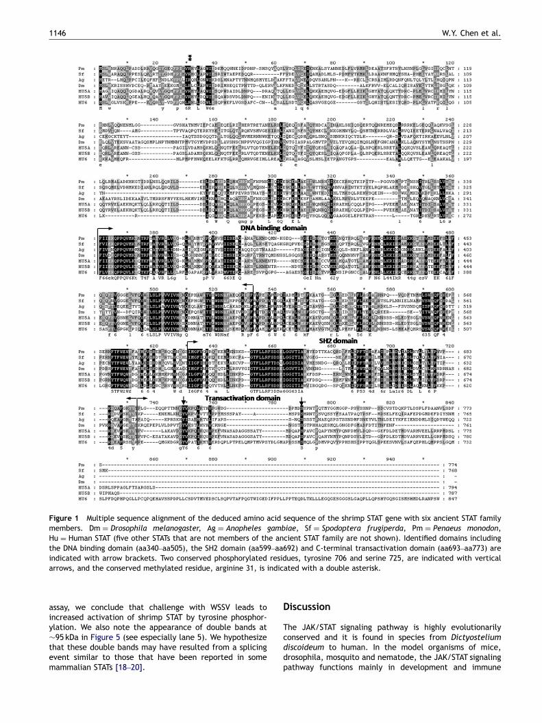
Altogether, the full-length cDNA of shrimp STAT (Accessionnumber: AY327491) contains a 2235 nucleotide coding regionthat encodes 744 amino acid residues with a predictedmolecular weight of 88.4 kDa. The cDNA sequence of shrimpSTAT suggested that the transcription initiation site is 134nucleotides upstream of the start codon, ATG, and that thepolyA signal sequence, AATAAA, is located between 49 and54 nucleotides downstream of the stop codon. Using multi-ple sequence alignment, several functional domains, includ-ing the DNA binding domain (aa340–aa505), SH2 domain(aa599–aa692) and C-terminal transactivation domain(aa693–aa773), were identified in shrimp STAT (Figure 1).Several conserved amino acid residues that provide im-portant regulation mechanisms in known STATs were alsofound in shrimp STAT (i.e. C-terminal phosphorylation sitesat Y706 and S725, and methylation site at R31).
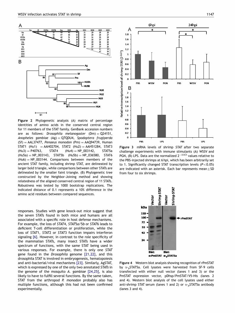
To investigate the evolutionary relatedness betweenshrimp STAT and other STATs, the conserved central regionfrom the DNA binding domain to the SH2 domain(aa340–aa692) was subjected to phylogenetic analysis.The resulting identity matrix (Figure 2A) and dendrogram(Figure 2B) suggest that shrimp STAT is relatively close to the
insect STATs (Spodoptera frugiperda STAT, Anopheles gam-biae STAT, drosophila STAT) and to human STAT5a/5b andhuman STAT6.
Transcription level of shrimp STAT decreased afterWSSV infection
Real-time RT-PCR was used to test whether the transcriptionlevel of shrimp STAT was changed after challenge by WSSVand two bacterial immune stimulants, PGN and LPS. Datawere expressed as 2�DDCt and changes in the transcriptionlevels of the shrimp STAT target gene relative to the PBS-injected control are shown in Figure 3. The transcriptionlevels of shrimp STAT were decreased after challenge witheither PGN or WSSV (Figure 3A). At 6 hpi, the decrease wasonly statistically significant in the PGN challenged group butat 24 hpi, statistically significant decreases were observed inboth of the challenged groups. In a separate study using LPSas the stimulant, there was no statistically significantdifference from the control group (Figure 3B).
Anti-phosphorylated human STAT5a antibodyrecognized shrimp STAT
To determine whether anti-phosphorylated human STAT5aantibody could recognize shrimp STAT, we transientlytransfected SF-9 cells with a shrimp STAT gene under thecontrol of a Drosophila heat shock protein 70 promoter.After heat shock at 16–18 h post transfection, the recombi-nant shrimp STATwas detected by both the anti-shrimp STATserum and the anti-phosphorylated human STAT5a antibody(Figure 4, lanes 2 and 4). No recombinant PmSTAT signal wasdetected in the Sf9 cells transfected with control plasmid(Figure 4, lanes 1 and 3). We concluded that the anti-phosphorylated human STAT5a antibody was successfullyable to recognize shrimp STAT.
Shrimp STAT was activated in response to WSSVinfection
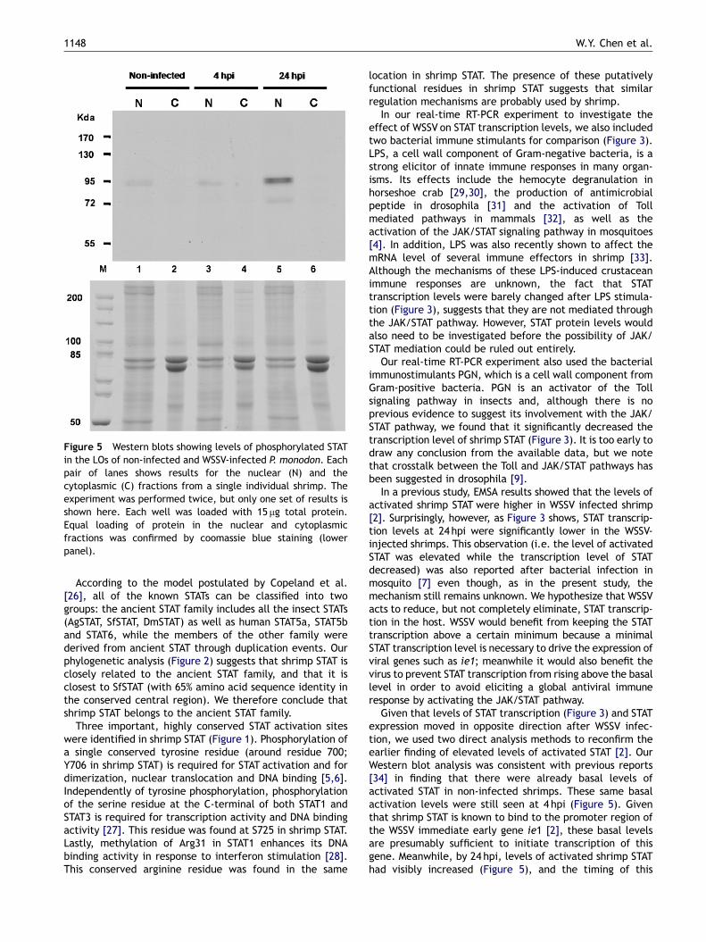
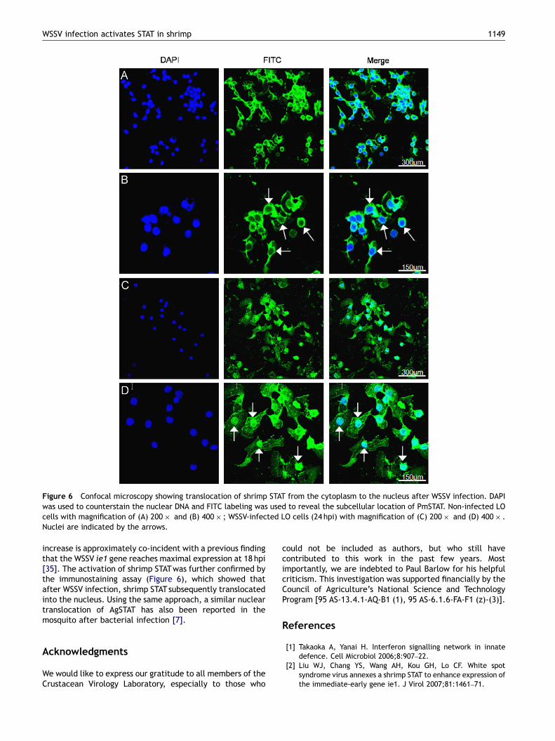
To investigate whether the shrimp STAT was activated afterWSSV infection, cytoplasmic and nuclear extracts of the LOsfrom non-infected and WSSV-infected shrimp were analyzedby Western blot using polyclonal anti-phosphorylated humanSTAT5a antibodies. Activated shrimp STATwas detected onlyin the nuclear fractions (Figure 5). Lane 1 shows the basallevel of activated shrimp STAT in non-infected shrimp, andat 4 hpi levels of activated STAT were approximately thesame (Figure 5, lane 3). However, by 24 hpi, the levels ofactivated shrimp STATwere visibly increased (Figure 5, lane5). Further evidence of the activation of shrimp STAT in vivowas provided by an immunostaining assay. In this assay,green fluorescence from the FITC-labeled shrimp STATprotein was visible in both the uninfected and WSSV-infected primary culture LO cells (Figure 6). However, inthe uninfected cells, the fluorescence was absent in thenucleus (Figure 6A and B) whereas after WSSV infection(24 hpi) the signals were stronger and the protein was mostlylocated in the nucleus region (Figure 6C and D). Based onboth the Western blot analysis and the immunostaining
ARTICLE IN PRESS
Figure 1 Multiple sequence alignment of the deduced amino acid sequence of the shrimp STAT gene with six ancient STAT familymembers. Dm ¼ Drosophila melanogaster, Ag ¼ Anopheles gambiae, Sf ¼ Spodoptera frugiperda, Pm ¼ Penaeus monodon,Hu ¼ Human STAT (five other STATs that are not members of the ancient STAT family are not shown). Identified domains includingthe DNA binding domain (aa340–aa505), the SH2 domain (aa599–aa692) and C-terminal transactivation domain (aa693–aa773) areindicated with arrow brackets. Two conserved phosphorylated residues, tyrosine 706 and serine 725, are indicated with verticalarrows, and the conserved methylated residue, arginine 31, is indicated with a double asterisk.
W.Y. Chen et al.1146
assay, we conclude that challenge with WSSV leads toincreased activation of shrimp STAT by tyrosine phosphor-ylation. We also note the appearance of double bands at�95 kDa in Figure 5 (see especially lane 5). We hypothesizethat these double bands may have resulted from a splicingevent similar to those that have been reported in somemammalian STATs [18–20].
Discussion
The JAK/STAT signaling pathway is highly evolutionarilyconserved and it is found in species from Dictyosteliumdiscoideum to human. In the model organisms of mice,drosophila, mosquito and nematode, the JAK/STAT signalingpathway functions mainly in development and immune
ARTICLE IN PRESS
Figure 2 Phylogenetic analysis (A) matrix of percentageidentities of amino acids in the conserved central regionfor 11 members of the STAT family. GenBank accession numbersare as follows: Drosophila melanogaster (Dm) ¼ Q24151,Anopheles gambiae (Ag) ¼ Q7QDU4, Spodoptera frugiperda(Sf) ¼ AAL37477, Penaeus monodon (Pm) ¼ AAQ94739, HumanSTAT1 (Hu1) ¼ AAH02704, STAT2 (Hu2) ¼ AAH51284, STAT3(Hu3) ¼ P40763, STAT4 (Hu4) ¼ NP_003142, STAT5a(Hu5a) ¼ NP_003143, STAT5b (Hu5b) ¼ NP_036580, STAT6(Hu6) ¼ NP_003144. Comparisons between members of theancient STAT family, including shrimp STAT, are delineated bylarger bold triangle, while comparisons between other STATs aredelineated by the smaller faint triangle. (B) Phylogenetic treeconstructed by the Neighbor-Joining method and showingrelatedness of the aligned conserved central region of 11 STATs.Robustness was tested by 1000 bootstrap replications. Theindicated distance of 0.1 represents a 10% difference in theamino acid residues between compared sequences.
Figure 3 mRNA levels of shrimp STAT after two separatechallenge experiments with immune stimulants (A) WSSV andPGN, (B) LPS. Data are the normalized 2�DDCt values relative tothe PBS-injected shrimps at 6 hpi, which has been arbitrarily setto 1. Significantly changed STAT transcription levels (Po0.05)are indicated with an asterisk. Each bar represents mean7SDfrom four to six shrimps.
Figure 4 Western blot analysis showing recognition of rPmSTATby a-pSTAT5a. Cell lysates were harvested from SF-9 cellstransfected with either null vector (lanes 1 and 3) or thePmSTAT expression vector, pDhsp/PmSTAT/V5-His (lanes 2and 4). Western blot analysis of the cell lysates used eitheranti-shrimp STAT serum (lanes 1 and 2) or a-pSTAT5a antibody(lanes 3 and 4).
WSSV infection activates STAT in shrimp 1147
responses. Studies with gene knock-out mice suggest thatthe seven STATs found in both mice and humans are allassociated with a specific role in host defense mechanisms.For example, the loss of STAT4, STAT5a/5b or STAT6 leads todeficient T-cell differentiation or proliferation, while theloss of STAT1, STAT2 or STAT3 function impairs interferonsignaling [6]. However, in contrast to the role specificity ofthe mammalian STATs, many insect STATs have a widerspectrum of functions, with the same STAT being used invarious responses. For example, there is only one STATgene found in the Drosophila genome [21,22], and thisdrosophila STAT is involved in embryogenesis, hematopoiesisand anti-bacterial/viral mechanisms [23]. Similarly, AgSTAT,which is expressed by one of the only two annotated STATs inthe genome of the mosquito A. gambiae [24,25], is alsolikely to have to fulfill several functions. By the same taken,STAT from the arthropod P. monodon probably also hasmultiple functions, although this had not been confirmedexperimentally.
ARTICLE IN PRESS
Figure 5 Western blots showing levels of phosphorylated STATin the LOs of non-infected and WSSV-infected P. monodon. Eachpair of lanes shows results for the nuclear (N) and thecytoplasmic (C) fractions from a single individual shrimp. Theexperiment was performed twice, but only one set of results isshown here. Each well was loaded with 15 mg total protein.Equal loading of protein in the nuclear and cytoplasmicfractions was confirmed by coomassie blue staining (lowerpanel).
W.Y. Chen et al.1148
According to the model postulated by Copeland et al.[26], all of the known STATs can be classified into twogroups: the ancient STAT family includes all the insect STATs(AgSTAT, SfSTAT, DmSTAT) as well as human STAT5a, STAT5band STAT6, while the members of the other family werederived from ancient STAT through duplication events. Ourphylogenetic analysis (Figure 2) suggests that shrimp STAT isclosely related to the ancient STAT family, and that it isclosest to SfSTAT (with 65% amino acid sequence identity inthe conserved central region). We therefore conclude thatshrimp STAT belongs to the ancient STAT family.
Three important, highly conserved STAT activation siteswere identified in shrimp STAT (Figure 1). Phosphorylation ofa single conserved tyrosine residue (around residue 700;Y706 in shrimp STAT) is required for STAT activation and fordimerization, nuclear translocation and DNA binding [5,6].Independently of tyrosine phosphorylation, phosphorylationof the serine residue at the C-terminal of both STAT1 andSTAT3 is required for transcription activity and DNA bindingactivity [27]. This residue was found at S725 in shrimp STAT.Lastly, methylation of Arg31 in STAT1 enhances its DNAbinding activity in response to interferon stimulation [28].This conserved arginine residue was found in the same
location in shrimp STAT. The presence of these putativelyfunctional residues in shrimp STAT suggests that similarregulation mechanisms are probably used by shrimp.
In our real-time RT-PCR experiment to investigate theeffect of WSSV on STAT transcription levels, we also includedtwo bacterial immune stimulants for comparison (Figure 3).LPS, a cell wall component of Gram-negative bacteria, is astrong elicitor of innate immune responses in many organ-isms. Its effects include the hemocyte degranulation inhorseshoe crab [29,30], the production of antimicrobialpeptide in drosophila [31] and the activation of Tollmediated pathways in mammals [32], as well as theactivation of the JAK/STAT signaling pathway in mosquitoes[4]. In addition, LPS was also recently shown to affect themRNA level of several immune effectors in shrimp [33].Although the mechanisms of these LPS-induced crustaceanimmune responses are unknown, the fact that STATtranscription levels were barely changed after LPS stimula-tion (Figure 3), suggests that they are not mediated throughthe JAK/STAT pathway. However, STAT protein levels wouldalso need to be investigated before the possibility of JAK/STAT mediation could be ruled out entirely.
Our real-time RT-PCR experiment also used the bacterialimmunostimulants PGN, which is a cell wall component fromGram-positive bacteria. PGN is an activator of the Tollsignaling pathway in insects and, although there is noprevious evidence to suggest its involvement with the JAK/STAT pathway, we found that it significantly decreased thetranscription level of shrimp STAT (Figure 3). It is too early todraw any conclusion from the available data, but we notethat crosstalk between the Toll and JAK/STAT pathways hasbeen suggested in drosophila [9].
In a previous study, EMSA results showed that the levels ofactivated shrimp STAT were higher in WSSV infected shrimp[2]. Surprisingly, however, as Figure 3 shows, STAT transcrip-tion levels at 24 hpi were significantly lower in the WSSV-injected shrimps. This observation (i.e. the level of activatedSTAT was elevated while the transcription level of STATdecreased) was also reported after bacterial infection inmosquito [7] even though, as in the present study, themechanism still remains unknown. We hypothesize that WSSVacts to reduce, but not completely eliminate, STAT transcrip-tion in the host. WSSV would benefit from keeping the STATtranscription above a certain minimum because a minimalSTAT transcription level is necessary to drive the expression ofviral genes such as ie1; meanwhile it would also benefit thevirus to prevent STAT transcription from rising above the basallevel in order to avoid eliciting a global antiviral immuneresponse by activating the JAK/STAT pathway.
Given that levels of STAT transcription (Figure 3) and STATexpression moved in opposite direction after WSSV infec-tion, we used two direct analysis methods to reconfirm theearlier finding of elevated levels of activated STAT [2]. OurWestern blot analysis was consistent with previous reports[34] in finding that there were already basal levels ofactivated STAT in non-infected shrimps. These same basalactivation levels were still seen at 4 hpi (Figure 5). Giventhat shrimp STAT is known to bind to the promoter region ofthe WSSV immediate early gene ie1 [2], these basal levelsare presumably sufficient to initiate transcription of thisgene. Meanwhile, by 24 hpi, levels of activated shrimp STAThad visibly increased (Figure 5), and the timing of this
ARTICLE IN PRESS
Figure 6 Confocal microscopy showing translocation of shrimp STAT from the cytoplasm to the nucleus after WSSV infection. DAPIwas used to counterstain the nuclear DNA and FITC labeling was used to reveal the subcellular location of PmSTAT. Non-infected LOcells with magnification of (A) 200� and (B) 400� ; WSSV-infected LO cells (24 hpi) with magnification of (C) 200� and (D) 400� .Nuclei are indicated by the arrows.
WSSV infection activates STAT in shrimp 1149
increase is approximately co-incident with a previous findingthat the WSSV ie1 gene reaches maximal expression at 18 hpi[35]. The activation of shrimp STATwas further confirmed bythe immunostaining assay (Figure 6), which showed thatafter WSSV infection, shrimp STATsubsequently translocatedinto the nucleus. Using the same approach, a similar nucleartranslocation of AgSTAT has also been reported in themosquito after bacterial infection [7].
Acknowledgments
We would like to express our gratitude to all members of theCrustacean Virology Laboratory, especially to those who
could not be included as authors, but who still havecontributed to this work in the past few years. Mostimportantly, we are indebted to Paul Barlow for his helpfulcriticism. This investigation was supported financially by theCouncil of Agriculture’s National Science and TechnologyProgram [95 AS-13.4.1-AQ-B1 (1), 95 AS-6.1.6-FA-F1 (z)-(3)].
References
[1] Takaoka A, Yanai H. Interferon signalling network in innatedefence. Cell Microbiol 2006;8:907–22.
[2] Liu WJ, Chang YS, Wang AH, Kou GH, Lo CF. White spotsyndrome virus annexes a shrimp STAT to enhance expression ofthe immediate-early gene ie1. J Virol 2007;81:1461–71.
ARTICLE IN PRESS
W.Y. Chen et al.1150
[3] Dostert C, Jouanguy E, Irving P, Troxler L, Galiana-Arnoux D,Hetru C, et al. The JAK-STAT signaling pathway is required butnot sufficient for the antiviral response of drosophila. NatImmunol 2005;6:946–53.
[4] Lin CC, Chou CM, Hsu YL, Lien JC, Wang YM, Chen ST, et al.Characterization of two mosquito STATs, AaSTAT and CtSTAT:differential regulation of tyrosine phosphorylation and DNAbinding activity by lipopolysaccharide treatment and byJapanese encephalitis virus infection. J Biol Chem 2004;279:3308–17.
[5] Levy DE, Darnell Jr. JE. Stats: transcriptional control andbiological impact. Nat Rev Mol Cell Biol 2002;3:651–62.
[6] Shuai K, Liu B. Regulation of JAK-STAT signalling in the immunesystem. Nat Rev Immunol 2003;3:900–11.
[7] Barillas-Mury C, Han YS, Seeley D, Kafatos FC. Anophelesgambiae Ag-STAT, a new insect member of the STAT family, isactivated in response to bacterial infection. EMBO J 1999;18:959–67.
[8] Agaisse H, Petersen U-M, Boutros M, Mathey-Prevot B,Perrimon N. Signaling role of hemocytes in Drosophila JAK/STAT-dependent response to septic injury. Dev Cell 2003;5:441–50.
[9] Lagueux M, Perrodou E, Levashina EA, Capovilla M, HoffmannJA. Constitutive expression of a complement-like protein inToll and JAK gain-of-function mutants of Drosophila. Proc NatlAcad Sci USA 2000;97:11427–32.
[10] Agaisse H, Perrimon N. The roles of JAK/STAT signaling inDrosophila immune responses. Immunol Rev 2004;198:72–82.
[11] Didcock L, Young DF, Goodbourn S, Randall RE. The V proteinof simian virus 5 inhibits interferon signalling by targetingSTAT1 for proteasome-mediated degradation. J Virol 1999;73:9928–33.
[12] Miller DM, Rahill BM, Boss JM, Lairmore MD, Durbin JE,Waldman JW, et al. Human cytomegalovirus inhibits majorhistocompatibility complex class II expression by disruption ofthe Jak/Stat pathway. J Exp Med 1998;187:675–83.
[13] Abendroth A, Slobedman B, Lee E, Mellins E, Wallace M,Arvin AM. Modulation of major histocompatibility class IIprotein expression by varicella-zoster virus. J Virol 2000;74:1900–7.
[14] He Y, Katze MG. To interfere and to anti-interfere: theinterplay between hepatitis C virus and interferon. ViralImmunol 2002;15:95–119.
[15] Tsai JM, Wang HC, Leu JH, Hsiao HH, Wang AH, Kou GH, et al.Genomic and proteomic analysis of thirty-nine structuralproteins of shrimp white spot syndrome virus. J Virol 2004;78:11360–70.
[16] Livak KJ, Schmittgen TD. Analysis of relative gene expressiondata using real-time quantitative PCR and the 2�DDCT method.Methods 2001;25:402–8.
[17] Wang CH, Yang HN, Tang CY, Lu CH, Kou GH, Lo CF.Ultrastructure of white spot syndrome virus development inprimary lymphoid organ cell cultures. Dis Aquat Organ 2000;41:91–104.
[18] Azam M, Lee C, Strehlow I, Schindler C. Functionally distinctisoforms of STAT5 are generated by protein processing.Immunity 1997;6:691–701.
[19] Caldenhoven E, van Dijk TB, Solari R, Armstrong J, RaaijmakersJA, Lammers JW, et al. STAT3b, a splice variant of transcriptionfactor STAT3, is a dominant negative regulator of transcription.J Biol Chem 1996;271:13221–7.
[20] Schaefer TS, Sanders LK, Park OK, Nathans D. Functionaldifferences between Stat3alpha and Stat3beta. Mol Cell Biol1997;17:5307–16.
[21] Hou XS, Melnick MB, Perrimon N. Marelle acts downstream ofthe Drosophila HOP/JAK kinase and encodes a protein similarto the mammalian STATs. Cell 1996;84:411–9.
[22] Yan R, Small S, Desplan C, Dearolf CR, Darnell Jr. JE.Identification of a Stat gene that functions in Drosophiladevelopment. Cell 1996;84:421–30.
[23] Arbouzova NI, Zeidler MP. JAK/STAT signalling in Drosophila:insights into conserved regulatory and cellular functions.Development 2006;133:2605–16.
[24] Christophides GK, Zdobnov E, Barillas-Mury C, Birney E, BlandinS, Blass C, et al. Immunity-related genes and gene families inAnopheles gambiae. Science 2002;298:159–65.
[25] Zdobnov EM, von Mering C, Letunic I, Torrents D, Suyama M,Copley RR, et al. Comparative genome and proteome analysisof Anopheles gambiae and Drosophila melanogaster. Science2002;298:149–59.
[26] Copeland NG, Gilbert DJ, Schindler C, Zhong Z, Wen Z, DarnellJE, et al. Distribution of the mammalian Stat gene family inmouse chromosomes. Genomics 1995;29:225–8.
[27] Decker T, Kovarik P. Serine phosphorylation of STATs. Oncogene2000;19:2628–37.
[28] Mowen KA, Tang J, Zhu W, Schurter BT, Shuai K, Herschman HR,et al. Arginine methylation of STAT1 modulates IFNa/b-inducedtranscription. Cell 2001;104:731–41.
[29] Iwanaga S, Kawabata S-I, Muta T. New types of clotting factorsand defense molecules found in horseshoe crab hemolymph: theirstructures and functions. J Biochem (Tokyo) 1998;123:1–15.
[30] Kurata S, Ariki S, Kawabata S. Recognition of pathogens andactivation of immune responses in Drosophila and horseshoecrab innate immunity. Immunobiology 2006;211:237–49.
[31] Kim YS, Ryu JH, Han SJ, Choi KH, Nam KB, Jang IH, et al. Gram-negative bacteria-binding protein, a pattern recognitionreceptor for lipopolysaccharide and beta-1,3-glucan thatmediates the signaling for the induction of innate immunegenes in Drosophila melanogaster cells. J Biol Chem 2000;275:32721–7.
[32] Heumann D, Roger T. Initial responses to endotoxins and Gram-negative bacteria. Clin Chim Acta 2002;323:59–72.
[33] Okumura T. Effects of lipopolysaccharide on gene expression ofantimicrobial peptides (penaeidins and crustin), serine protei-nase and prophenoloxidase in haemocytes of the Pacific whiteshrimp, Litopenaeus vannamei. Fish Shellfish Immunol 2007;22:68–76.
[34] Vinkemeier U. Getting the message across, STAT! Designprinciples of a molecular signaling circuit. J Cell Biol 2004;167:197–201.
[35] Liu WJ, Chang YS, Wang CH, Kou GH, Lo CF. Microarray andRT-PCR screening for white spot syndrome virus immediate-early genes in cycloheximide-treated shrimp. Virology 2005;334:327–41.