Embed Size (px)
Citation preview
Congenital heart disease in adults
Heart DiseaseBraunwald
CV R4 李威廷醫師Supervisor: 詹世鴻醫師
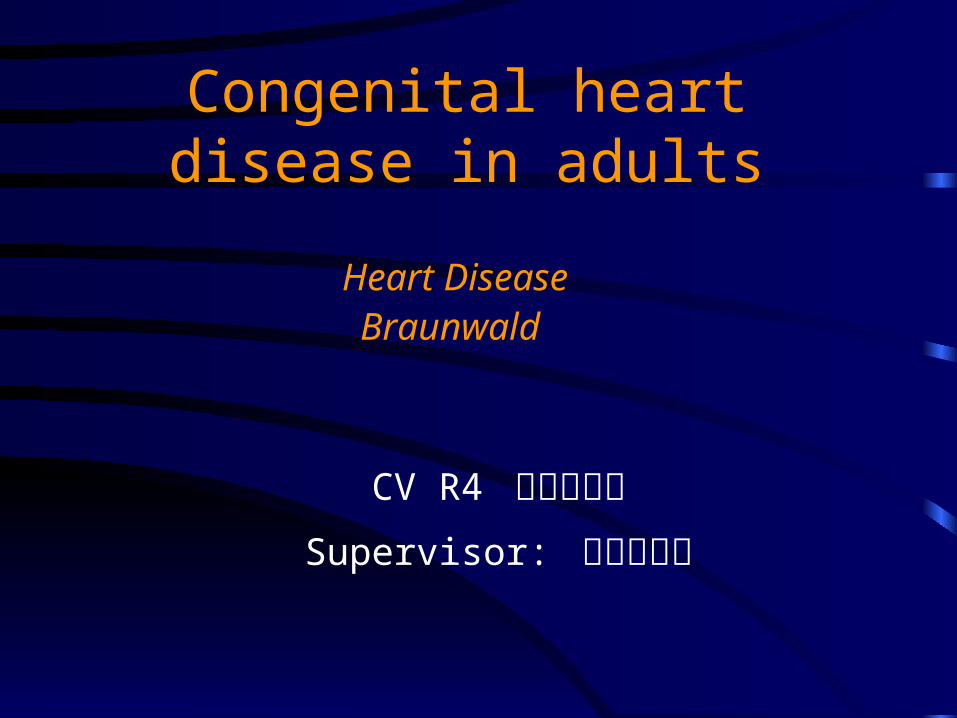
Pregnancy
Maternal risk:• Pulmonary hypertension• Obstructive lesions (LVOTO, RVOTO)• Ventricular dysfunction• Heart failure• Ascending aortic aneurysm (Marfan syndrome)
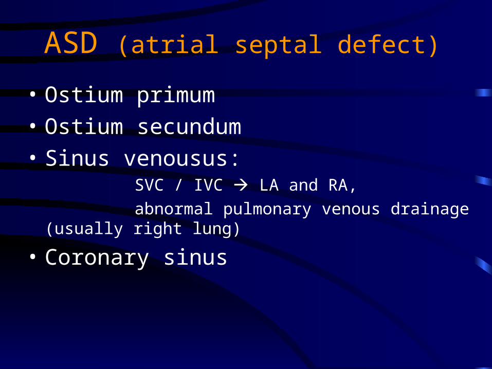
ASD (atrial septal defect)
• Ostium primum
• Ostium secundum
• Sinus venousus: SVC / IVC LA and RA,
abnormal pulmonary venous drainage (usually right lung)
• Coronary sinus
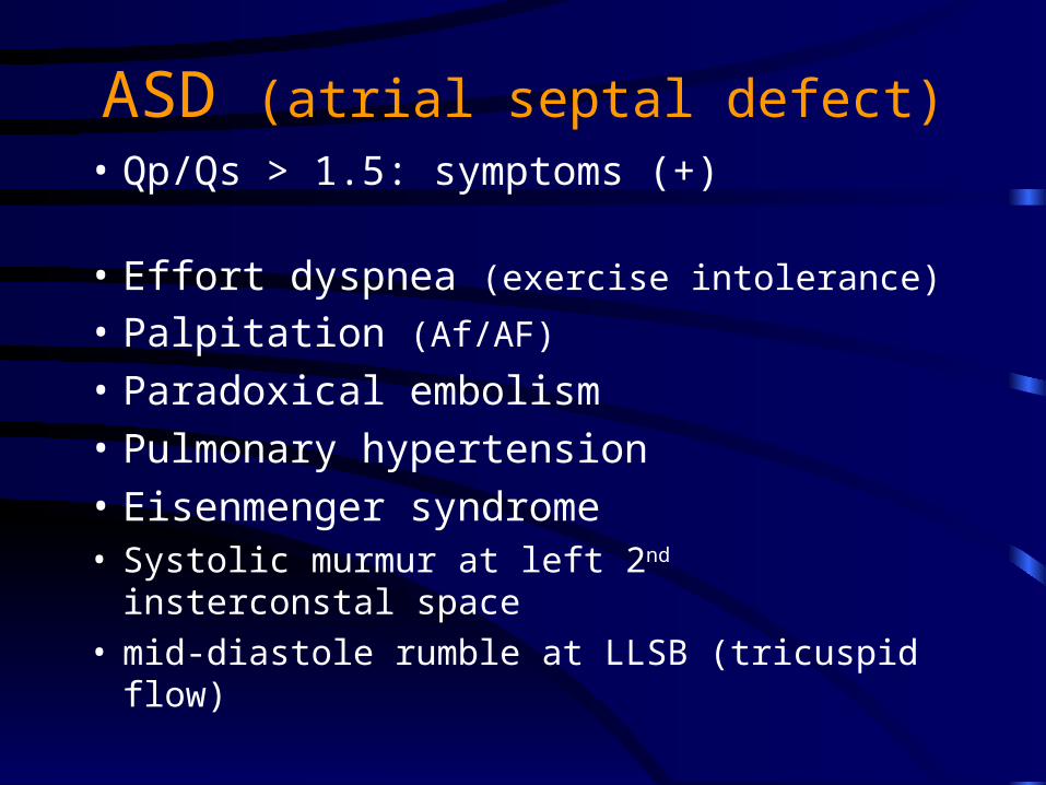
ASD (atrial septal defect)• Qp/Qs > 1.5: symptoms (+)
• Effort dyspnea (exercise intolerance)
• Palpitation (Af/AF)
• Paradoxical embolism
• Pulmonary hypertension
• Eisenmenger syndrome• Systolic murmur at left 2nd insterconstal space• mid-diastole rumble at LLSB (tricuspid flow)
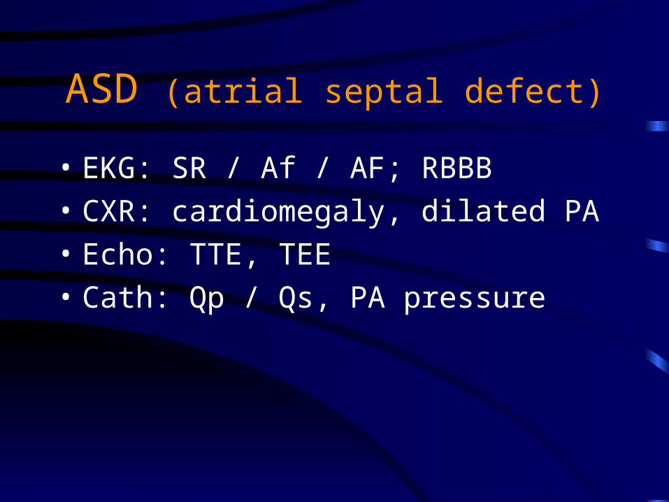
ASD (atrial septal defect)
• EKG: SR / Af / AF; RBBB
• CXR: cardiomegaly, dilated PA
• Echo: TTE, TEE
• Cath: Qp / Qs, PA pressure
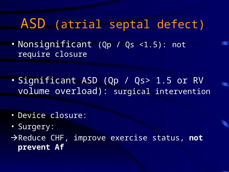
ASD (atrial septal defect)
• Nonsignificant (Qp / Qs <1.5): not require closure
• Significant ASD (Qp / Qs> 1.5 or RV volume overload): surgical intervention
• Device closure: • Surgery:
Reduce CHF, improve exercise status, not prevent Af
Isolated ventricular septal defect
• Muscular VSD
• Membranous VSD
• Doubly committed subarterial or outlet VSD
Isolated ventricular septal defect
• Restrictive VSD: Qp/Qs <1.5, no s/s
• Moderately restrictive VSD: Qp/Qs =1.5-2.5,
with s/s (LA and LV dilate, elevated PA, dyspnea, S3, holosystolic murmur)
• Nonrestrictive VSD: Eisenmenger syndrome
• Apical diastolic rumble and S3
Isolated ventricular septal defect
• EKG: Af, deep Q+ tall R + tall T in V5, V6 (LV overload)
• CXR: dilated PA
• Echo: TTE
• Cath: Qp / Qs, PA pressure
Isolated ventricular septal defect
Interventional indication:• Significant VSD (Qp/Qs >1.5, PA> 50mmHg, LA,
LV dilate, LV function deterioration)• Absence of irreversible pulmonary HTN
• Reduce AR, IE, slightly increase intraventricular conduction delay
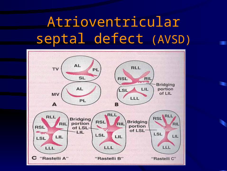
Atrioventricular septal defect (AVSD)
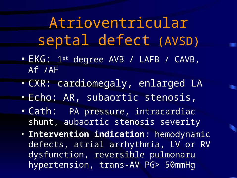
Atrioventricular septal defect (AVSD)
• EKG: 1st degree AVB / LAFB / CAVB, Af /AF
• CXR: cardiomegaly, enlarged LA
• Echo: AR, subaortic stenosis,• Cath: PA pressure, intracardiac shunt, aubaortic
stenosis severity• Intervention indication: hemodynamic defects,
atrial arrhythmia, LV or RV dysfunction, reversible pulmonaru hypertension, trans-AV PG> 50mmHg
Patent ductus arteriosus
• 1: 2000—1:5000
• 6th primitive aortic arch
• Proximal PA descending AO
• Increase IE (murmur)
• LA and LV volume overload
• Continuous “machinery” murmur
Patent ductus arteriosus
• EKG: deep Q+ tall R + tall T in V5, V6 (LV volume overload)
• CXR: cardiomegaly, LA, LV dilate• Echo: TTE (suprasternal or parasternal short axis),
systolic and diastolic flow toward the pulmonary valve in the main pulmonary artery
Patent ductus arteriosus
• Intervention: PAP > 2/3 SAP, PAR > 2/3 SAR, Qp/Qs >1.5, evidence of PA reactivity with challenged to vasodilator (O2, NO)
• Irreversible pulmonary HTN (x)• Active endarteritis (x)
• Transcatheter:
• Surgery:
Bicuspid aortic valve
• 1-2% of population
• Male: female= 4:1
• 20% with coarctation of AO and PDA
• Late aortic stenosis at 60+ y/o
• Increase IE
Bicuspid aortic valve
• EKG: from normal to LVH
• CXR: ascending aorta dilatation, valvular calcification
• Echo: concomitant defects such as coarctation or dissection of aorta
Bicuspid aortic valve
Interventional indication:• AS: dyspnea, angina, syncope, <0.6cm2,• AR:• AO root >55mm: prophylactic surgery
• Balloon valvuloplasty
• surgery
Subaortic stenosis
• AS, LVH, mild-moderate AR
• IE
• Systolic murmur at mid-left sternal edge
• Indication for intervention: • pressure gradient >50mmHg, • progressive or moderate to severe AR
Coarctation of the aorta
• Simple coarctation:• Complex coarctation: combine other lesions
(bicuspid aortic valve, intracranial aneurysm, and acquired intercostal artery aneurysm)
• Significant coarctation: trans-lesion pressure gradient >20mmHg or proximal hypertension
Coarctation of the aorta
• Epistaxis, headache, leg weakness on exertion• Leg claudication is rare (in abdominal coarctation)• Brachial pressure> popliteal pressure 10mmHg• Widespread cresendo-decresendo systolic murmur at
chest wall• “Corkscrew” torturosity of retinal artery
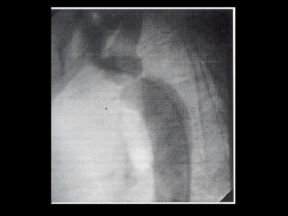
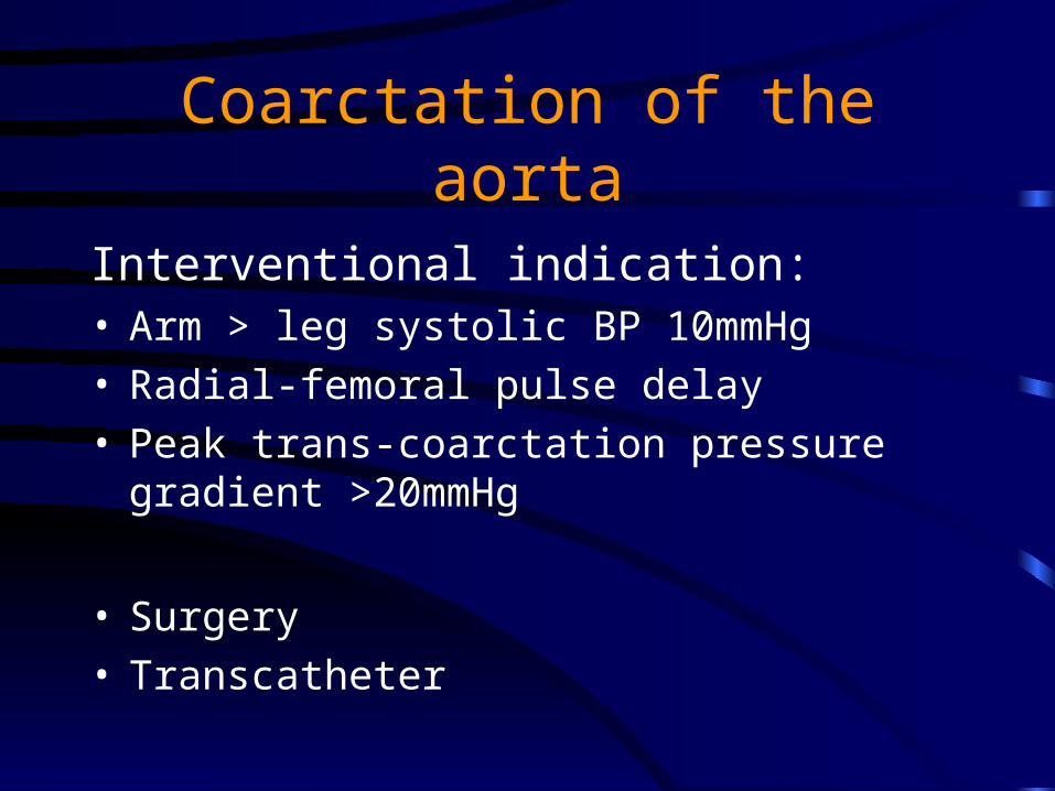
Coarctation of the aorta
Interventional indication:• Arm > leg systolic BP 10mmHg • Radial-femoral pulse delay• Peak trans-coarctation pressure gradient >20mmHg
• Surgery• Transcatheter
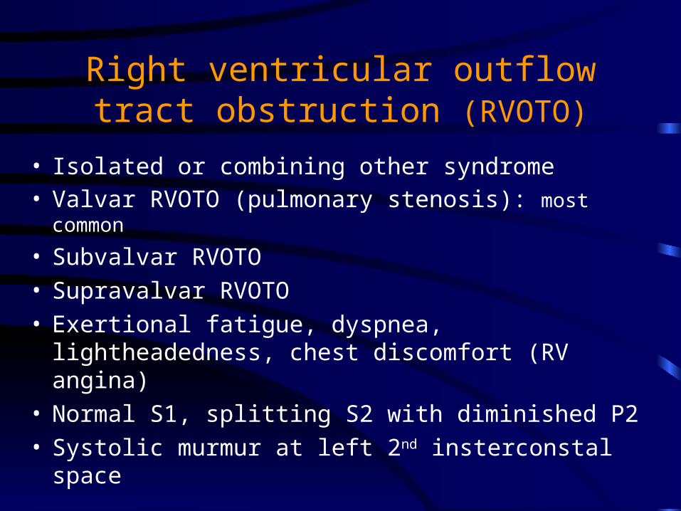
Right ventricular outflow tract obstruction (RVOTO)
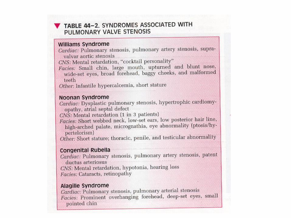
• Isolated or combining other syndrome• Valvar RVOTO (pulmonary stenosis): most common
• Subvalvar RVOTO• Supravalvar RVOTO• Exertional fatigue, dyspnea, lightheadedness, chest
discomfort (RV angina)• Normal S1, splitting S2 with diminished P2• Systolic murmur at left 2nd insterconstal space
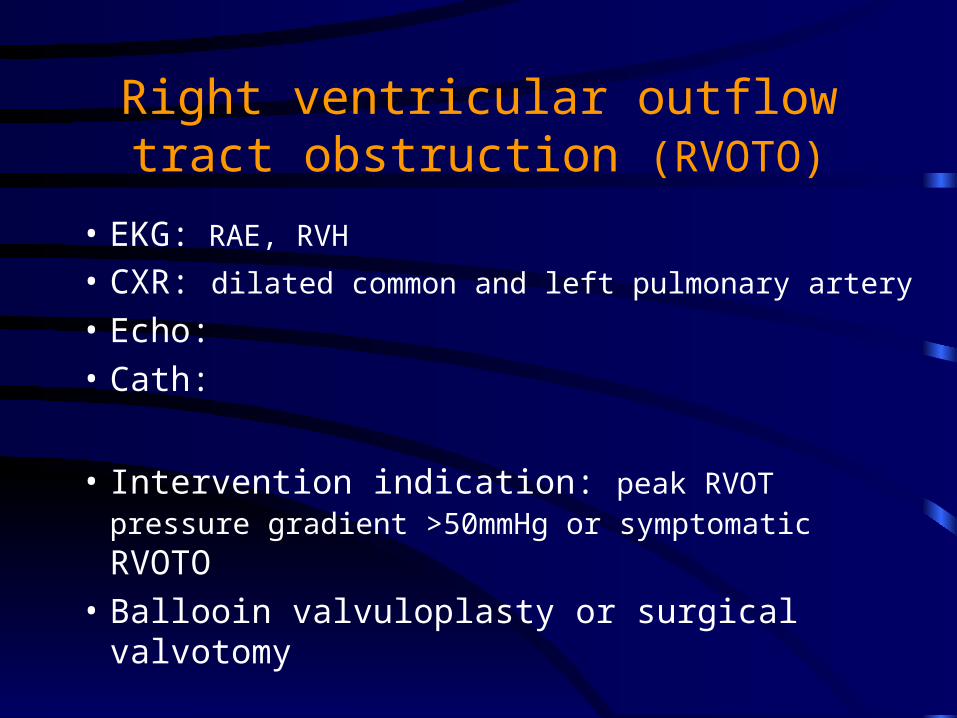
Right ventricular outflow tract obstruction (RVOTO)
• EKG: RAE, RVH
• CXR: dilated common and left pulmonary artery
• Echo:
• Cath:
• Intervention indication: peak RVOT pressure gradient >50mmHg or symptomatic RVOTO
• Ballooin valvuloplasty or surgical valvotomy
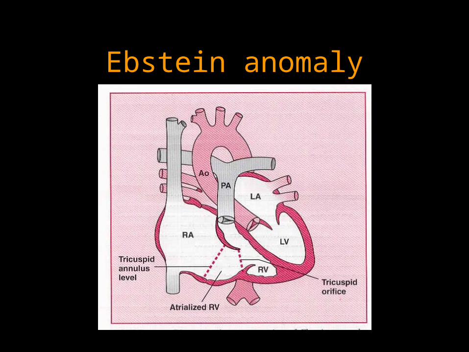
Ebstein anomaly
Ebstein anomaly
• Apical displacement of the septal , posterior, or (rarely) anterior leaflet of the tricuspid valve
• “Atrialization” of the inflow tract of RV• Small functional RV• Asymtomatic all the life in some patients
• Exercise intolerance, supraventricular arrhythmia, cyanosis (RL shunt at atrial level)
Ebstein anomaly
• Wide splitting S1 (sail-sound), • Wide splitting S2 (RBBB), • Right-sided S3
• EKG: Af / AF, RAE, RBBB
• CXR: “water-bottle” appearance• Echo: apical displacement of tricuspid valve
(8mm/m2)
Ebstein anomalyInterventional indication:• NYHA class III or IV• Progressive cyanosis• Right heart failure• Paradoxical embolite• Recurrent SVT or asymptomatic cardiomegaly (C/T ratio
>65%) (relative indication)
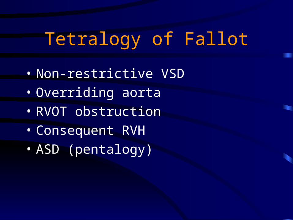
Tetralogy of Fallot
• Non-restrictive VSD
• Overriding aorta
• RVOT obstruction
• Consequent RVH
• ASD (pentalogy)

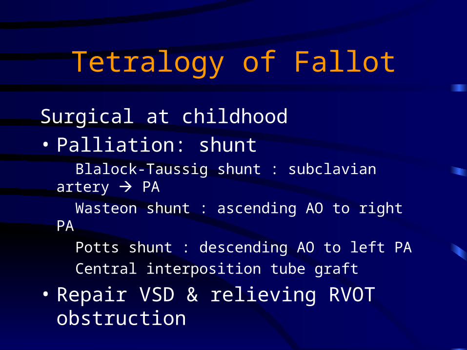
Tetralogy of Fallot
Surgical at childhood
• Palliation: shunt Blalock-Taussig shunt : subclavian artery PA
Wasteon shunt : ascending AO to right PA
Potts shunt : descending AO to left PA
Central interposition tube graft
• Repair VSD & relieving RVOT obstruction
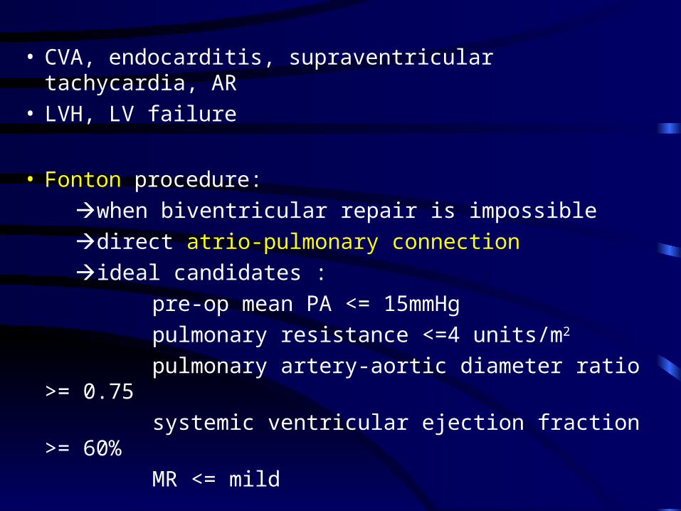
• CVA, endocarditis, supraventricular tachycardia, AR• LVH, LV failure
• Fonton procedure:
when biventricular repair is impossible
direct atrio-pulmonary connection
ideal candidates :
pre-op mean PA <= 15mmHg
pulmonary resistance <=4 units/m2
pulmonary artery-aortic diameter ratio >= 0.75
systemic ventricular ejection fraction >= 60%
MR <= mild
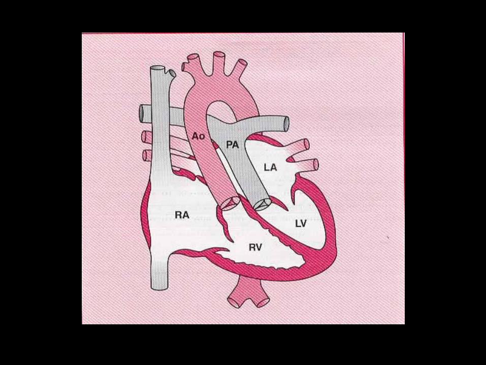
Complete transposition of the great arteries
• Simple transposition
• Complex transposition: VSD, PS
• Survival before surgical repair is dependent on mixing of the circulations at one level by natural (ASD, VSD, PDA) or intervention
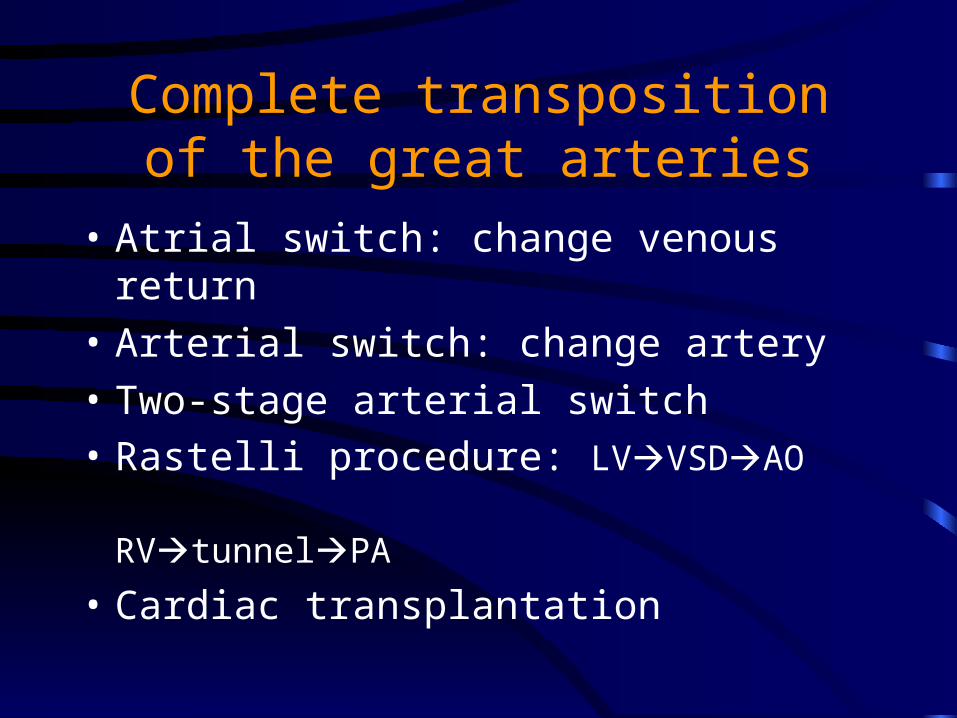
Complete transposition of the great arteries
• Atrial switch: change venous return
• Arterial switch: change artery
• Two-stage arterial switch• Rastelli procedure: LVVSDAO
RVtunnelPA
• Cardiac transplantation
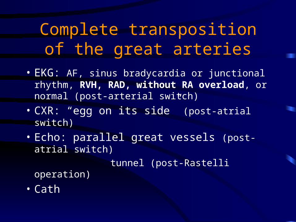
Complete transposition of the great arteries
• EKG: AF, sinus bradycardia or junctional rhythm, RVH, RAD, without RA overload, or normal (post-arterial switch)
• CXR: “egg on its side” (post-atrial switch)
• Echo: parallel great vessels (post-atrial switch)
tunnel (post-Rastelli operation)
• Cath
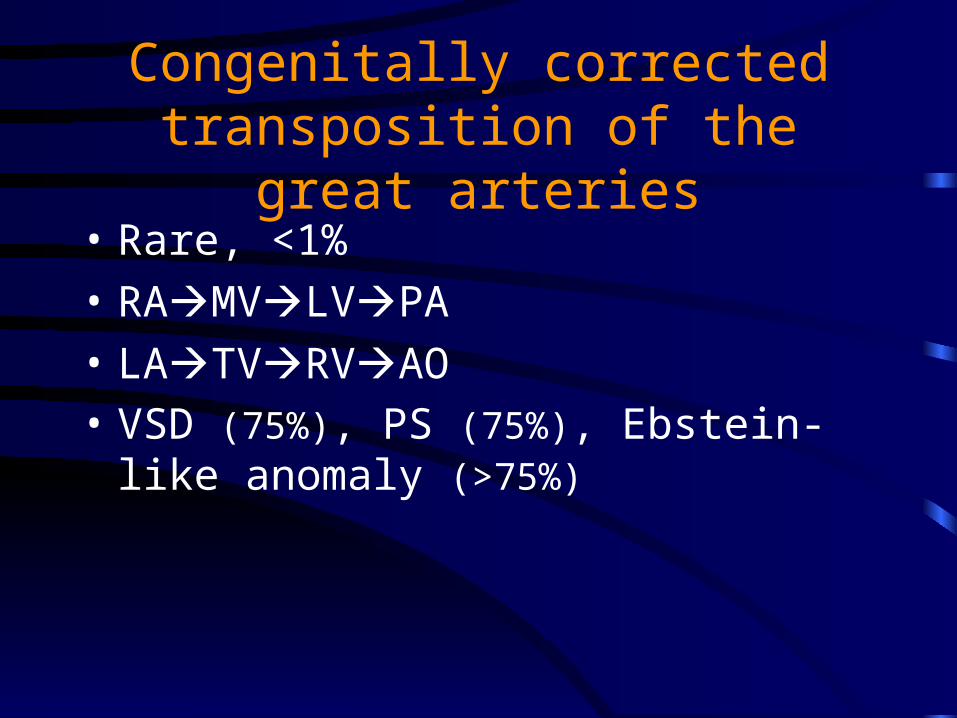
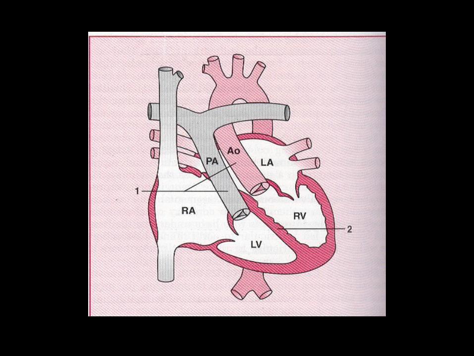
Congenitally corrected transposition of the great arteries
• Rare, <1%
• RAMVLVPA
• LATVRVAO• VSD (75%), PS (75%), Ebstein-like anomaly
(>75%)
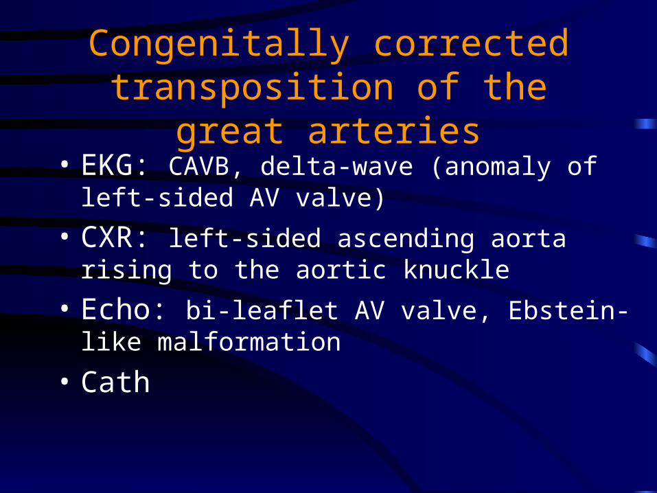
Congenitally corrected transposition of the great arteries
• EKG: CAVB, delta-wave (anomaly of left-sided AV valve)
• CXR: left-sided ascending aorta rising to the aortic knuckle
• Echo: bi-leaflet AV valve, Ebstein-like malformation
• Cath
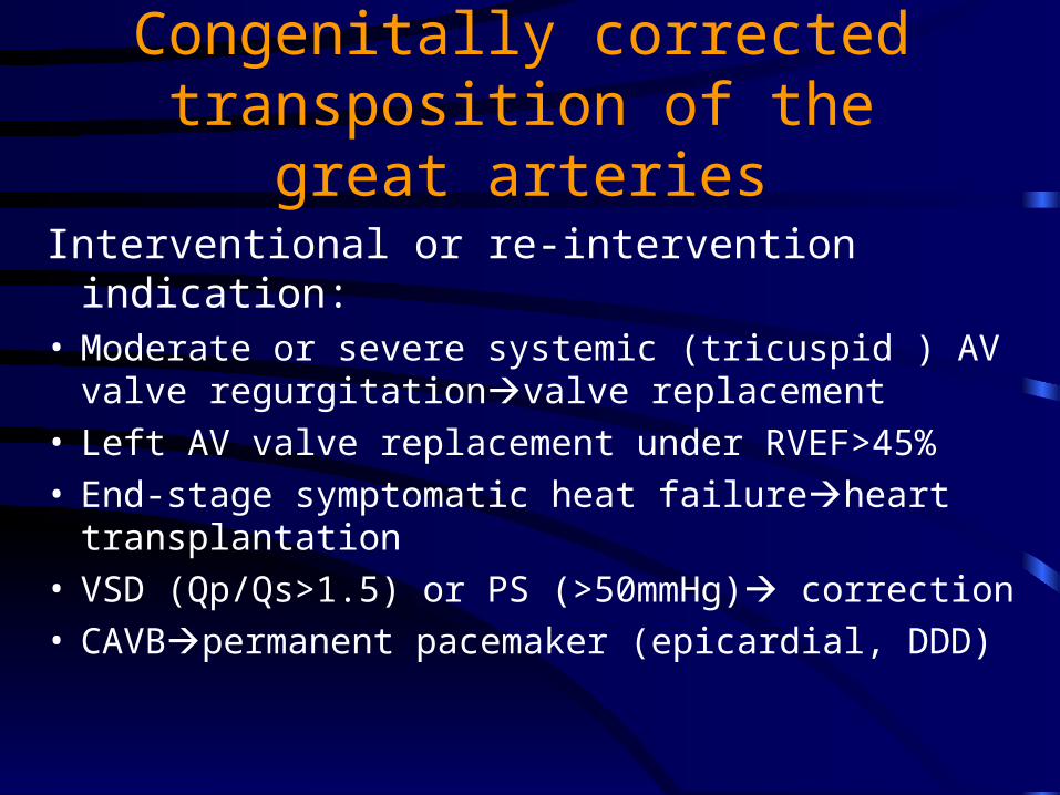
Congenitally corrected transposition of the great arteries
Interventional or re-intervention indication:• Moderate or severe systemic (tricuspid ) AV valve
regurgitationvalve replacement• Left AV valve replacement under RVEF>45%• End-stage symptomatic heat failureheart
transplantation• VSD (Qp/Qs>1.5) or PS (>50mmHg) correction• CAVBpermanent pacemaker (epicardial, DDD)
Eisenmenger syndrome
• R L shunt or bidirectional shunts
• “Differnetial cyanosis”
• Complication: CHF (most often), sudden death, hemoptysis
• EKG: RAE, RVH, RAD, atrial arrhythmia
• Restore physiological balance
• Avoid unnecessary surgery
• Heart-lung transplantation
Eisenmenger syndrome
• CCB
• ACEI
• Prostacyclin
• Pulmonary artery banding
Medical managekent of cyanotic congenital heart disease
• Hyperviscosity syndrome
• Iron deficiency
• Hemostatic abnormalities• Cerebrovascular events: infarction or hemorrhage
• Renal dysfunction