Embed Size (px)
Citation preview
Bioactive Compounds in Health and Disease 2020; 3(8):141-153 www.ffhdj.com Page 141 of 153
Research Article Open Access
Ultrafine Bubble Water Usefulness in Fecal Microbiota Transplantation: Recognition of Transplanted Microbiota in
Intestinal Epithelial Cells
Shin Shimizu1, 2, 3*, Katsuaki Dan4, Chihiro Tanaka1, 2, 3, Mikiko Tanaka1, 2, 3, Yoshimu Tanaka1, 5,
Masahiko Shirotani1, 6, Kunihiro Kitamura1, 7, Kensho Yorozu1, 8, Masayuki Oehorumu1, 9, Goro
Tsukamoto1, 2
1Intestinal Microbiota Transplantation Clinical Study Group, Osaka;2Symbiosis Inc., Osaka; 3 Intestinal Microbiota
Transplantation Clinical Research Inc., Osaka; 4Division of Research and Development, Research Organization of
Biological Activity, Tokyo; 5Tanaka Clinic, Osaka;6Luke’s ashiya Clinic, Hyogo; 7Kitamura Clinic, Fukuoka; 8Yorozu Clinic,
Tottori; 9LIFE Clinic Tateshina, Nagano
Corresponding author: Shin Shimizu, Intestinal Microbiota Transplantation Clinical Study Group, Osaka, Japan.
Submission Date: July 27th, 2020; Acceptance Date: August 24th, 2020; Publication Date: August 31st, 2020
Please cite this article as: Shimizu S., Dan K., Tanaka C., Tanaka M., Tanaka Y., Shirotani M., Kitamura K., et al. Ultrafine
bubble water usefulness in fecal microbiota transplantation recognition of transplanted microbiota in intestinal
epithelial cells. 2020; 3(8): 141-153 DOI: https://www.doi.org/10.31989/bchd.v3i8.735
ABSTRACT
Background: Indications for various diseases such as diabetes mellitus and metabolic syndrome, etc. of Fecal
Microbiota Transplantation (FMT) have been investigated. For the establishment of the FMT therapy, the
examination of the solvent has not been carried out, though the use of the physiological saline is fixed, at present.
We have produced the Mr. Shimizu made ultrafine bubble water (UFB) that produces a larger number of bubbles
than existing UFB.
Objective: To verify the usefulness of UFB, we prepared a conventional Saline and UFB-prepared microbial
preparation (Bio3); (Bio3/Saline) and (Bio3/UFB). The bacterial preparations, Bio3, contain glycated bacteria, lactic
acid bacteria, and butyric acid bacteria. FMT was carried out to the diabetic model mouse using these microbial
preparations, and whether the disease state was improved was examined. Cultured intestinal epithelial cells
(CaCO-2) were also used to test for differentiation by UFB and Saline from variations in several receptors that
recognize microbiota-mRNA. In addition, glucose uptake into cells was measured.
Bioactive Compounds in Health and Disease 2020; 3(8):141-153 www.ffhdj.com Page 142 of 153
Methods: FMT with (Bio3/UFB) and (Bio3/Saline) was performed in streptozotocin-induced diabetic mice (STZ-
mice) in duplicate at 0, 7 days. Blood glucose levels (7, 14 days) and blood insulin levels (14 days) were measured.
Cultured intestinal epithelial cells were also used to test for differentiation by UFB and Saline from change in
several receptors that recognize microbiota-mRNA (Toll-like receptors, NOD-like receptors, and RIG-I-like
receptors). In addition, glucose uptake into cells was measured using fluorescently labeled glucose analog (NBDG).
Results: UFB could reduce the blood glucose level of STZ-induced diabetic model mice. However, no such effects
were observed in Saline. Stimulation of serially diluted Bio3 with UFB-suspension were showed significant
alteration in TLR4 and IL-− The amount of glucose uptake in the (Bio3/UFB) group was significantly
different at 30 min, inhibited or delayed.
Conclusion: It is concluded that UFB-mediated cross-talk between intestinal bacteria and intestinal epithelial cells
and inhibition or delay of intestinal epithelial glucose uptake may have been associated with the reduction of
blood glucose levels in diabetic model mice. The superiority of UFB as a suspension used for the transfer of
bacteria has been suggested.
INTRODUCTION
It has become clear that disturbed intestinal microbiota
(dysbiosis) is involved not only in digestive diseases such
as inflammatory bowel disease and irritable bowel
syndrome, but also in various diseases such as metabolic
diseases such as diabetes mellitus and metabolic
syndrome, autoimmune diseases such as rheumatoid
arthritis and multiple sclerosis, and psychiatric disorders
such as autism and depression [1, 2]. Fecal Microbiota
Transplantation (FMT) has attracted attention as a
bacteriological treatment with fewer side effects in an
attempt to improve this dysbiosis.
FMT was shown to have a significantly higher
recurrence-free cure rate for recurrent Clostridium
difficile infection (CDI) in 1958 [3], and in 2013 compared
with the conventional treatment, oral vancomycin [4].
Currently, the Infectious Diseases Society of America
(IDSA) guideline strongly recommends FMT for recurrent
CDI [5]. Ulcerative colitis (UC): New protocols for UC have
been investigated, and combinatorial therapy
(Antibiotics-FMT: A-FMT) with allergic antimicrobial
therapy (Amoxicillin, Fosfomycin, Metronidazole: AFM)
and FMT has also been advocated [6, 7].
For the establishment of the FMT therapy, there is a
room for examination such as administration route,
dosage, administration frequency, selection method of
the donor, etc.. Especially, on the preparation of the
bacterial solution, the examination of the solvent, etc.
has not been carried out, though the use of the
physiological saline is fixed, at present.
Ultrafine bubble water is a technology that generates
foam of 1 or less into the liquid. Because of the fine
volume of the foam, it continues to exist in the liquid for
a long period of time, and its application such as cleaning
ability and preservation of food is spreading from the
potential characteristics of the foam surface. We have
produced the Mr. Shimizu made ultrafine bubble water
(UFB; International Public Number: WO2019/168034 A1
and trade mark registration number: 2020-57176) that
produces a larger number of bubbles than existing UFB,
and have obtained some evidence assuming that the
bacteria can be drawn into the epithelial lining of the
intestinal mucosa by using it as a solvent for the
Bioactive Compounds in Health and Disease 2020; 3(8):141-153 www.ffhdj.com Page 143 of 153
implanted bacterial suspension (in preparation for
posting).
In the present study, we investigated the changes in
blood glucose levels and the blood insulin levels at the
end of treatment in streptozotocin-induced diabetic mice
(STZ-mice) after enema with conventional Saline and
UFB-prepared microbial preparation (Bio3) [8];
hereinafter referred to as (Bio3/Saline) and (Bio3/UFB),
respectively. The bacterial preparations, Bio3, used here
contain glycated bacteria, lactic acid bacteria, and butyric
acid bacteria.
Enterobacteria are foreign to the organism but are not
eliminated by the intestinal immune system and are
symbiotic. Much remains unknown as to how intestinal
epithelial cells recognize and accept and eliminate
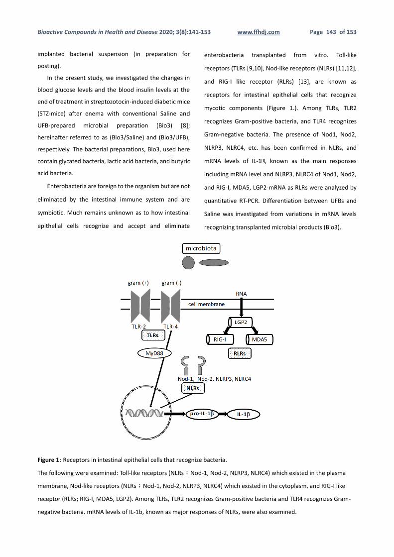
enterobacteria transplanted from vitro. Toll-like
receptors (TLRs [9,10], Nod-like receptors (NLRs) [11,12],
and RIG-I like receptor (RLRs) [13], are known as
receptors for intestinal epithelial cells that recognize
mycotic components (Figure 1.). Among TLRs, TLR2
recognizes Gram-positive bacteria, and TLR4 recognizes
Gram-negative bacteria. The presence of Nod1, Nod2,
NLRP3, NLRC4, etc. has been confirmed in NLRs, and
mRNA levels of IL-1 , known as the main responses
including mRNA level and NLRP3, NLRC4 of Nod1, Nod2,
and RIG-I, MDA5, LGP2-mRNA as RLRs were analyzed by
quantitative RT-PCR. Differentiation between UFBs and
Saline was investigated from variations in mRNA levels
recognizing transplanted microbial products (Bio3).
Figure 1: Receptors in intestinal epithelial cells that recognize bacteria.
The following were examined: Toll-like receptors (NLRs;Nod-1, Nod-2, NLRP3, NLRC4) which existed in the plasma
membrane, Nod-like receptors (NLRs;Nod-1, Nod-2, NLRP3, NLRC4) which existed in the cytoplasm, and RIG-I like
receptor (RLRs; RIG-I, MDA5, LGP2). Among TLRs, TLR2 recognizes Gram-positive bacteria and TLR4 recognizes Gram-
negative bacteria. mRNA levels of IL-1b, known as major responses of NLRs, were also examined.
Bioactive Compounds in Health and Disease 2020; 3(8):141-153 www.ffhdj.com Page 144 of 153
In addition, it is assumed that the uptake of glucose is
inhibited or delayed in intestinal epithelial cells, etc.,
when the disease state improvement effect is observed
in STZ mouse in which the insulin production function is
almost destroyed.
Therefore, the uptake of glucose was also examined
when (Bio3/UFB) and (Bio3/Saline) were added to
cultured intestinal epithelial cells.
A part of the usefulness of UFB as a preparation solvent
of the microbiota was recognized under the condition of
diabetes mellitus disease state model mouse and
cultured intestine epithelial cell.
MATERIALS & METHODS
Materials: UFB was granted by the Intestinal Flora
Transplantation Study Group. Saline purchased the
Japanese Physiological Saline (NISSIN PHARMACEUTICAL)
solution. Bacterial formulations (Bio Three®;
manufactured by Takeda Consumer Healthcare Co., Ltd.)
were donated by the Intestinal Flora Transplantation
Clinical Study Group. Glucose CII-test Wako (FUJIFILM
Wako Pure Chemical Corporation) was used to measure
blood glucose levels. Ultra Sensitive Mouse Insulin ELISA
Kit (Morinaga Institute of Biological Science, Inc.) was
used to measure blood insulin levels. 2-NBDG(2-N-(7-
Nitrobenz-2-oxa-1,3-diazol-4-yl)Amino)-2-Deoxyglucose)
(NBDG) was purchased from ThermoFisher (Carlsbad, CA).
Mouse insulin, streptozotocin (STZ), and other molecular
biological reagents were purchased from FUJIFILM Wako
Pure Chemical Corporation and used.
Animals: CD-1 (ICR) mice (male, 4 weeks old) purchased
from Japanese Charles River Inc. were previously housed
for 1 week and then used for experiments. Experiments
were conducted in accordance with the Animal
Experiment Guideline of the Biological Activity Research
Organization. Cell culture: Intestinal epithelial cells
(CaCO-2): RCB0988 were imported from European
Collection of Cell Cultures (ECACC) via a distributor
(K.A.C.) and cultured in a 5% CO2, 37°C incubator by
designated dedicated medium (MEM + 20% FBS + 0. 1mM
NEAA; non-essential amino acids) for use.
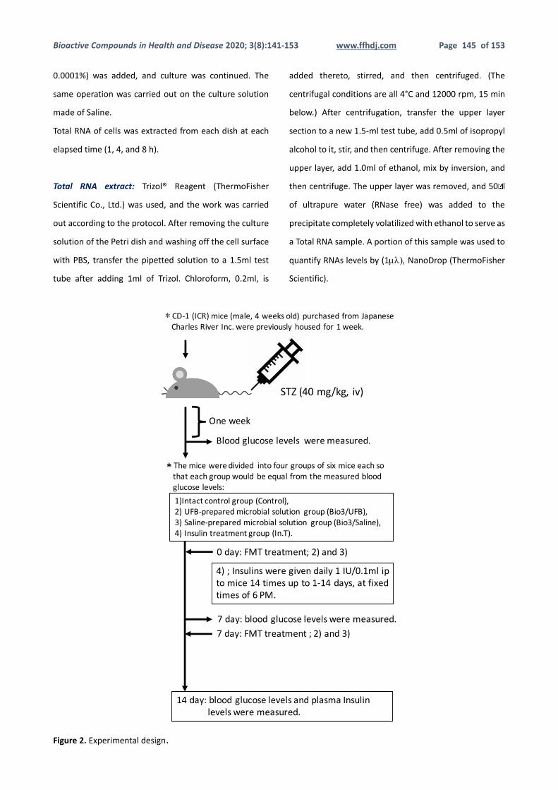
Preparation of model animals for diabetes mellitus and
FMT experiment: 100 mmol/L citrate buffer (pH 4.5) STZ
was administered to mice at 40mg/kg, iv. One week later,
blood was drawn from the mouse ocular venous plexus
using a capillary, and the mice were divided into four
groups of six mice each so that each group would be
equal from the measured blood glucose levels:
1) Intact control group (Control), 2) UFB-prepared
microbial solution group (Bio3/UFB), 3) Saline-prepared
microbial solution group (Bio3/Saline), and 4) Insulin
treatment group (In.T). The bacterial fluid was injected
using a single-dose needle from the anus to the large
intestine with 1ml per mouse on the first day of the
experiment and twice on the seventh day. Insulins were
given daily 1IU/0.1ml ip to mice 14 times up to 1-14 days,
at fixed times of 6 PM. Blood glucose levels were
collected twice on days 7 and 14 using a capillary from
the mouse ophthalmic venous plexus, and blood insulin
levels were measured in the jugular vein under ether
anesthesia on day 14, respectively. The experimental
protocol is shown in Figure 2.
Recognition of Bacterial Products in Intestinal Epithelial
Cells: CaCO-2 at a concentration of 50000 cells/ml and
2ml (a total of 0.1 million cells) were pre-cultured in 35
mm-dish for 48 hours before being used for the
experiment. After removing the old culture solution in
dish and washing the cell surface with phosphate
buffered saline (PBS), 2ml of a culture solution made of
UFB in which Bio3 powder was added at various
concentrations (10-fold serial dilutions from 1% to
Bioactive Compounds in Health and Disease 2020; 3(8):141-153 www.ffhdj.com Page 145 of 153
0.0001%) was added, and culture was continued. The
same operation was carried out on the culture solution
made of Saline.
Total RNA of cells was extracted from each dish at each
elapsed time (1, 4, and 8 h).
Total RNA extract: Trizol® Reagent (ThermoFisher
Scientific Co., Ltd.) was used, and the work was carried
out according to the protocol. After removing the culture
solution of the Petri dish and washing off the cell surface
with PBS, transfer the pipetted solution to a 1.5ml test
tube after adding 1ml of Trizol. Chloroform, 0.2ml, is
added thereto, stirred, and then centrifuged. (The
centrifugal conditions are all 4°C and 12000 rpm, 15 min
below.) After centrifugation, transfer the upper layer
section to a new 1.5-ml test tube, add 0.5ml of isopropyl
alcohol to it, stir, and then centrifuge. After removing the
upper layer, add 1.0ml of ethanol, mix by inversion, and
then centrifuge. The upper layer was removed, and 50 l
of ultrapure water (RNase free) was added to the
precipitate completely volatilized with ethanol to serve as
a Total RNA sample. A portion of this sample was used to
quantify RNAs levels by (1) NanoDrop (ThermoFisher
Scientific).
Figure 2. Experimental design.
Bioactive Compounds in Health and Disease 2020; 3(8):141-153 www.ffhdj.com Page 146 of 153
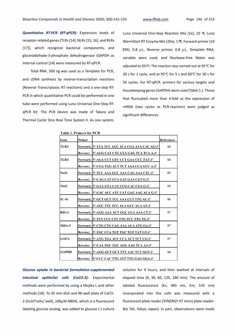
Quantitative RT-PCR (RT-qPCR): Expression levels of
receptor-related genes (TLRs [14], NLRs [15, 16], and RLRs
[17]), which recognize bacterial components, and
glyceraldehyde-3-phosphate dehydrogenase (GAPDH as
Internal control [14] were measured by RT-qPCR.
Total RNA, 500 ng was used as a Template for PCR,
and cDNA synthesis by reverse-transcription reactions
(Reverse Transcriptase; RT reactions) and a one-step RT-
PCR in which quantitative PCR could be performed in one
tube were performed using Luna Universal One-Step RT-
qPCR Kit. The PCR device was made of Takara and
Thermal Cycler Dice Real Time System II. As one system,
Luna Universal One-Step Reaction Mix (2x), 10 l, Luna
WarmStart RT Enzyme Mix (20x), 1 l, Forward primer (10
M), 0.8 , Reverse primer, 0.8 , Template RNA,
variable were used, and Nuclease-free Water was
adjusted to 20 l. The reaction was carried out at 95℃ for
30 s for 1 cycle, and at 95℃ for 5 s and 60℃ for 30 s for
50 cycles. For RT-qPCR, primers for various targets and
housekeeping genes (GAPDH) were used (Table 1.). Those
that fluctuated more than 4-fold as the expression of
mRNA (two cycles as PCR-reaction) were judged as
significant differences.
Glucose uptake in bacterial formulation-supplemented
intestinal epithelial cells (CaCO-2): Experimental
methods were performed by using a Mojika L and other
methods [18]. To 35 mm-dish and 96-well plate of CaCO-
2 (5x10⁴cells/ well), 100M NBDG, which is a fluorescent
labeling glucose analog, was added to glucose (-) culture
solution for 4 hours, and then washed at intervals of
elapsed time (0, 30, 60, 120, 180 min). The amount of
labeled fluorescence (Ex; 485 nm, Em; 535 nm)
incorporated into the cells was measured with a
fluorescent plate reader (SYNERGY HT micro plate reader:
Bio Tek, Tokyo, Japan). In part, observations were made
Bioactive Compounds in Health and Disease 2020; 3(8):141-153 www.ffhdj.com Page 147 of 153
under fluorescent microscopy to acquire fluorescent
images (BZ-X710, Keyence, Osaka, Japan) and joined its
software BZ-analyzer (Keyence). Experiments were
performed under identical conditions with n=4, and
results are presented as mean values ± SD (standard
deviation) for each group. Significant differences were
tested by One way ANOVA.
RESULTS
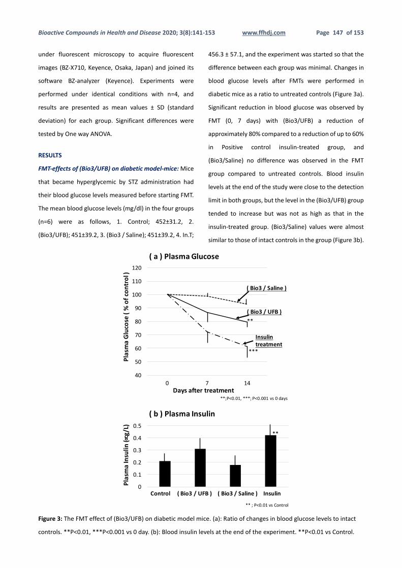
FMT-effects of (Bio3/UFB) on diabetic model-mice: Mice
that became hyperglycemic by STZ administration had
their blood glucose levels measured before starting FMT.
The mean blood glucose levels (mg/dl) in the four groups
(n=6) were as follows, 1. Control; 452±31.2, 2.
(Bio3/UFB); 451±39.2, 3. (Bio3 / Saline); 451±39.2, 4. In.T;
456.3 ± 57.1, and the experiment was started so that the
difference between each group was minimal. Changes in
blood glucose levels after FMTs were performed in
diabetic mice as a ratio to untreated controls (Figure 3a).
Significant reduction in blood glucose was observed by
FMT (0, 7 days) with (Bio3/UFB) a reduction of
approximately 80% compared to a reduction of up to 60%
in Positive control insulin-treated group, and
(Bio3/Saline) no difference was observed in the FMT
group compared to untreated controls. Blood insulin
levels at the end of the study were close to the detection
limit in both groups, but the level in the (Bio3/UFB) group
tended to increase but was not as high as that in the
insulin-treated group. (Bio3/Saline) values were almost
similar to those of intact controls in the group (Figure 3b).
Figure 3: The FMT effect of (Bio3/UFB) on diabetic model mice. (a): Ratio of changes in blood glucose levels to intact
controls. **P<0.01, ***P<0.001 vs 0 day. (b): Blood insulin levels at the end of the experiment. **P<0.01 vs Control.
0
0.1
0.2
0.3
0.4
0.5
(b)PlasmaInsulin
40
50
60
70
80
90
100
110
120
0 7 14
(a)PlasmaGlucose
Plasm
aGlucose(%ofcontrol)
Plasm
aInsulin(µg/L)
(Bio3/Saline)
(Bio3/UFB)
Insulintreatment
Control(Bio3/UFB)(Bio3/Saline)Insulin
Daysaftertreatment
Figure3.
**
**;P<0.01vsControl
**
***
**;P<0.01,***;P<0.001vs0days
Bioactive Compounds in Health and Disease 2020; 3(8):141-153 www.ffhdj.com Page 148 of 153
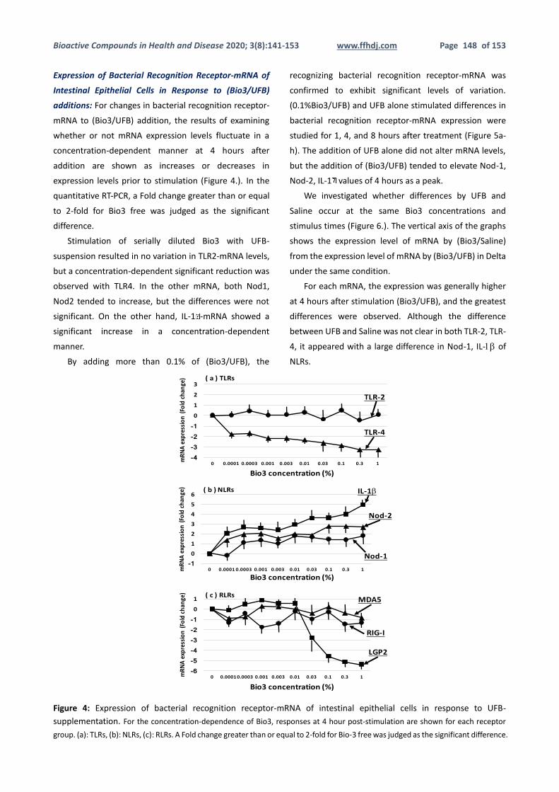
Expression of Bacterial Recognition Receptor-mRNA of
Intestinal Epithelial Cells in Response to (Bio3/UFB)
additions: For changes in bacterial recognition receptor-
mRNA to (Bio3/UFB) addition, the results of examining
whether or not mRNA expression levels fluctuate in a
concentration-dependent manner at 4 hours after
addition are shown as increases or decreases in
expression levels prior to stimulation (Figure 4.). In the
quantitative RT-PCR, a Fold change greater than or equal
to 2-fold for Bio3 free was judged as the significant
difference.
Stimulation of serially diluted Bio3 with UFB-
suspension resulted in no variation in TLR2-mRNA levels,
but a concentration-dependent significant reduction was
observed with TLR4. In the other mRNA, both Nod1,
Nod2 tended to increase, but the differences were not
significant. On the other hand, IL-1 -mRNA showed a
significant increase in a concentration-dependent
manner.
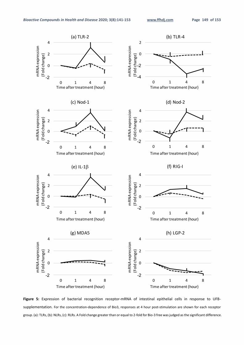
By adding more than 0.1% of (Bio3/UFB), the
recognizing bacterial recognition receptor-mRNA was
confirmed to exhibit significant levels of variation.
(0.1%Bio3/UFB) and UFB alone stimulated differences in
bacterial recognition receptor-mRNA expression were
studied for 1, 4, and 8 hours after treatment (Figure 5a-
h). The addition of UFB alone did not alter mRNA levels,
but the addition of (Bio3/UFB) tended to elevate Nod-1,
Nod-2, IL-1 values of 4 hours as a peak.
We investigated whether differences by UFB and
Saline occur at the same Bio3 concentrations and
stimulus times (Figure 6.). The vertical axis of the graphs
shows the expression level of mRNA by (Bio3/Saline)
from the expression level of mRNA by (Bio3/UFB) in Delta
under the same condition.
For each mRNA, the expression was generally higher
at 4 hours after stimulation (Bio3/UFB), and the greatest
differences were observed. Although the difference
between UFB and Saline was not clear in both TLR-2, TLR-
4, it appeared with a large difference in Nod-1, IL- of
NLRs.
Figure 4: Expression of bacterial recognition receptor-mRNA of intestinal epithelial cells in response to UFB-
supplementation. For the concentration-dependence of Bio3, responses at 4 hour post-stimulation are shown for each receptor
group. (a): TLRs, (b): NLRs, (c): RLRs. A Fold change greater than or equal to 2-fold for Bio-3 free was judged as the significant difference.
Bioactive Compounds in Health and Disease 2020; 3(8):141-153 www.ffhdj.com Page 149 of 153
Figure 5: Expression of bacterial recognition receptor-mRNA of intestinal epithelial cells in response to UFB-
supplementation. For the concentration-dependence of Bio3, responses at 4 hour post-stimulation are shown for each receptor
group. (a): TLRs, (b): NLRs, (c): RLRs. A Fold change greater than or equal to 2-fold for Bio-3 free was judged as the significant difference.
Bioactive Compounds in Health and Disease 2020; 3(8):141-153 www.ffhdj.com Page 150 of 153
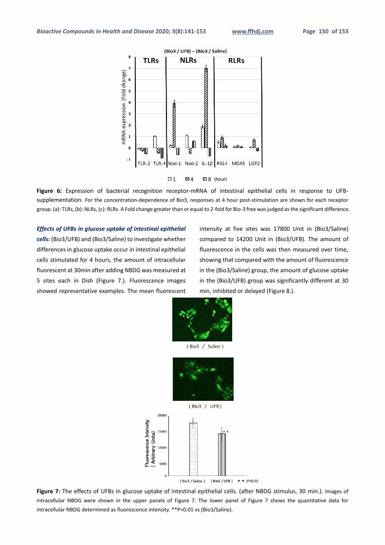
Figure 6: Expression of bacterial recognition receptor-mRNA of intestinal epithelial cells in response to UFB-
supplementation. For the concentration-dependence of Bio3, responses at 4 hour post-stimulation are shown for each receptor
group. (a): TLRs, (b): NLRs, (c): RLRs. A Fold change greater than or equal to 2-fold for Bio-3 free was judged as the significant difference.
Effects of UFBs in glucose uptake of intestinal epithelial
cells: (Bio3/UFB) and (Bio3/Saline) to investigate whether
differences in glucose uptake occur in intestinal epithelial
cells stimulated for 4 hours, the amount of intracellular
fluorescent at 30min after adding NBDG was measured at
5 sites each in Dish (Figure 7.). Fluorescence images
showed representative examples. The mean fluorescent
intensity at five sites was 17800 Unit in (Bio3/Saline)
compared to 14200 Unit in (Bio3/UFB). The amount of
fluorescence in the cells was then measured over time,
showing that compared with the amount of fluorescence
in the (Bio3/Saline) group, the amount of glucose uptake
in the (Bio3/UFB) group was significantly different at 30
min, inhibited or delayed (Figure 8.).
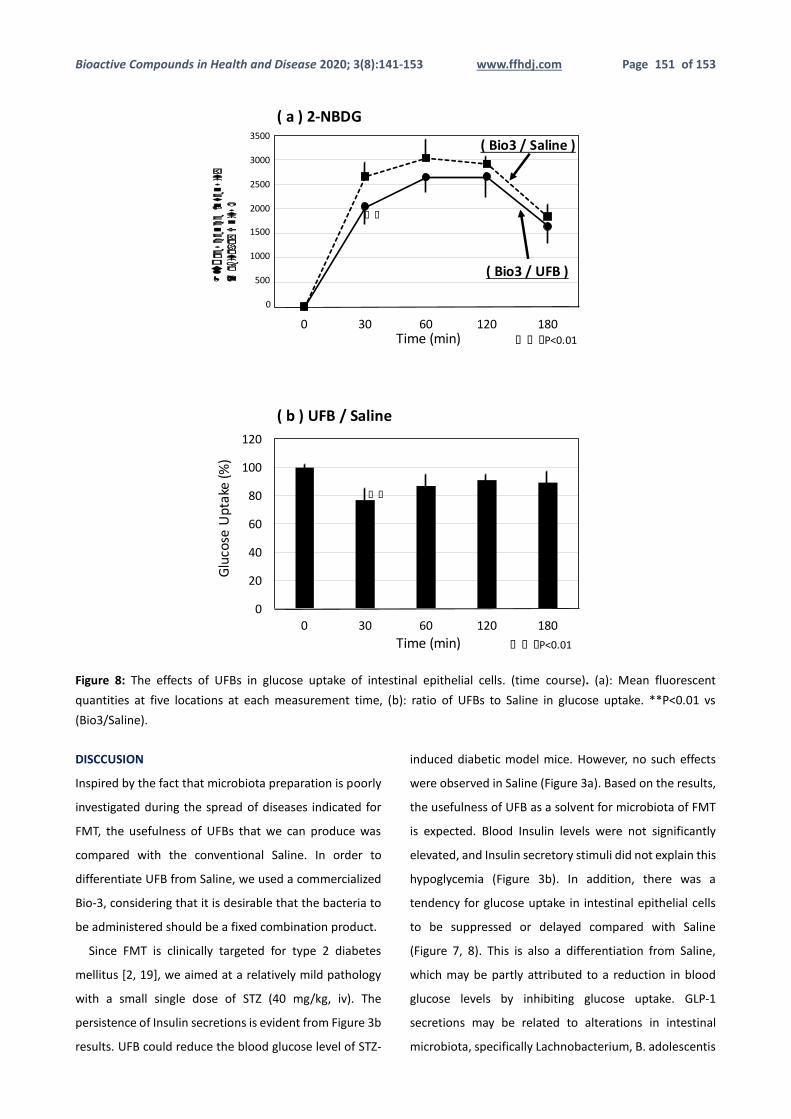
Figure 7: The effects of UFBs in glucose uptake of intestinal epithelial cells. (after NBDG stimulus, 30 min.). Images of
intracellular NBDG were shown in the upper panels of Figure 7. The lower panel of Figure 7 shows the quantitative data for
intracellular NBDG determined as fluorescence intensity. **P<0.01 vs (Bio3/Saline).
Bioactive Compounds in Health and Disease 2020; 3(8):141-153 www.ffhdj.com Page 151 of 153
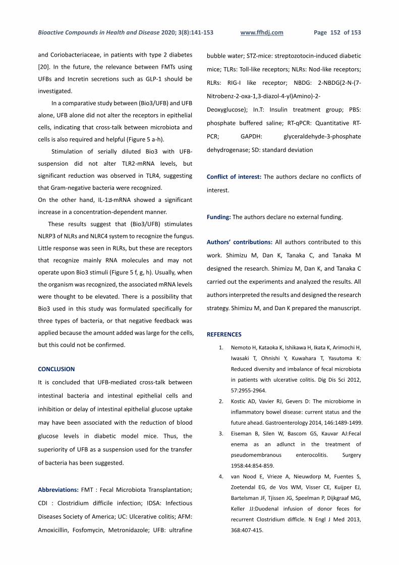
Figure 8: The effects of UFBs in glucose uptake of intestinal epithelial cells. (time course). (a): Mean fluorescent
quantities at five locations at each measurement time, (b): ratio of UFBs to Saline in glucose uptake. **P<0.01 vs
(Bio3/Saline).
DISCCUSION
Inspired by the fact that microbiota preparation is poorly
investigated during the spread of diseases indicated for
FMT, the usefulness of UFBs that we can produce was
compared with the conventional Saline. In order to
differentiate UFB from Saline, we used a commercialized
Bio-3, considering that it is desirable that the bacteria to
be administered should be a fixed combination product.
Since FMT is clinically targeted for type 2 diabetes
mellitus [2, 19], we aimed at a relatively mild pathology
with a small single dose of STZ (40 mg/kg, iv). The
persistence of Insulin secretions is evident from Figure 3b
results. UFB could reduce the blood glucose level of STZ-
induced diabetic model mice. However, no such effects
were observed in Saline (Figure 3a). Based on the results,
the usefulness of UFB as a solvent for microbiota of FMT
is expected. Blood Insulin levels were not significantly
elevated, and Insulin secretory stimuli did not explain this
hypoglycemia (Figure 3b). In addition, there was a
tendency for glucose uptake in intestinal epithelial cells
to be suppressed or delayed compared with Saline
(Figure 7, 8). This is also a differentiation from Saline,
which may be partly attributed to a reduction in blood
glucose levels by inhibiting glucose uptake. GLP-1
secretions may be related to alterations in intestinal
microbiota, specifically Lachnobacterium, B. adolescentis
0 30 60 120 180
(a)2-NBDG
0
20
40
60
80
100
120
0 30 60 120 180
(b)UFB/Saline
Fluorescence Intensity
(Arbitrary Units)
Glu
cose
Upt
ake
(%)
3500
3000
2500
2000
1500
1000
500
0
* *
Time(min)
Time(min)
* *
* * *P<0.01
* * *P<0.01
(Bio3/Saline)
(Bio3/UFB)
Figure8.
Bioactive Compounds in Health and Disease 2020; 3(8):141-153 www.ffhdj.com Page 152 of 153
and Coriobacteriaceae, in patients with type 2 diabetes
[20]. In the future, the relevance between FMTs using
UFBs and Incretin secretions such as GLP-1 should be
investigated.
In a comparative study between (Bio3/UFB) and UFB
alone, UFB alone did not alter the receptors in epithelial
cells, indicating that cross-talk between microbiota and
cells is also required and helpful (Figure 5 a-h).
Stimulation of serially diluted Bio3 with UFB-
suspension did not alter TLR2-mRNA levels, but
significant reduction was observed in TLR4, suggesting
that Gram-negative bacteria were recognized.
On the other hand, IL-1 -mRNA showed a significant
increase in a concentration-dependent manner.
These results suggest that (Bio3/UFB) stimulates
NLRP3 of NLRs and NLRC4 system to recognize the fungus.
Little response was seen in RLRs, but these are receptors
that recognize mainly RNA molecules and may not
operate upon Bio3 stimuli (Figure 5 f, g, h). Usually, when
the organism was recognized, the associated mRNA levels
were thought to be elevated. There is a possibility that
Bio3 used in this study was formulated specifically for
three types of bacteria, or that negative feedback was
applied because the amount added was large for the cells,
but this could not be confirmed.
CONCLUSION
It is concluded that UFB-mediated cross-talk between
intestinal bacteria and intestinal epithelial cells and
inhibition or delay of intestinal epithelial glucose uptake
may have been associated with the reduction of blood
glucose levels in diabetic model mice. Thus, the
superiority of UFB as a suspension used for the transfer
of bacteria has been suggested.
Abbreviations: FMT : Fecal Microbiota Transplantation;
CDI : Clostridium difficile infection; IDSA: Infectious
Diseases Society of America; UC: Ulcerative colitis; AFM:
Amoxicillin, Fosfomycin, Metronidazole; UFB: ultrafine
bubble water; STZ-mice: streptozotocin-induced diabetic
mice; TLRs: Toll-like receptors; NLRs: Nod-like receptors;
RLRs: RIG-I like receptor; NBDG: 2-NBDG(2-N-(7-
Nitrobenz-2-oxa-1,3-diazol-4-yl)Amino)-2-
Deoxyglucose); In.T: Insulin treatment group; PBS:
phosphate buffered saline; RT-qPCR: Quantitative RT-
PCR; GAPDH: glyceraldehyde-3-phosphate
dehydrogenase; SD: standard deviation
Conflict of interest: The authors declare no conflicts of
interest.
Funding: The authors declare no external funding.
Authors’ contributions: All authors contributed to this
work. Shimizu M, Dan K, Tanaka C, and Tanaka M
designed the research. Shimizu M, Dan K, and Tanaka C
carried out the experiments and analyzed the results. All
authors interpreted the results and designed the research
strategy. Shimizu M, and Dan K prepared the manuscript.
REFERENCES
1. Nemoto H, Kataoka K, Ishikawa H, Ikata K, Arimochi H,
Iwasaki T, Ohnishi Y, Kuwahara T, Yasutoma K:
Reduced diversity and imbalance of fecal microbiota
in patients with ulcerative colitis. Dig Dis Sci 2012,
57:2955-2964.
2. Kostic AD, Vavier RJ, Gevers D: The microbiome in
inflammatory bowel disease: current status and the
future ahead. Gastroenterology 2014, 146:1489-1499.
3. Eiseman B, Silen W, Bascom GS, Kauvar AJ:Fecal
enema as an adlunct in the treatment of
pseudomembranous enterocolitis. Surgery
1958:44:854-859.
4. van Nood E, Vrieze A, Nieuwdorp M, Fuentes S,
Zoetendal EG, de Vos WM, Visser CE, Kuijper EJ,
Bartelsman JF, Tjissen JG, Speelman P, Dijkgraaf MG,
Keller JJ:Duodenal infusion of donor feces for
recurrent Clostridium difficle. N Engl J Med 2013,
368:407-415.
Bioactive Compounds in Health and Disease 2020; 3(8):141-153 www.ffhdj.com Page 153 of 153
5. McDonald LC, Gerding DN, Johnson S, Bakken JS,
Carroll KC, Coffin SE, Dubberke ER, Garey KW, Gould
CV, Kelly C, Loo V, Shaklee Sammons J, Sandora TJ,
Wilcox MH: Clinical Practice Guidelines for
Clostridium difficile Infection in Adults and Children:
2017 Update by the Infectious Diseases Society of
America (IDSA) and Society for Healthcare
Epidemiology of America (SHEA) . Clin Infect Dis. 2018,
19;66(7):e1-e48.
6. Ohkusa T, Kato K, Terao S, Chiba T, Mabe K, Murakami
K, Mizokami Y, Sugiyama T, Yanaka A, Takeuchi Y,
Yamato S, Yokoyama T, Okayasu I, Watanabe S, Tajiri H,
Sato N; Japan UC Antibiotic Therapy Study Group:
Newly developed antibiotic combination therapy for
ulcerative colitis: a double-blind placebo-controlled
multicenter trial. Am J Gastroenterol 2010, 105:1820-
1829.
7. Ishikawa D, Sasaki T, Osada T, Kuwahara-Arai K, Haga
K, Shibuya T, Hiramatsu K, Watanabe S: Changes in
intestinal microbiota following combination therapy
with fecal microbial transplantation and antibiotics
for ulcerative colitis. Inflamm Bowel Dis 2017, 23:
116-125.
8. BIO-THREE H Powder TOA BIOPHARMA CO., LTD.
[http://www.toabio.co.jp/cms/toa/forlign/item_inde
x_e.html]
1. 9. Imler J L, Hoffmann J A: Toll receptors in innate
immunity. Trends Cell Biol 2001, 11(7):304-311.
9. Kawai T, Akira S: Toll-like receptors and their
crosstalk with other innate receptors in infection and
immunity. Immunity 2011, 34(5):637-650.
10. Ting J P-Y, Lovering R C, Alnemri E S, Bertin J, Boss J M,
Davis B K, Flavell R A, Girardin S E, Godzik A, Harton J
A, Hoffman H M, Hugot J P, Inohara N, Mackenzie A,
Maltais L J, Nunez G, Ogura Y, Otten L A, Philpott D,
Reed J C, Reith W, Schreiber S, Steimle V, Ward P A:
The NLR gene family: a standard nomenclature 2008,
28(3): 285-287.
11. Inohara N, Chamaillard M, McDonald C, Nuñez G:
NOD-LRR proteins: role in host-microbial interactions
and inflammatory disease. Annu Rev Biochem 2005,
74:355-383.
12. Yoneyama M, Fujita T: RNA recognition and signal
transduction by RIG-I-like receptors. 2009, 227(1): 54-
65.
13. Badi S A, Khatami S, Irani S, Siadat S D: Induction
effects of bacteroides fragilis derived outer
membrane vesicles on toll like receptor 2, Toll like
receptor 4 genes expression and cytokines
concentration in human intestinal epithelial cells. Cell
J 2018, 21(1): 57-61.
14. Kim J G, Lee S J, Kagnoff M F: Nod1 is an essential
signal transducer in intestinal epithelial cells infected
with bacteria that avoid recognition by toll-like
receptors. Infect Immun 2004, 72(3): 1487-1495.
15. Xu Y W, Xing R X, Zhang W H, Li L, Wu Y, Hu J, Wang C,
Luo Q L, Shen J L, Chen X: Toxoplasma ROP16I/III
ameliorated inflammatory bowel diseases via
inducing M2 phenotype of macrophages. World J
Gastroenterol 2019, 25(45): 6634-6652.
16. Ohta K, Fukui A, Shigeishi H, Ishida Y, Nishi H, Tobiume
K, Takechi M, Kamata N: Expression and function of
RIG-I in oral keratinocytes and fibroblasts. Cell Physiol
Biochem 2014, 34:1556-1565.
17. Mojica L, Luna-Vital D A, Mejia E G: Black bean
peptides inhibit glucose uptake in CaCO-2
asenocarcinoma cells by blocking the expression and
translocation pathway of glucose transporters.
Toxicology Reports 2018, 5:552-560.
18. Kang Y, Cai Y: Gut microbiota and obesity: implications
for fecal microbiota transplantation therapy.
Hormones 2017: 16(3): 223-234.
19. Cornejo-Pareja I, Martín-Núñez GM, Roca-Rodríguez
MM, Cardona F, Coin-Aragüez L, Sánchez-Alcoholado
L, Gutiérrez-Repiso C, Muñoz-Garach A, Fernández-
García JC, Moreno-Indias I, Tinahones FJ: H. pylori
Eradication Treatment Alters Gut Microbiota and GLP-
1 Secretion in Humans. J Clin Med 2019, 8(4): 451-467.