Embed Size (px)
Citation preview
Hearing ImpairmentDr Ahmad Alamadi FRCS
Consultant Otolaryngologist
Al Baraha Hospital
Objectives Basic anatomy Types of hearing losses History & Examination Etiology Investigations Management
Introduction
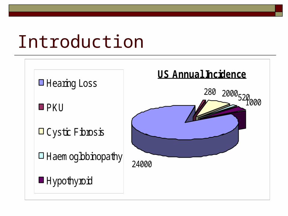
US Annual Incidence
280 20005201000
24000
Hearing Loss
PKU
Cystic Fibrosis
Haemoglobinopathy
Hypothyroid
IntroductionEffects of Hearing Impairment on Development
Has life long effect on Language Delay Literacy Educational achievement Vocational Opportunities Academic difficulties Psychosocial adjustments and/or difficulties
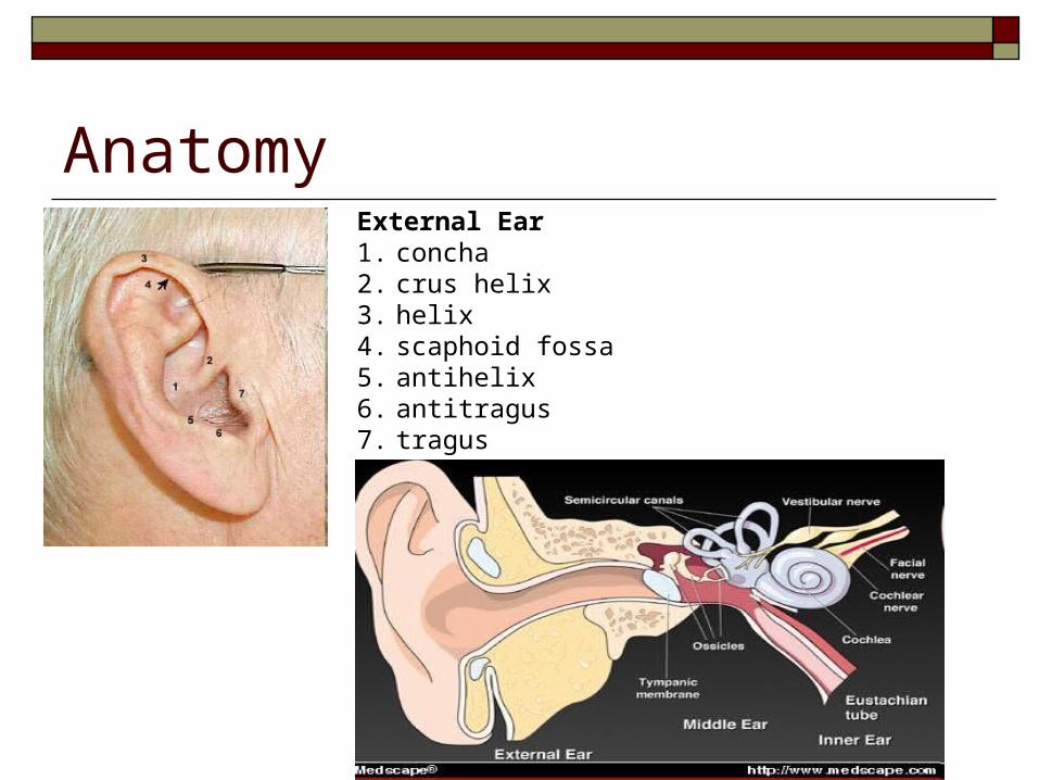
AnatomyExternal Ear 1. concha 2. crus helix 3. helix 4. scaphoid fossa 5. antihelix 6. antitragus 7. tragus lobule not labeled
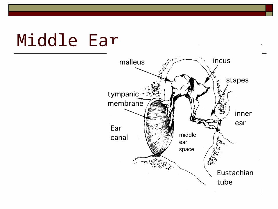
Middle Ear
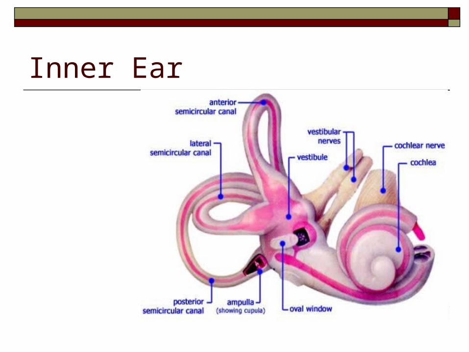
Inner Ear
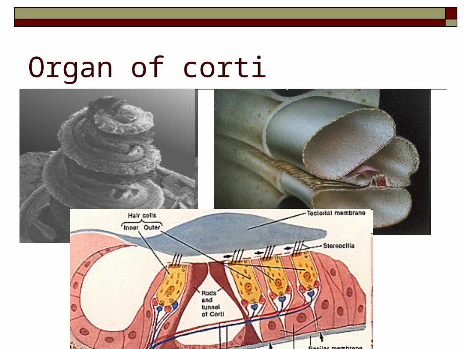
Organ of corti
Pathophysiology
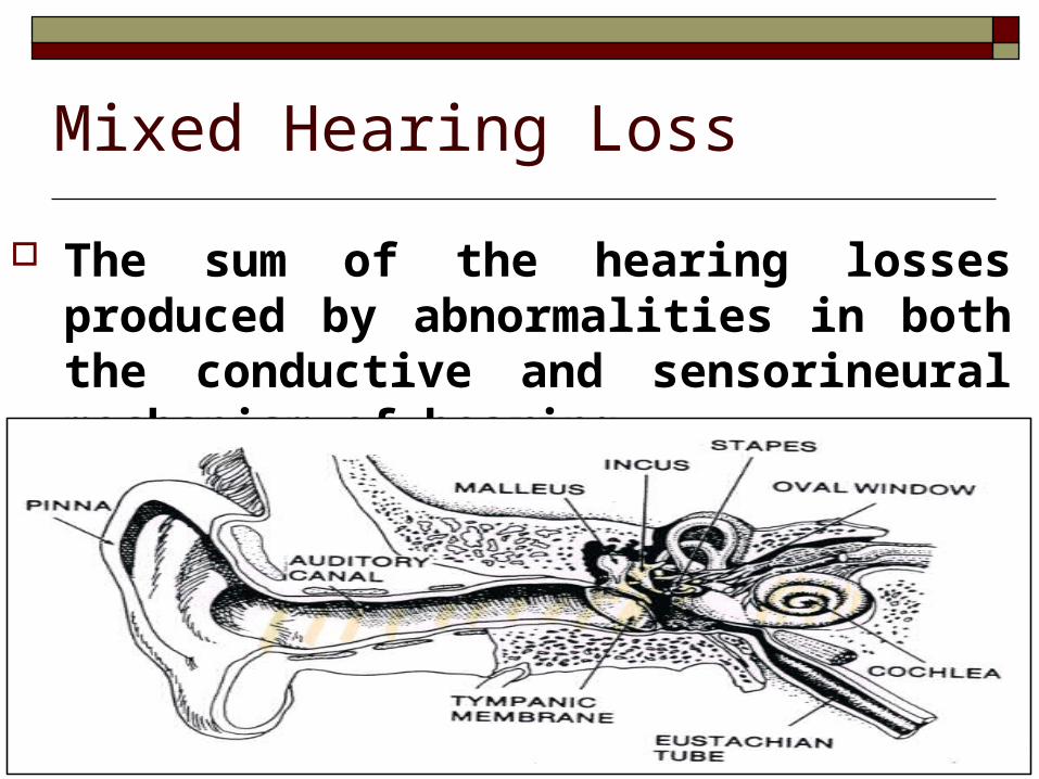
Sound waves to the auricle through the external auditory canal to the tympanic membrane. When they strike the tympanic membrane, the waves cause it to vibrate, setting off a chain of vibrations along the ossicles (malleus, incus, and stapes) to the membrane of the oval window at the entrance to the cochlea. This process amplifies the environment sound by approximately 20-fold.
The cochlea is the end organ of hearing and is shaped like a snail shell with 2.5 turns. Inside, 2 membranes longitudinally divide the cochlea into 3 sections: the scala tympani, the scala vestibuli, and the scala media. All 3 are filled with fluid of various ion concentrations (similar to intracellular and extracellular constituents).
Along one of the membranes in the scala media, or cochlear duct, lie the internal and external hair cells. Movement of the stapes on the oval window creates a wave or vibration in the perilymph fluid of the cochlea. This fluid movement, which opens ion channels in the hair cells, displaces the hair cells, triggering an action potential and causing a nerve in the cochlea to fire to the brain.
Thousands of nerves representing more than 20,000 frequencies are located along the length of the cochlea; these nerves account for the hearing range. The microscopic nerves culminate in the cochlear portion of the eighth cranial nerve. The location of the vibration in the cochlea is correlated with the frequency of the original pitch. Low-frequency sounds are near the apex, and high-frequency sounds are near the base.
Types of Hearing Losses
Conductive hearing loss Sensori-neural hearing loss Mixed hearing loss Central Auditory disorders or Neural hearing
loss
Conductive Hearing Loss
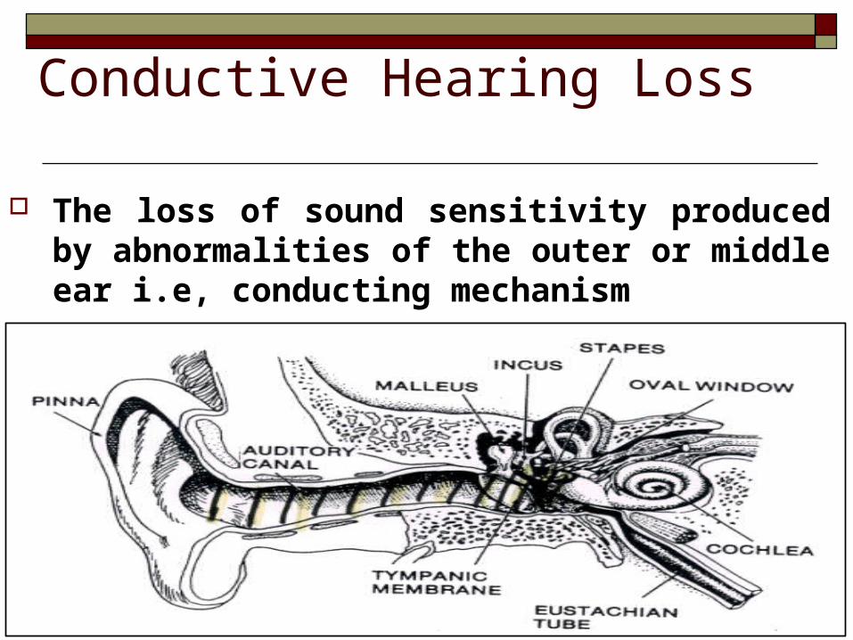
The loss of sound sensitivity produced by abnormalities of the outer or middle ear i.e, conducting mechanism
Sensori-Neural Hearing Loss
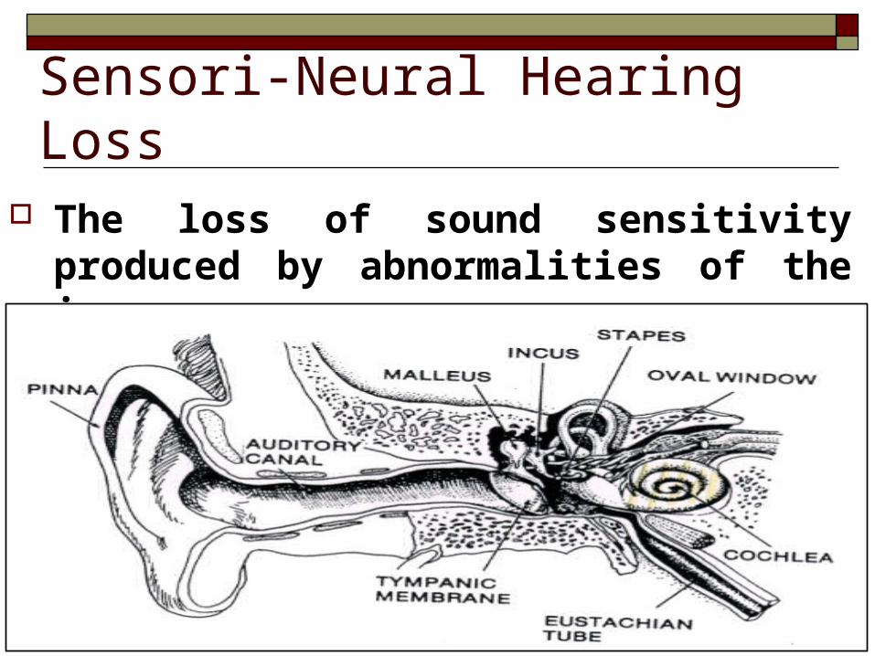
The loss of sound sensitivity produced by abnormalities of the inner ear.
Mixed Hearing Loss
The sum of the hearing losses produced by abnormalities in both the conductive and sensorineural mechanism of hearing.
Central OR Neural Hearing Loss
The loss or impairment in processing of information produced by abnormality in neural system i.e, Nerve fiber or central auditory cortex.
Clinical history Hearing loss
Age of onset Severity Risk factors
Family history of congenital or early SNHL Congenital infection known to be associated with SNHL Craniofacial anomalies Birth weight of less than 1500 g (<3.3 lb) Hyperbilirubinemia over the exchange level Exposure to ototoxic medications Bacterial meningitis Low Apgar scores at birth Prolonged mechanical ventilation Findings of a syndrome associated with SNHL
Syndromic Deafness DiGeorge sequelae CHLYesBranchio-oto-facial syndrome Townes-Brocks syndrome Miller syndrome Bixler syndrome CHARGE syndrome Jervell Lange-Nielson Limb-oto-cardiac syndrome Alport syndrome Branchio-oto-renal syndrome Kearns-Sayre syndrome Epstein syndrome Barakat syndrome Noonan syndrome Killian/Teschler-Nicola syndrome
Clinical history Hearing Impairment Pain Discharge Tinnitus Vertigo
Examination Otoscopy Nose PNS Facial nerve Free field test Tuning fork tests
Weber Test Rinne Test
Investigations Lab Studies
TFT, BUN, CR ECG ESR, Rhf, ANA Connexin 26
Imaging Studies Tests for Hearing Loss
PTA ABR ASSR OAE
Etiology Genetic causes connexion 26 Syndromic associations Congenital infections
cytomegaloviral herpes Rubella Syphilis Toxoplasmosis varicella
Postnatal Prematurity low birth weight anoxia hyperbilirubinemia sepsis
meningitis mumps ototoxic medications
aminoglycosides furosemide
major head injury
Management Medical: antibiotics
Surgical: Ventilation tubes, reconstruction of middle ear
Amplification Technology: Analog & digital hearing aids + Speech and language therapy.
Surgical+ Amplification Technology: Cochlear Implants (Severe Sensorineural loss or greater)